
Abstract
Purpose
Improvements of heat-delivery systems have led to hyperthermia (HT) being increasingly recognized as an adjunct treatment modality also for brain tumors. But how HT affects the immune phenotype of glioblastoma cells is only scarcely known.
Materials and Methods
We therefore investigated the effect of in vitro HT, radiotherapy (RT), and the combination of both (RHT) on cell death modalities, immune checkpoint molecule (ICM) expression and release of the danger signal HSP70 of two human glioblastoma cell lines (U87 and U251) by using multicolor flow cytometry and ELISA. Hyperthermia was performed once or twice for 60-minute sessions reaching temperatures of 39 °C, 41 °C, and 44 °C, respectively. RT was administered with 5 x 2 Gy.
Results
A hyperthermia chamber for cell culture t-flasks regulating the temperature via a contact sensor was developed. While the glioblastoma cells were rather radioresistant, particularly in U251 cells, the combination of RT with HT significantly increased the percentage of apoptotic and necrotic cells for all temperatures examined and for both, single and double HT application. In line with that, an increased release of HSP 70 was seen only in U251 cells, mainly following treatment with HT at temperatures of 44 °C alone or in combination with RT. In contrast, immune suppressive (PD-L1, PD-L2, HVEM) and immune stimulatory (ICOS-L, CD137-L and Ox40-L) ICMs were significantly increased mostly on U87 cells, and particularly after RHT with 41 °C.
Conclusions
Individual assessment of the glioblastoma immune cell phenotype with regard to the planned treatment is mandatory to optimize multimodal radio-immunotherapy protocols including HT.
1. Introduction
The most common and most aggressive primary malignant brain tumor is glioblastoma (GBM), which accounts for nearly 60% of brain tumors in adults [Citation1]. Despite advances in glioblastoma therapy, which consists of surgery followed by radiotherapy and concomitant chemotherapy [Citation2,Citation3], the overall prognosis remains poor, with a 1-year survival rate of 71.0% and a 5-year survival rate of 10.0% [Citation4,Citation5].
To prolong survival and kill tumor cells of this very diffuse infiltrating brain tumor, radiotherapy after surgical tumor resection is regarded as standard. It is usually administered to a total dose of 60 Gy with 2 Gy per fraction over a 6-week period, in combination with temozolomide [Citation3,Citation6]. Other irradiation protocols have been studied, but no beneficial outcomes for total doses higher than 60 Gy have been found [Citation7]. Rather, the risk of radiation necrosis in combination with chemotherapy has to be considered for healthy tissue. The current standard of care for GBM patients consists of concomitant administration of temozolomide at a dose of 75 mg/m2 during RT, followed by another 6 cycles of temozolomide (150–200 mg/m2 on days 1–5 every 28 days). Nausea, thrombocytopenia, and neutropenia are considered to be the most common side effects associated with temozolomide during adjuvant therapy [Citation3,Citation8–10].
Challenges to the development of optimized therapies arise from the unique tumor and immune microenvironment of glioblastoma and the blood-brain barrier [Citation11]. As immunotherapy has transformed the treatment of many solid tumors, extensive studies and research have been conducted on immune-based therapeutic approaches for glioblastoma. Key focus has been set on immune checkpoint inhibitors (ICIs) that are antibodies that bind immune checkpoint molecules (ICMs) on tumor or immune cells and thereby restore or activate T cell-mediated anti-tumor immune responses. Besides inhibition of the prominent programmed cell death protein 1 (PD-1)/PD-L1 and cytotoxic T lymphocyte antigen (CTLA)-4 axis, further immune suppressive and immune stimulatory ICMs play a role also in the human glioma microenvironment [Citation12]. This might be one reason why reported randomized phase 2 and 3 clinical trials of ICIs, as single therapy or in combination with standard therapies such as RT and CT, have not demonstrated a survival benefit to date in either newly diagnosed or recurrent glioblastoma [Citation13]. Another prominent immune suppressive ICM, the herpes virus entry mediator (HVEM), was just recently discovered to correlate with poor prognosis when being overexpressed in human glioblastoma [Citation14]. That`s why we focused particularly on the analyses how the treatment of glioblastoma cells with RT and hyperthermia (see below) affects the expression of the prominent immune suppressive ICMs of the PD-1 and HVEM axis.
Nevertheless, further ICMs might contribute to the immune microenvironment in glioblastoma and are currently to be identified, as these of the immune stimulatory axis. Here the inducible T cell co-stimulator (ICOS) and 4-1BB (CD137) as an activation-induced costimulatory molecule that is expressed on activated immune cells were just recently identified by machine learning methods of immunotherapy targets in gliomas [Citation15]. Therefore, we included the expression analyses of ICOS-L and CD137-L on glioblastoma cells in our study as well. Jahan et al. recently demonstrated in a preclinical model system that combination of vaccination with PD-1 inhibition and agonist anti-Ox40 therapy is effective against murine glioma [Citation16]. Seyfrid and colleagues showed for the first time that another stimulatory ICM, namely CD70, is elevated in recurrent glioblastoma and that CD70 knockdown reduced the immunogenicity of the tumor [Citation17]. However, knowledge about the expression of the latter-named ICMs during and following standard treatment of glioblastoma is scarce.
There is undoubted evidence that specific cancer-related immunosuppressive mechanisms play a central role in glioblastoma [Citation18]. To address these manifold features of GBM, other and additional treatment modalities need to be explored to find the best multimodal therapy tailored to the patient. Here, knowledge about the expression of ICMs is important to optimize multimodal glioblastoma therapies including the targeting of novel ICMs.
Historically, different forms of hyperthermia (HT) were first studied in the 18th to 19th centuries in attempts to cure cancer [Citation19,Citation20]. Hyperthermia describes a form of treatment in which the temperature in local parts of the body is raised above the physiological range to produce therapeutic effects [Citation21]. Increasing local temperature from 40 °C to 44 °C has been described to reduce cancer growth [Citation22,Citation23]. Additionally, positive immunobiological interactions of HT with RT or chemotherapy have been reported [Citation24–27].
In randomized controlled trials comparing radiotherapy with and without hyperthermia in the treatment of cervical cancer, it was described that additional HT application was able to significantly improve 12-year overall survival rate from 20% to 37% [Citation28]. In non-muscle invasive bladder cancer the disease-free 10-year survival rate could be significantly improved from 15% to 53% [Citation29]. The clinical application of hyperthermia in glioblastoma [Citation30–32] has also been able to show first promising results. Thus, for glioblastoma, a 2-year survival rate of 31% was achieved in the treatment arm employing hyperthermia, twice as much as when radiotherapy was used on its own without additional HT [Citation33]. Although there are recent studies demonstrating hyperthermia as a promising adjunct to conventional brain tumor therapy, large-scale randomized controlled trials are still lacking, but should be possible in the near future due to improvements in heat-delivery applicators and nanoparticle-based HT techniques [Citation30,Citation34,Citation35]. Therefore, preclinical research is needed to get first hints about the immune phenotype of glioblastoma cells after exposure to HT, RT and RHT, to elucidate multimodal tumor therapies for glioblastoma in the future with additional combination of distinct ICM antagonist or agnostic antibodies.
2. Materials and methods
2.1. Cell lines and cultivation
Two human glioblastoma cell lines, cultured at 37 °C, 5% CO2, and 90% humidity under sterile conditions, were used in the experiments. U251 and U87 were grown in DMEM medium (PAN-Biotech GmbH, Aidenbach, Germany) supplemented with 10% fetal bovine serum (FBS; Biochrome AG, Berlin, Germany) and 100 U/mL PenStrep (100 U/mL penicillin and 100 µg/mL streptomycin).
2.2. Treatments and sampling
For the experiments, cells were seeded in 75 cm2 t-flasks one day before each treatment and cultured under standard conditions. On day 0, hyperthermia treatment was performed for 60 min at 39 °C, 41 °C, and 44 °C (d0 to 60′) () and after 60 min, portions of the samples were evaluated, or irradiation was started. Irradiation in the RT arm (RT) and the combined RHT arm was performed with normofractionation with a single dose per fraction of 2 Gy of X-rays (120 kV, 12.2 mA for 0.5 min) for 5 days using an X-ray system (Booth: Seifert; Generator: Isovolt Titan series - GE Technologies; Hürth; Germany) and in 75 cm2 t-flasks. According to the clinical hyperthermia guidelines, the time interval between RT and HT must be at least 1 h and at most 4 h [Citation7]. In our experimental setup, RT was performed 1 h after HT. As studies have shown, the tumor thermal enhancement ratio (TER) is almost the same when HT is applied before or after RT, if the above time interval is maintained [Citation36]. The experimental setup is shown in .
Figure 1. Experimental treatment set-up for analyses of cell death forms and immune checkpoint molecule expression and the release of the danger signal HSP70 of glioblastoma cells following RT and/or HT treatments. The glioblastoma cells were seeded one day prior to the treatment (d-1d). The experiment consists of a HT arm, a RT arm and a combination arm of HT and RT (RHT). For effective heating, the 75 cm2 t-flasks were placed in the heat chamber for 60 min. Irradiation was performed over 5 days with 2 Gy each and took place in the RHT arm 60 min after hyperthermia. Both RT and RHT samples were always irradiated at the same time. Standard sampling was performed in all arms on day 0 (d0), d3 (72 h) and d5 (120 h).
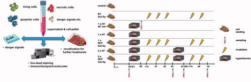
Cells were seeded one day before treatment (d-1d). The experiment consists of a HT arm, a RT arm, and a combination HT and RT (RHT) arm. For effective heating, the 75 cm2 t-flasks were placed in the heating chamber for 60 min (). Heating was performed at 39 °C, 41 °C, and 44 °C and the heating chamber was placed in the incubator at 5% CO2, and 90% humidity under sterile conditions. Irradiation was performed over 5 days at 2 Gy each and occurred 60 min after hyperthermia in the RHT arm. RT and RHT samples were always irradiated at the same time. Standard samples were collected in all treatment arms at day 0 (d0), d3 (72 h), and d5 (120 h) for further analysis.
Figure 2. Development of a heating chamber with accurate temperature control. By combining experimental measurements and simulation, a very accurate basis of heat application in the preclinical in vitro hyperthermia setting is assured: Design drawing of the heating chamber (a), simulation model within COMSOL® Multiphysics (b) and simulated temperature profile to achieve Ttarget of 39 °C (blue line), 41 °C (green line) and 44 °C (red line) starting from Tbase of 37 °C (c). The average temperature may deviate from the set temperature by a maximum of ± 0.1 °C and the heating power is regulated by a control unit itself which is connected to a thermocouple on the heating plate.
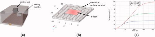
Since temperature monitoring in particular is a challenge and no commercial hyperthermia chambers are available, a hyperthermia chamber for culture t-flasks (up to 175 cm2) was developed and characterized based on simulations and validation experiments ().
2.3. Cell death detection by AnnexinV/PI staining
Multicolor flow cytometry (CytoFLEX S, Beckman Coulter, Fullerton, CA, USA) of cells stained with Annexin V/propidium iodide (PI) was used to analyze the different forms of cell death after RT and/or HT treatment. Staining of cells with propidium-iodide (PI) allows to identify necrotic cells, since PI penetrates only into cells that have lost their membrane integrity. By co-staining with FITC-conjugated AnnexinV as described in [Citation37], apoptotic cells that undergo controlled cell death can be differentiated from primary and secondary necrotic as they expose phosphatidylserine (PS) on the outer membrane leaflet but still show an intact membrane. 105 cells per 96-well were resuspended in Ringer’s solution (B. Braun, Melsungen, Germany) containing 1.0 µg/mL PI (Sigma Aldrich, Munich, Germany) and 0.5 µg/mL FITC-labeled AnnexinV (Geneart, life technologies, Regensburg, Germany). The gating strategy is shown in Suppl. Figure 1.
2.4. Detection of heat shock protein 70 (HSP70) by ELISA
A Sandwich DuoSet® IC ELISA kit (R&D Systems, DYC 1663-5; Minneapolis; MN, USA) was used to analyze the amount of heat shock protein 70 (HSP70) in tumor cell supernatant as described in the manufacturer's instructions. After each treatment, the cell culture supernatant for HSP70 ELISA was centrifuged at 300 × g for 5 min at room temperature and stored at −80 °C. The cell culture medium was added to the t-flasks at the beginning of each experiment and was neither refilled nor diluted or supplemented. Therefore, all samples for the HSP70 ELISA are to be considered with identical initial conditions.
2.5. Detection of immune checkpoint molecule and EGFR expression by multicolor flow cytometry
To quantify ICM expression, 1 × 10⁵ glioblastoma cells per 96-well were incubated with 100 µl of the antibody staining solution () for 30 min in the dark at 4 °C after harvesting. Zombie NIR was added at a concentration of 0.1 µl/well to distinguish viable from dead cells. Control samples were only stained with Zombie NIR. The mean fluorescence intensity (MFI) of the Zombie stained samples was subtracted from the fully stained samples.
Table 1. Antibodies used for the analyses of the surface expression of immune checkpoint molecules and of EGFR by multicolor flow cytometry [Citation27].
The gating strategy and fluorescence detection are shown in Suppl. Figure 2. Analysis was performed by multicolor flow cytometry (CytoFLEX S, Beckman Coulter, Fullerton, CA, USA), and data are presented as normalized expression: Change in mean fluorescence intensity compared with mock-treated cells.
2.6. Statistical analysis
Prism 7 and 8 software (Graph Pad; San Diego, CA, USA) was used to generate the graphs and statistics. A Kruskal-Wallis test was used for the analysis. Results were considered statistically significant at * p < 0.05, ** p < 0.01, *** p < 0.001 and **** p < 0.0001. Each experiment was performed at least four times in independent experiments, each measured in duplicate.
3. Results
3.1. Design and validation of a preclinical HT chamber
Since temperature monitoring during the experiment is essential and since hyperthermia chambers for preclinical examinations are not commercially available, a hyperthermia chamber for cell culture t-flasks (up to 175 cm2) was first developed and characterized, based on simulations and validation experiments. shows a design drawing of the heating chamber, shows the simulation model in COMSOL® Multiphysics of the heating plate including the specific ohmic resistance wire, and a dummy t-flask. As an example, shows the simulated heating process from 37 °C to Ttarget of 39 °C, 41 °C or 44 °C at a constant heating power of 50 W until stationary conditions are reached in the dummy t-flask (red box). In practice, the hyperthermia chamber is already preheated to the target temperature (TTarget) before the experiment. Both in practice and in the simulations, the average temperature may deviate from the set temperature by a maximum of ± 0.1 °C and the heating power is regulated by a control unit itself which is connected to a thermocouple on the heating plate.
To investigate the effect of HT at different temperatures and in combination with RT on cell death and immune phenotype of the glioblastoma cells, the tumor cells were subjected either to just one of the two treatments (HT, RT) or to both in combination (RHT). Specifically, the inactivation efficiency was analyzed by monitoring the percentage of apoptotic and necrotic tumor cells, the immunogenic potential of the treated tumor cells by detecting the immune activating danger signal (HSP70), and the surface expression of various immune-activating and immune suppressive immune checkpoint molecules.
3.2. Cell death induction by hyperthermia and radiotherapy in U87 and U251 glioblastoma cell lines as a function of temperature and number of HT applications
On day 0 (d0) and day 5 (d5) cell death modalities of U87 and U251 cells were examined using AnnexinV/PI staining and multicolor flow cytometry. The tumor cells were treated in the in-house developed heat chamber () at clinically relevant temperatures (39 °C, 41 °C and 44 °C) and the cells were exposed to heat for 60 min either once or twice.
As shown in , the viability of the tumor cells did not significantly decrease 60 min after HT (day 0). It has to be noted that the percentages of apoptotic tumor cells at day 0 is generally low (2–7%) and in the range of general cell culture conditions.
Figure 3. Glioblastoma cells are rather radioresistant, but combination with hyperthermia increases particularly apoptosis and at 44 °C also necrosis in U251 cells. Percentages of apoptotic and necrotic of U87 and U251 glioblastoma cells after normofractionated radiotherapy and hyperthermia treatment at different temperatures and after single (1x) and double (2x) application is displayed. Apoptosis of U87 (a), apoptosis of U251 (b), necrosis of U87 (c) and necrosis of U251 (d) were analyzed by AxV/PI-staining and multicolor flow cytometry measurement 60 min after treatment (d = 0) and at day 5. Standard deviation is derived from at least four independent experiments, each measured in duplicates. Statistics: Kruskal-Wallis test with uncorrected Dunn’s multiple comparison comparing the treatment-related total percentages of apoptotic and necrotic cells to the corresponding controls of untreated cells at the indicated time points (d0, d5); *(p < 0.05), **(p < 0.01), ***(p < 0.001), ****(p < 0.0001).
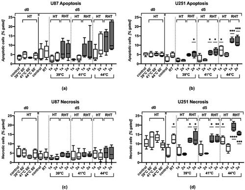
On day 5, HT also failed to induce significant apoptosis or necrosis in both glioblastoma cell lines when temperatures of 39 °C or 41 °C were applied. However, HT with 44 °C resulted in significantly increased percentages of apoptotic and necrotic U251 cells, but only when used once and not when used twice.
The glioblastoma cells were rather radioresistant, as RT did not affect apoptosis and necrosis, with the exception of U251 cells, where a slight but significant increase of necrosis was observed at day 5.
However, again particularly in U251 cells, combination of RT with HT significantly increased the percentages of apoptotic and necrotic cells for all temperatures examined and for both, single and double HT application. In U87 cells, a similar trend was seen, but only regarding apoptosis induction.
3.3. Release of danger signal HSP70 in the supernatant of the tumor cells following hyperthermia and/or radiotherapy
The task of intracellular HSPs is to monitor the correct folding of newly formed proteins and to stabilize the structure of proteins when cells are put under stress (e.g. at high temperatures or by radiotherapy). In contrast, the extracellular heat shock protein HSP70 acts as danger signal and is involved in stimulating immune cells. To investigate the extent of HSP70 release in our experimental setup, the release was quantitatively evaluated by sandwich ELISA from the supernatant of treated and untreated tumor cells, respectively ().
Figure 4. U251 cells that show necrosis induction after combined treatment with RHT and HT with 44 °C also release increased amounts of HSP70 under these conditions. Heatmaps of HSP 70 protein content (ng/106 cells) in supernatant of U87 and U251 tumor cells are shown. After the treatments with hyperthermia (HT), radiotherapy (RT) or combination (RHT), the danger signal HSP 70 was detected in the supernatant of U87 (a) and U251 (b) cells at the indicated time points (day 3 and 5). Additionally, HSP70 was analyzed 60 min after HT on day 0. Note the difference in scaling between the two cell lines: The maximum of total protein content is higher in U251 cells. Standard deviation is derived from at least four independent experiments, each measured in duplicates. Statistics: Kruskal-Wallis test with uncorrected Dunn’s multiple comparison comparing the protein content of the respective treated cells to the corresponding controls of untreated cells at the indicated time points (d0, d5); * (p < 0.05), **(p < 0.01), ***(p < 0.001), ****(p < 0.0001).
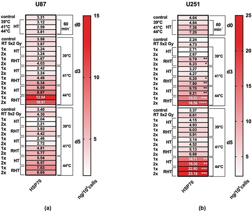
As revealed for cell death induction (), particularly U251 cells showed a significant increase of HSP70 in the cell supernatant. At day 3, HT at 39 °C, 41 °C and 44 °C in combination with fractionated RT and both as single and double application induced a significant release of HSP70, with the highest levels reached after HT at 44 °C. At that temperature, HT alone was sufficient to induce a significantly increased release of HSP70. At day 5, only HT with 44 °C alone and in combination with RT resulted in an increased HSP70 content in the tumor cell supernatant. As for U87 cells, a tendency toward increased HSP70 release can only be seen with 44 °C.
3.4. Impact of hyperthermia and radiotherapy on the expression of immune suppressive and immune stimulating immune checkpoint molecules
With the advent of immune checkpoint inhibitors (ICIs) in cancer treatment in recent years, studies have shown that clinical outcomes of cancer patients have improved significantly [Citation38,Citation39]. However, to date, only a small percentage of patients respond well to the aforementioned anti-PD-1/anti-PD-L1 therapies, so it is of great interest how RT, HT and in particular RHT modulate the expression of PD-L1 and of other immune checkpoint molecules (ICMs), which so far has not been explored in detail and remains largely unknown.
Therefore, the effects of HT, RT, and a combination of both treatment modalities on the expression of prominent ICMs of the immunological synapse in U87 and U251 human glioblastoma cells were investigated comparing expression before and after (day 5) the respective treatment. Immunosuppressive (PD-L1, PD-L2, HVEM), immunostimulatory (CD137-L, Ox40-L, CD70), and immunomodulatory (ICOS-L) checkpoint molecules as well as epidermal growth factor receptor (EGFR) expression, as an example of a prominent oncogenic factor, were analyzed.
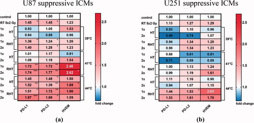
The most striking observation was that in contrast to cell death induction and release of HSP70, U87 but not U251 cells were significantly altered in their ICM expression particularly following RHT (). Expression of both ligands of the PD-1 receptor, namely PD-L1 and PD-L2, was significantly increased on U87 cells after RHT at 41 °C and 44 °C. At 44 °C, HT alone was sufficient to induce an increased expression. A tendency toward an increased expression of another immune suppressive ICM, HVEM was observed after RHT at 41 °C. In the case of U251 cells, a tendency for increased expression of PD-1, PD-L2, and HVME was measured only after RHT at 44 °C. Temperatures below this (39 °C and 41 °C) led to a partial decrease in expression.
Figure 5. Immune suppressive checkpoint molecules were significantly increased mostly on U87 cells, and particularly after RHT with 41 °C and 44 °C. Heatmaps of normalized expression (change in mean fluorescence intensity compared to untreated cells) of immunosuppressive checkpoint molecules (ICMs) on day 5 on the cell surface of U87 (a) and U251 (b) human glioblastoma cells after normofractionated radiotherapy and hyperthermia are shown. Standard deviation is derived from five independent experiments, each measured in duplicates. Statistics: Kruskal-Wallis test with uncorrected Dunn’s multiple comparison; *(p < 0.05), ** (p < 0.01), ***(p < 0.001).
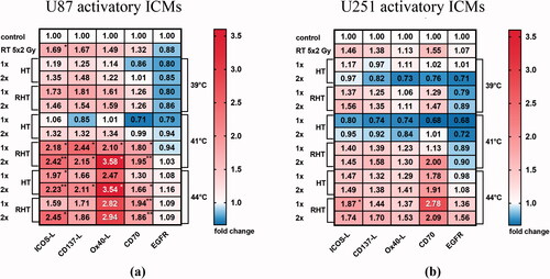
As observed for the immune suppressive ICMs, immune stimulatory ICM expression was also significantly increased mostly on U87 cells, and particularly after RHT at 41 °C (). The highest increase in expression on U87 cells after this treatment was seen for ICOS-L, CD137-L and Ox40-L. Of note, ICOS-L and Ox40-L were significantly increased at 44 °C, particularly after HT alone. On U251 cells, only minor alterations took place which were most pronounced for increased CD70 expression after RHT at 41 °C and 44 °C.
Figure 6. Immune stimulatory checkpoint molecules were significantly increased mostly on U87 cells, and particularly after RHT with 41 °C. Heatmaps of normalized expression (change in mean fluorescence intensity compared to untreated cells) of immune activating checkpoint molecules (ICMs) and of EGFR on day 5 on the cell surface of U87 (a) and U251 (b) human glioblastoma cells after normofractionated radiotherapy and hyperthermia are shown. Standard deviation is derived from five independent experiments, each measured in duplicates. Statistics: Kruskal-Wallis test with uncorrected Dunn’s multiple comparison; *(p < 0.05), ** (p < 0.01), ***(p < 0.001).
The expression of the oncogenic EGFR was not significantly altered in both glioblastoma cell lines following any treatment examined.
4. Discussion
Because of the additional impact of RT on the immune system, the combination of RT with immunotherapies is being explored in more and more clinical trials [Citation40]. The currently established treatment standard for glioblastoma includes no additional immune modulating approaches such as HT and the immunogenic potential of HT has not been studied at a level comparable to that of RT. It of great interest to see how the immunogenicity of glioblastoma cells can be altered. This might result in the development of more efficient combination therapies for glioblastoma, by using HT and ICIs in addition to surgery and RCT [Citation41–44].
The direct cytotoxic effect of hyperthermia is largely mediated by protein aggregation [Citation45]. This in turn leads to stimulated heat-induced synthesis of so-called heat shock proteins (HSPs), which, when released from the cell, act as danger signals for the adaptive and innate immune system [Citation46]. We revealed for the first time that HSP70 can be released by glioblastoma cells after HT with 44 °C alone and at even lower temperatures in combination with RT (). However, this is not a general feature, as U87 cells were seen to respond much less when compared to U251 cells. The latter show necrosis induction after combined treatment with RHT and HT with 44 °C along with release increased amounts of HSP70 under these conditions. It has already been shown, that the U251 and U87 cell lines differ in the expression of proteins that are linked to the regulation of metabolic pathways, e.g. glycolysis, purine metabolism, nicotinamide nucleotide metabolism and RNA splicing [Citation47]. We conclude that these cell lines also differ in their immune phenotype following RHT with 44 °C, with U251 having more immunogenic properties, reflected by HSP70 release and apoptosis and necrosis induction following RHT ( and Citation4). A mixture of apoptosis and necrosis is regarded to be particularly immunogenic [Citation48]. This may prove to be clinically relevant because on days with increased HSP70 expression, there is also a microenvironment through which immune cells may be attracted to the tumor area after irradiation, resulting also in increased activation [Citation49,Citation50].
Alongside extracellular danger signals, ICMs on the tumor cell surface strongly contribute to the immune properties of the tumor, particularly in the effector phase [Citation51]. Research has revealed that RT strongly affects the expression of PD-L1 [Citation52,Citation53,Citation54,Citation55,Citation56], but there is limited information on the expression of other ICMs after RT and/or HT exposure of tumor cells [Citation57]. Our studies show that U 87 cells, in which RHT did not trigger a significant release of HSP70, were much more affected by changes in expression of both, immune suppressive and immune stimulatory ICMs after these treatments ( and Citation6). Future studies employing functional assays, will have to elucidate whether the increased expression of Ox40-L, e.g. counterbalances the increased expression of the PD-1 ligands on U87 cells. We also revealed for the first time that HT at 44 °C as single treatment induces an increased expression of ICOS-L on U87 cells. This has previously only been observed for HPV positive head and neck cancer cells after RT with 5 × 3 Gy, but not after RT with 19.3 Gy [Citation58]. This suggests that the expression including its dynamics of ICMs after cell stressors such as RT and HT are depended on multiple factors such as dose and source of stress application, e.g. heat versus ionizing radiation. Mechanistic studies have to be performed in the future to reveal respective mechanisms. Our data demonstrate that there are strong differences in ICM regulation between different glioblastoma cell lines. This calls for a more individualized screening in future in order to define clinically relevant immune parameters in glioblastoma cells that might guide multimodal therapy including RT, HT and ICIs [Citation42,Citation59].
Finally, our preclinical setting showed that even HT at temperatures in the range of 39 °C can increase HSP70 release and cell death when being combined with RT. However, temperatures of 41 °C and 44 °C are more effective in the combined setting, and HT at 44 °C is effective even when used on its own. Further, whether one or two sessions of HT are used seems to be of minor importance when it comes to the immune phenotype of glioblastoma cells. This is worth knowing, since in different countries rather one or two HT treatments per week are given in addition to RT [Citation60] and the timing should always be considered when combining RT with HT and ICIs [Citation61]. For example, our data suggest that on day 3, an immune stimulatory microenvironment can be created by RT plus HT at 41 °C, whereas later on only 44 °C is effective in this setting ().
The heating temperature, the combination with RT, the individual properties of glioblastoma tumor cells, the dynamics of HSP70 release, cell death induction and the expression of various immune checkpoint molecules should be considered as novel ‘thermic’ parameters which affect the design of multimodal therapies for glioblastoma including RT, HT and ICIs in the future. Long-term assessment of each patient's immunological phenotype using tissue biopsies, blood samples, clinical data (MRI, CT) and computer-based techniques such as deep-learning and artificial intelligence would be an important aid in optimizing patient-adapted multimodality radioimmunotherapy, including HT [Citation42,Citation62–64]. This has already been proven for radiochemotherapy settings in combination with ICIs in other tumor entities such as lung or head and neck cancers. Zhou et al. demonstrated that immune signatures in the peripheral blood considering also the expression of ICMs such as PD-1 on immune cells can predict therapy responses [Citation65]. Further, immune cell-based patient selection for consecutive radio-immunotherapies is on the way into clinics, again also considering ICM expressions, e.g. for prediction of treatment failure [Citation66]. These analyses are lacking for multimodal therapies of glioblastoma. Recent glioblastoma trials such as the Checkmate-498 or Checkmate-548 did not show significant improvements and their primary endpoint was missed in each case [Citation67–69]. This might suggest that additional immune therapies targeting more individually other ICMs than that of the PD-1/PD-L1 axis should be taken into consideration for the design of new clinical trials for glioblastoma, as well as the addition of HT as immune modulator.
Author contributions
Experiments were performed by ES, MiHa and MR. The manuscript was written by ES, MiHa, and USG. BF, TW and AR helped with data analysis and the conceptional design. The structure of the manuscript was compiled by all authors. All authors reviewed and approved of the final version of the manuscript.
Supplemental Material
Download PDF (614.6 KB)Acknowledgements
The present work was performed by Eileen Stoll in (partial) fulfilment of the requirements for obtaining the degree Dr. med. dent. at the Friedrich-Alexander-Universität Erlangen-Nürnberg (FAU).
Disclosure statement
The authors report there are no competing interests to declare.
Additional information
Funding
References
- Jemal A, Siegel R, Xu J, et al. Cancer statistics, 2010. CA Cancer J Clin. 2010;60(5):796–300.
- Davis ME. Glioblastoma: Overview of disease and treatment. Clin J Oncol Nurs. 2016;20(5 Suppl):S2–S8.
- Stupp R, Mason WP, van den Bent MJ, et al. Radiotherapy plus concomitant and adjuvant temozolomide for glioblastoma. N Engl J Med. 2005;352(10):987–996. 043330.
- Stupp R, Taillibert S, Kanner AA, et al. Maintenance therapy with Tumor-Treating fields plus temozolomide vs temozolomide alone for glioblastoma: a randomized clinical trial. JAMA. 2015;314(23):2535–2543.
- Ostrom QT, Gittleman H, Fulop J, et al. CBTRUS statistical report: Primary brain and Central nervous system tumors diagnosed in the United States in 2008-2012. Neuro Oncol. 2015;17(suppl 4):iv1–62.
- Kocher M, Frommolt P, Borberg SK, et al. Randomized study of postoperative radiotherapy and simultaneous temozolomide without adjuvant chemotherapy for glioblastoma. Strahlenther Onkol. 2008;184(11):572–579.
- Lawrence YR, Li XA, el Naqa I, et al. Radiation dose-volume effects in the brain. Int. J. Radiat. Oncol. Biol. Phys. 2010;76(3):S20–S27.
- Straube C, Combs SE. [Combining radiation plus temozolomide in glioblastoma patients older than 65 years of age]. Strahlenther Onkol. 2017;193(6):510–512.
- Fietkau R. [Concurrent radiochemotherapy for the treatment of solid tumors]. Strahlenther Onkol. 2012;188(Suppl 3):263–271.
- Putz F, Putz T, Goerig N, et al. Improved survival for elderly married glioblastoma patients : Better treatment delivery, less toxicity, and fewer disease complications. Strahlenther Onkol. 2016;192(11):797–805.
- Nabors L, Horbinski C, Robins I. NCC guidelines in oncology (NCCN guidelines®), national comprehensive cancer network. 2019.
- Ghouzlani A, Kandoussi S, Tall M, et al. Immune checkpoint inhibitors in human glioma microenvironment. Front Immunol. 2021;12:679425.
- Lee EQ. Immune checkpoint inhibitors in GBM. J Neurooncol. 2021;155(1):1–11.
- Han M-Z, Wang S, Zhao W-B, et al. Immune checkpoint molecule herpes virus entry mediator is overexpressed and associated with poor prognosis in human glioblastoma. EBioMedicine. 2019;43:159–170.
- Agarwal P, Beale OM, Zhang X, et al. Machine learning identification of immunotherapy targets in Low-Grade glioma using RNA sequencing expression data. World Neurosurg. 2022. DOI:10.1016/j.wneu.2022.03.123
- Jahan N, Talat H, Alonso A, et al. Triple combination immunotherapy with GVAX, anti-PD-1 monoclonal antibody, and agonist anti-OX40 monoclonal antibody is highly effective against murine intracranial glioma. Oncoimmunology. 2019;8(5):e1577108.
- Seyfrid M, Maich WT, Shaikh VM, et al. CD70 as an actionable immunotherapeutic target in recurrent glioblastoma and its microenvironment. J Immunother Cancer. 2022; 10(1):e003289.
- Nduom EK, Weller M, Heimberger AB. Immunosuppressive mechanisms in glioblastoma. Neuro Oncol. 2015;17(Suppl 7):vii9–14.
- Busch W. Über den einfluss welche heftigere erysipeln zuweilig auf organisierte neubildungen ausüben. Verhandlungen Des Naturhistorischen Vereines Der Preussischen Rheinlande Und Westphalens. 1866;23:28–30.
- Rosenblum AS. Relation of febrile diseases to the psychoses: translation from trudi vrach and odessk G. Boln, 1876-77, vol. 2, part B, by zakon S. J. with comments by neymann C. A. Arch Dermatol Res. 1943;48:52–58.
- Toraya-Brown S, Fiering S. Local tumour hyperthermia as immunotherapy for metastatic cancer. Int J Hyperthermia. 2014;30(8):531–539.
- van der Zee J. Heating the patient: a promising approach? Ann Oncol. 2002;13(8):1173–1184.
- Kalamida D, Karagounis IV, Mitrakas A, et al. Fever-range hyperthermia vs. hypothermia effect on cancer cell viability, proliferation and HSP90 expression. PLoS ONE. 2015;10(1):e0116021.
- Lee TW, Murad GJA, Hoh BL, Rahman M. fighting fire with fire: the revival of thermotherapy for gliomas. Anticancer Res. 2014;34:565–574.
- Kampinga HH. Cell biological effects of hyperthermia alone or combined with radiation or drugs: a short introduction to newcomers in the field. Int J Hyperthermia. 2006;22(3):191–196.
- Hader M, Streit S, Rosin A, et al. In vitro examinations of cell death induction and the immune phenotype of cancer cells following Radiative-Based hyperthermia with 915 MHz in combination with radiotherapy. Cells. 2021;10(6):1436.
- Hader M, Savcigil DP, Rosin A, et al. Differences of the immune phenotype of breast cancer cells after ex vivo hyperthermia by Warm-Water or microwave radiation in a Closed-Loop system alone or in combination with radiotherapy. Cancers. 2020;12(5):1082.
- Franckena M, Stalpers LJA, Koper PCM, et al. Long-term improvement in treatment outcome after radiotherapy and hyperthermia in locoregionally advanced cervix cancer: an update of the dutch deep hyperthermia trial. Int J Radiat Oncol Biol Phys. 2008;70(4):1176–1182.
- Colombo R, Salonia A, Leib Z, et al. Long-term outcomes of a randomized controlled trial comparing thermochemotherapy with mitomycin-C alone as adjuvant treatment for non-muscle-invasive bladder cancer (NMIBC). BJU Int. 2011;107(6):912–918.
- Hulshof M, Raaymakers BW, Lagendijk JJW, et al. A feasibility study of interstitial hyperthermia plus external beam radiotherapy in glioblastoma multiforme using the multi ELectrode current source (MECS) system. Int J Hyperthermia. 2004;20(5):451–463.
- Maier-Hauff K, Ulrich F, Nestler D, et al. Efficacy and safety of intratumoral thermotherapy using magnetic iron-oxide nanoparticles combined with external beam radiotherapy on patients with recurrent glioblastoma multiforme. J Neurooncol. 2011;103(2):317–324.
- Mahmoudi K, Bouras A, Bozec D, et al. Magnetic hyperthermia therapy for the treatment of glioblastoma: a review of the therapy's history, efficacy and application in humans. Int J Hyperthermia. 2018;34(8):1316–1328.
- Sneed PK, Stauffer PR, McDermott MW, et al. Survival benefit of hyperthermia in a prospective randomized trial of brachytherapy boost ± hyperthermia for glioblastoma multiforme. Inter J Rad Oncol Biol Phys. 1998;40(2):287–295.
- Oberacker E, Kuehne A, Oezerdem C, et al. Radiofrequency applicator concepts for thermal magnetic resonance of brain tumors at 297 MHz (7.0 Tesla)). Int J Hyperthermia. 2020;37(1):549–563.
- Alphandéry E, Grand-Dewyse P, Lefèvre R, et al. Cancer therapy using nanoformulated substances: scientific, regulatory and financial aspects. Expert Rev Anticancer Ther. 2015;15(10):1233–1255.
- Stupp R, Taillibert S, Kanner A, et al. Effect of Tumor-Treating fields plus maintenance temozolomide vs maintenance temozolomide alone on survival in patients with glioblastoma: a randomized clinical trial. JAMA. 2017;318(23):2306–2316.
- Henry CM, Hollville E, Martin SJ. Measuring apoptosis by microscopy and flow cytometry. Methods. 2013;61(2):90–97.
- Michot JM, Bigenwald C, Champiat S, et al. Immune-related adverse events with immune checkpoint blockade: a comprehensive review. Eur J Cancer. 2016;54:139–148.
- D'Incecco A, Andreozzi M, Ludovini V, et al. PD-1 and PD-L1 expression in molecularly selected non-small-cell lung cancer patients. Br J Cancer. 2015;112(1):95–102.
- Deloch L, Derer A, Hartmann J, et al. Modern radiotherapy concepts and the impact of radiation on immune activation. Front Oncol. 2016;6:141.
- Rückert M, Deloch L, Fietkau R, et al. Immune modulatory effects of radiotherapy as basis for well-reasoned radioimmunotherapies. Strahlenther Onkol. 2018;194(6):509–519.
- Hader M, Frey B, Fietkau R, et al. Immune biological rationales for the design of combined radio- and immunotherapies. Cancer Immunol Immunother. 2020;69(2):293–306.
- van Leeuwen CM, Crezee J, Oei AL, et al. The effect of time interval between radiotherapy and hyperthermia on planned equivalent radiation dose. Int J Hyperthermia. 2018;34(7):901–909.
- Mei X, Cate R t, van Leeuwen CM, et al. Radiosensitization by hyperthermia: the effects of temperature, sequence, and time interval in cervical cell lines. Cancers. 2020;12(3):582.
- Barnes JA, Dix DJ, Collins BW, et al. Expression of inducible Hsp70 enhances the proliferation of MCF-7 breast cancer cells and protects against the cytotoxic effects of hyperthermia. Cell Stress Chaper. 2001;6(4):316–325. > 2.0.co;2.
- Multhoff G, Pockley AG, Streffer C, et al. Dual role of heat shock proteins (HSPs) in anti-tumor immunity. Curr Mol Med. 2012;12(9):1174–1182.
- Li H, Lei B, Xiang W, et al. Differences in protein expression between the U251 and U87 cell lines. Turk Neurosurg. 2017;27(6):894–903.
- Euw E v, Barrio MM, Furman D, et al. Monocyte-derived dendritic cells loaded with a mixture of apoptotic/necrotic melanoma cells efficiently cross-present gp100 and MART-1 antigens to specific CD8(+) T lymphocytes. J Transl Med. 2007;5:19.
- Frey B, Rückert M, Weber J, et al. Hypofractionated irradiation has immune stimulatory potential and induces a timely restricted infiltration of immune cells in Colon cancer tumors. Front Immunol. 2017;8:231.
- Linder M, Pogge von Strandmann E. The role of extracellular HSP70 in the function of Tumor-Associated immune cells. Cancers. 2021;13(18):4721.
- Andersen MH. The balance players of the adaptive immune system. Cancer Res. 2018;78(6):1379–1382.
- Derer A, Spiljar M, Bäumler M, et al. Chemoradiation increases PD-L1 expression in certain melanoma and glioblastoma cells. Front Immunol. 2016;7:610.
- Buchwald ZS, Wynne J, Nasti TH, et al. Radiation, immune checkpoint blockade and the abscopal effect: a critical review on timing, dose and fractionation. Front Oncol. 2018;8:612.
- Lim YJ, Koh J, Kim S, et al. Chemoradiation-Induced alteration of programmed Death-Ligand 1 and CD8+ Tumor-Infiltrating lymphocytes identified patients with poor prognosis in rectal cancer: a matched comparison analysis. Int J Radiat Oncol Biol Phys. 2017;99(5):1216–1224.
- Dovedi SJ, Adlard AL, Lipowska-Bhalla G, et al. Acquired resistance to fractionated radiotherapy can be overcome by concurrent PD-L1 blockade. Cancer Res. 2014;74(19):5458–5468.
- Hecht M, Büttner-Herold M, Erlenbach-Wünsch K, et al. PD-L1 is upregulated by radiochemotherapy in rectal adenocarcinoma patients and associated with a favourable prognosis. Eur J Cancer. 2016;65:52–60.
- Frey B, Weiss E-M, Rubner Y, et al. Old and new facts about hyperthermia-induced modulations of the immune system. Int J Hyperthermia. 2012;28(6):528–542.
- Wimmer S, Deloch L, Hader M, et al. Hypofractionated radiotherapy upregulates several immune checkpoint molecules in head and neck squamous cell carcinoma cells independently of the HPV status while ICOS-L is upregulated only on HPV-Positive cells. Int J Mol Sci. 2021;22(17):9114.
- Hall PE, Schmid P. Emerging drugs for the treatment of triple-negative breast cancer: a focus on phase II immunotherapy trials. Expert Opin Emerg Drugs. 2021;26(2):131–147.
- Datta NR, Ordóñez SG, Gaipl US, et al. Local hyperthermia combined with radiotherapy and-/or chemotherapy: recent advances and promises for the future. Cancer Treat Rev. 2015;41(9):742–753.
- Williamson CW, Sherer MV, Zamarin D, et al. Immunotherapy and radiation therapy sequencing: State of the data on timing, efficacy, and safety. Cancer. 2021;127(10):1553–1567.
- Zhou J-G, Donaubauer A-J, Frey B, et al. Development of a flow cytometry-based whole-blood prognostic immune signature in metastatic cancer patients treated with immune checkpoint inhibitors 2020.
- Huang Y, Bert C, Sommer P, et al. Deep learning for brain metastasis detection and segmentation in longitudinal MRI data. 2021. http://arxiv.org/pdf/2112.11833v2.
- Scheidegger S, Mingo Barba S, Gaipl US. Theoretical evaluation of the impact of hyperthermia in combination with radiation therapy in an artificial Immune-Tumor-Ecosystem. Cancers. 2021;13(22):5764.
- Zhou J-G, Donaubauer A-J, Frey B, et al. Prospective development and validation of a liquid immune profile-based signature (LIPS) to predict response of patients with recurrent/metastatic cancer to immune checkpoint inhibitors. J Immunother Cancer. 2021;9(2):e001845.
- Hecht M, Eckstein M, Rutzner S, et al. Induction chemoimmunotherapy followed by CD8+ immune cell-based patient selection for chemotherapy-free radioimmunotherapy in locally advanced head and neck cancer. J Immunother Cancer. 2022;10(1):e003747.
- Platten M. Vergleichbare effekte auf das gesamtüberleben. InFo Neurologie. 2021;23(3):18–18.
- Persico P, Lorenzi E, Dipasquale A, et al. Checkpoint inhibitors as High-Grade gliomas treatment: State of the art and future perspectives. J Clin Med. 2021;10(7):1367.
- Weller M, Lim M, Idbaih A, et al. CTIM-25. A randomized phase 3 study of nivolumab or placebo combined with radiotherapy plus temozolomide in patients with newly diagnosed glioblastoma with methylated mgmt promotor: checkmate 548. Neuro Oncol. 2021;23:55–56.