
ABSTRACT
Primary Objective
Traumatic brain injury (TBI) and sports-related concussion (SRC) may result in chronic functional and neuroanatomical changes. We tested the hypothesis that neuroimaging findings (cerebral blood flow (CBF), cortical thickness, and 1H-magnetic resonance (MR) spectroscopy (MRS)) were associated to cognitive function, TBI severity, and sex.
Research Design
Eleven controls, 12 athletes symptomatic following ≥3SRCs and 6 patients with moderate-severe TBI underwent MR scanning for evaluation of cortical thickness, brain metabolites (MRS), and CBF using pseudo-continuous arterial spin labeling (ASL). Cognitive screening was performed using the RBANS cognitive test battery.
Main Outcomes and Results
RBANS-index was impaired in both injury groups and correlated with the injury severity, although not with any neuroimaging parameter. Cortical thickness correlated with injury severity (p = 0.02), while neuronal density, using the MRS marker ((NAA+NAAG)/Cr, did not. On multivariate analysis, injury severity (p = 0.0003) and sex (p = 0.002) were associated with CBF. Patients with TBI had decreased gray (p = 0.02) and white matter (p = 0.02) CBF compared to controls. CBF was significantly lower in total gray, white matter and in 16 of the 20 gray matter brain regions in female but not male athletes when compared to female and male controls, respectively.
Conclusions
Injury severity correlated with CBF, cognitive function, and cortical thickness. CBF also correlated with sex and was reduced in female, not male, athletes. Chronic CBF changes may contribute to the persistent injury mechanisms in TBI and rSRC.
Introduction
Despite decades of research, the pathophysiology of traumatic brain injury (TBI) is to a large extent unknown, and current treatment options are limited. It is however well established that after the initial impact an ongoing, progressive disease process may ensue leading to cognitive decline and an increased risk for the development of neurodegenerative disorders (Citation1). Chronically, TBI is a common cause of long-term cognitive impairment, functional disability, and a decreased quality of life. Effective treatments for these persistent disabilities are scarce. Accelerated brain atrophy (Citation2), and persistent energy metabolic disturbances (Citation3,Citation4), have been observed, as well as TBI-induced injury to the blood–brain barrier and alterations of cerebrovascular reactivity and cerebral blood flow (CBF). These factors may be key contributors to post-injury functional deficits (Citation5–8), and pose alternative treatment targets. While CBF changes have received most attention (Citation9) to date, other mechanisms may contribute to the post-injury sequelae. To what extent these factors persist post-injury, how they relate to CBF changes, and how they are influenced by e.g. injury severity and sex remain to be established.
Rotational head impact during sports may lead to a sports-related concussion (SRC), defined as a mild TBI characterized by a transient disturbance of central nervous system function (Citation10). Full recovery within the normal range (in adults ≤ 14 days) occurs in a majority following a single SRC (Citation10). However, athletes sustaining repeated SRCs (rSRCs) are at risk for prolonged recovery (Citation11,Citation12). Furthermore, females commonly display a higher symptom load following SRC, and a longer duration of symptoms than males (Citation13,Citation14). At least 10–15% of concussed athletes have prolonged duration of symptoms (Citation15), and an increasing number of SRC athletes show persistent post-concussion symptoms, the mechanisms of which are unknown (Citation16).
While the biomechanics and pathophysiology of SRC and TBI show some differences, they also share certain similarities. In chronic TBI, CBF may be persistently reduced and be associated with cognitive function and affective symptoms (Citation5,Citation17–19). Post-concussion symptom burden may correlate with CBF alterations (Citation20), and if CBF normalizes, symptoms also tend to improve (Citation21). Following a single SRC, CBF was decreased in widespread brain regions up to 2 years post-injury (Citation22–24). CBF alterations following rSRC have not been thoroughly investigated, although it was globally decreased in retired male American football players (Citation25), and chronically altered cerebrovascular reactivity was observed in athletes with 1–4 previous SRCs (Citation6,Citation26). Furthermore, increasing age following repeated mild TBIs was associated with lower CBF (Citation6).
At least a few percent of concussed athletes develop persistent post-concussion symptoms, commonly referring to a constellation of prolonged symptom that include headache, dizziness, fatigue, anxiety, and cognitive impairment (Citation27–29). Of these, cognitive impairment (that encompasses impaired memory and attention) may be the most important factor determining overall outcome (Citation30,Citation31). In patients with moderate-severe TBI, or athletes with persisting post-concussion symptoms, disturbance of memory and attention may be related to structural neuroimaging findings that include reduced cortical thickness and energy metabolic disturbances (Citation32–34). Such findings, of which CBF alterations may be the most important, may also be related to the observed risk increase for early-onset dementias (Citation10,Citation35,Citation36).
In the present study, we hypothesized that decreased CBF is a key pathomechanism following rSRC and TBI, that is related to other neuroimaging findings such as a decrease of the proton (1H) magnetic resonance spectroscopy (1H-MRS) neuronal density marker (N-acetylaspartate + N-acetylaspartylglutamate)-to-creatine concentration ratio, (NAA+NAAG)/Cr), and decreased cortical thickness as well as to cognitive impairment. Sex-specific comparisons were included since differences in behavioral outcome between males and females have been reported in both TBI and SRC (Citation37,Citation38) and since non-injured, healthy females have a higher CBF when compared to healthy males (Citation39–41).
Material and methods
Ethics
The Regional Research Ethics Committee in Uppsala granted permission for the study. Written informed consent was obtained from all included subjects. All research was conducted in accordance with the ethical standards given in the Helsinki Declaration of 1975, as revised in 2008.
Patients with TBI, rSRC athletes, and controls
The rSRC athletes were recruited by e-mail notifications and personal contacts within the Swedish sports societies from across the country. The inclusion criteria were a minimum of 3 previous SRCs, persistent symptoms, defined as preventing them from return to athletic activity for at least 6 months since their last SRC (). The SRCs were diagnosed at time of injury by a team physician or team physiotherapist, trained in SRC diagnosis, using the criteria set forth at the Concussion in Sport consensus conferences (Citation10), including a disturbance of central nervous system function and/or brief loss of consciousness. All participants were also interviewed at time of study inclusion regarding their previous SRC history to confirm their initial diagnosis. Moreover, all participants had undergone previous routine clinical neuroimaging following their SRC, to rule out a more severe TBI.
Table 1. Characteristics of included controls, athletes with repetitive sports-related concussion (rSRC) and traumatic brain injury (TBI) patients. Mean values, standard deviations and p-values are shown. CI = confidence interval; SCAT = sport concussion assessment tool; # = number; SSS = symptom severity score; NOS = number of Symptoms; RBANS = repeatable battery for the assessment of neuropsychological status; CT = computed tomography; MR = magnetic resonance; DAI = diffuse axonal injury; NAA = N-acetylaspartate; NAAG = N-acetylaspartylglutamate; Cr = creatine; GM = gray matter; WM = white matter; a.u. = arbitrary unit. *significantly lower when compared to controls.
Any neuropathological finding unrelated to the SRC on magnetic resonance imaging (MRI) was a pre-specified exclusion criterion. An SRC was defined as a mild TBI (Citation10).
Patients with a moderate-severe TBI (moderate TBI defined as admission Glasgow Coma Scale (GCS) 9–12, loss of consciousness ≥5 min or focal neurological deficit (Citation42) and severe TBI defined as GCS 3–8, evaluated at hospital admission), admitted to the neurocritical care unit at Uppsala University Hospital, Sweden, ≥ 6 months previously were conveniently recruited.
Previously healthy, age- and sex-matched controls without history of previous TBI, SRC or neurological disorder, and without previous or present activity in any contact sport were recruited. Exclusion criteria were previous post-traumatic or neuropathological findings on MRI. The study contrls except two male participants were also part of a recent PET Imaging study (Citation43).
Evaluation of neuro-cognition and symptoms
A neuropsychologist (S.S) performed the Repeatable Battery for the Assessment of Neuropsychological Status (RBANS) (Citation44), an objective test to screen neurocognitive functions including attention, verbal functions, visuospatial, immediate, and delayed memory. Scores were acquired from all cases except for two male controls.
In rSRC athletes, the symptom part of the Sport Concussion Assessment Tool 3 (SCAT) (Citation45) was also used to evaluate symptom number and severity in each athlete. In SCAT, the athlete self-evaluates 22 symptoms with a severity from 0 to 6 (Citation46), where 0 is no symptom and 6 is the worst imaginable. The Symptom Severity Score (SSS) is calculated by adding all symptom severities, giving a SSS with a range between 0 and 132.
Each symptom with a severity >0, thus from 1 to 6, was counted resulting in a Number of Symptoms (NOS) score ranging from 0 to 22.
The RBANS and the symptom part of SCAT (in athletes) was evaluated in the morning prior to the MR scanning (vide infra).
MR acquisition and evaluation
The MR examinations were performed with a 3 T PET/MR scanner (Signa PET/MR, GE Healthcare, Milwaukee, USA). The MR protocol contained the following acquisition sequences: high-resolution 3D T1-weighted (T1w) imaging, 3D T2 fluid attenuated inversion recovery (T2-FLAIR), susceptibility-weighted angiography (SWAN), arterial spin labeling (ASL) and 1H-MRS. The images were acquired for morphological evaluation of all subjects and for co-registration of CBF maps. Diagnostic evaluation of structural MRI was performed independently by two neuroradiologists blinded to the injury status of each subject, using the criteria manifestation of posttraumatic lesions.
Cortical thickness
Cortical thickness measurements were made for left and right cerebral hemisphere using an automated software, estimating the gray/white matter border and the pial surface to calculate the shortest distance between these boundaries. From these values a mean global cortical thickness value was achieved, according to previously published methods (Citation47).
MR spectroscopy
Whole-brain 3D T2-weighted fast spin echo images (TR/TEeff 2500/88 ms) were used to guide the positioning of the two voxels (volumes of interest) (), one in predominantly white matter and one in gray matter.
Figure 1. The location of the examined voxels using 3 T magnetic resonance spectroscopy. A-C show position of the white matter voxel and D-F shows voxel position in the gray matter.
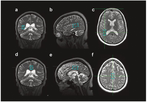
The spectra were acquired with PRESS sequence using the following parameters: TR/TE 5000/30 ms, spectral bandwidth 5000 Hz, 4096 time domain points, 8-phase cycles. Four dummy excitations were followed by 16 non-water-suppressed and 64 water-suppressed scans. Typical voxel size 1 × 1.5x2.5 cm3 and 1 × 1.5x3.5 cm3 was applied for white and gray matter, respectively. Spectrum processing was performed using LCModel (Sup. Figure S1) (Citation48). In the present work we evaluated the neuronal density marker (NAA+NAAG)/Cr concentration ratio.
Arterial spin labeling (ASL) for evaluation of CBF
Two 3D FSE Pseudo-Continuous ASL (pCASL) scans with spiral read-out and background suppression were acquired with a post-labeling delay of 2000 ms. There was a mean time interval of 127 (range: 72–339) minutes between the two ASL acquisitions. All acquisition parameters for the MR imaging are presented in Suppl. Table 1. CBF-maps, derived from pCASL, were calculated according the model previously recommended (Citation48), including a correction term for full proton density reference (Citation49–53). CBF-maps were co-registered to each subject’s corresponding T1w-image using the SPM12 toolbox (Welcome Trust Center for Neuroimaging, London, UK).
Cortical regions were segmented using the Freesurfer processing pipeline (version 6.0, http://surfer.nmr.mgh.harvard.edu) (Citation54) using T1w- and T2-FLAIR images. Cortical regions were combined in order to create bilateral frontal, parietal, occipital and temporal lobe regions. Furthermore, the amygdalae, caudate, putamen, pallidum, hippocampi and thalami bilaterally were also included (; ). The average regional CBF value derived from the two pCASL acquisitions was calculated and analyzed.
Figure 2. illustration of the cerebral parcellation using the software freesurfer (http://surfer.nmr.mgh.harvard.edu) used to divide the pseudo-continuous ASL(pCASL) data into total gray and white, the former divided into 20 gray matter regions, providing a mean cerebral blood flow (CBF) value for each region. Two scans were performed on each study participant, and the mean of the two scans was calculated for each region in each subject. The mean value, standard deviation and confidence interval for each region and each group are shown in . a. amygdala, b. total grey matter, c. total white matter, d. caudate nucleus, e. frontal lobe gray mater, f. hippocampus, g. occipital lobe gray matter, h. globus pallidum, i. parietal lobe gray matter, j. putamen, k. temporal lobe gray matter, l. thalamus.
Table 2. Comparison between male and female controls, rSRC athletes and TBI patients. No statistical comparisons of male and female TBI patients were made due to the low number of cases. In all measured aspects, there were no sex differences in the control and rSRC groups. Means and standard deviations are shown. rSRC = repeated sports-related concussions; TBI = traumatic brain injury; # = number; RBANS = Repeated battery assessment of neurological status; MRS = magnetic resonance spectroscopy; GM = grey matter; WM = white matter; CR = creatine; NAA = N-Acetylaspartate; NAAG = N-acetylaspartylglutamate; a.u. = arbitrary unit; mm = millimeter; § = due to low number of female TBI patients median and range is shown.
Table 3. Cerebral blood flow (CBF; in ml/100 gm/min) in total gray and white matter, the gray matter divided into 20 different brain regions, presented for all controls and TBI patients. For the rSRC athletes and controls, males and females are presented separately. Each evaluated brain region corresponds to a gray matter area except the total white matter region. rSRC = repeated sports-related concussions. L = left; R = right, CI = confidence interval, F = females, M = males.
Statistics
Statistical analysis was performed using GraphPad Prism version 8.1.0 for Windows, GraphPad Software, San Diego, California, USA. To evaluate the distribution of data D’Agostino & Pearson was used. Factors having normal distribution were CBF, RBANS index score, cortical thickness, MRS (NAA+NAAG)/Cre in both gray and white matter and TBI severity, whereas sex and time since the last SRC/TBI were not normally distributed. Normal data were described with mean values and standard deviation and non-normal data with median and range. To calculated differences between three groups, one-way ANOVA was used followed by Tukey’s multiple comparisons test. When factors were divided depending on sex, and difference between controls and athletes with rSRC were calculated, an unpaired t-test was used. The Spearman univariate correlation matrix was used to calculate the correlation variable between the above mentioned factors, except sex. Those correlations significant at the p ≤ 0.05 level were included in the multivariate analysis performed using multiple linear regression. Injury severity was used to divide our cohort in different groups to calculate group differences, using one-way ANOVA followed by Tukey’s multiple comparison test. Furthermore, injury severity was used as a continuous variable in a Spearman analysis followed by linear logistic regression.
Reliability of 1H-MRS data were assessed by standard deviations (Cramer-Rao lower bounds, CRLB), expressed in percent of the considered metabolite concentration. CRLB < 20% was used as a criterion of acceptable reliability (Citation39). The MR-S data used was CRLB SD <40%. Derived P-values are two-sided and presented as exact values, p-values ≤0.05 were considered significant. Due to low number of female patients with TBI, we refrained from performing sex-specific comparisons in the moderate-severe TBI cohort.
Results
Demographics for the included controls, rSRC athletes (mTBI) and patients with moderate-severe TBI are shown in .
The rSRC athletes included six females (25 ± 4.5 years old) and six males (30.5 ± 9.2 years old), and all had persistent post-concussive symptoms for ≥ 6 months following the last SRC (). The included athletes were at either amateur or professional level, although none was active at the time of study participation. One male and one female athlete were prescribed tricyclic antidepressants, and one female athlete a serotonin-selective reuptake inhibitor (SSRI). The time between injury and MR scanning varied between 6 and 132 months post-injury in the rSRC and TBI cohorts but CBF did not correlate to time since injury (p = 0.18; Suppl. Figure S2).
The female and male athletes had similar Symptom Severity Score (SSS), Number of Symptoms (NOS), number of SRCs and RBANS-index ().
The patients with TBI (27 ± 7 years old; 4 males, 2 females, ) had on admission a Glasgow Coma Scale (GCS) score of 12 (range 5–14), and at discharge from the neurosurgical department the GCS score was 14 (range 8–15). At time of MR scanning, all patients with TBI had a GCS score of 15. The injury mechanisms were fall injury (n = 3) or motor vehicle accidents (n = 3). The injury severity was moderate (defined as GCS 9–12, loss of consciousness ≥5 min or focal neurological deficit (Citation42) in 3, or severe (GCS 3–8) in 3. Four had a focal TBI (contusions, subdural hematomas) and 2 had diffuse axonal injury ().
Six male controls (25.5 ± 4.4 years old) and five female controls (26 ± 5.2 years old) were included. Two female controls were excluded from pre-specified criteria, one due to the finding of a clinically unknown posttraumatic lesion in the medial temporal lobe and one being a statistical outlier (± 2.5 SD compared to mean CBF values) in all measured regions.
Cognitive evaluation and symptoms scores
Cognition was evaluated with the RBANS test battery. As a group, patients with TBI and rSRC athletes performed significantly worse than controls (controls: 105.5 ± 21; rSRC: 80 ± 17; Patients with TBI: 75 ± 24; Controls to rSRC p = 0.01; Controls to TBI p = 0.01; rSRC to TBI p = 0.87; ). No differences were observed between rSRC athletes and patients with TBI, nor between male and female athletes. Male rSRC athletes performed worse than male controls (74 ± 9 vs. 113 ± 13, respectively; p = 0.0005), while females athletes had RBANS scores similar to those of the uninjured, healthy female controls (86 ± 22 vs. 104 ± 27, respectively; p = 0.25; ). Using the SCAT symptom evaluation no differences in Symptom Severity Score and Number of Symptoms between male or female athletes were observed ().
Structural MRI
Investigation of structural lesions in the included subjects was made by evaluation of the MRI sequences SWAN and T2 FLAIR. One nonspecific small white matter lesion without clinical significance was found in one male control and in two male rSRC athletes. One female athlete had a minor pituitary lesion without clinical importance. In the patients with TBI, residual contusions (n = 4) and diffuse axonal injury (DAI; n = 2) were observed (not shown).
Cortical thickness
The global mean cortical thickness was evaluated using MRI for each individual. The mean cortical thickness was similar in the controls (2.72 ± 0.08 mm) and rSRC (2.71 ± 0.11 mm) groups (p = 0.95), while it was significantly decreased in patients with TBI (2.60 ± 0.07 mm) when compared to controls (p = 0.05) but not rSRC athletes (p = 0.07); . The cortical thickness was similar in male and female rSRC athletes (p = 0.45) ()
Figure 3. Global mean cortical thickness, measured in millimeter (mm). There were a significant difference in thickness between controls and TBI patients (p = 0.05 CI = 0.002–0.23), not between rSRC and TBI (p = 0.07 CI = 0.007–0.22) and not between controls and rSRC (p = 0.96 CI = −0.08–0.11). rSRC = repeated sportsrelated concussions; TBI = traumatic brain injury.
MR spectroscopy
We analyzed (NAA+NAAG)/Cr concentration ratios from one voxel in gray and one in white matter. There was no difference between groups nor any sex differences within the control or SRC groups (; for p-values see ).
CBF
First, we evaluated global white and gray matter CBF in the control, rSRC and TBI groups. The CBF was similar in the rSRC and control groups, while patients with TBI had lower CBF compared to controls (gray matter p = 0.01; white matter p = 0.007) and rSRC athletes (gray matter p = 0.02; white matter p = 0.03; ).
Figure 4. Mean and individual global changes in cerebral blood flow (CBF) in traumatic brain injury (TBI) patients and athletes symptomatic following repeated sport related concussion (rSRC) showing decreased CBF in both white and gray matter. furthermore, CBF is lower in female athletes with rSRC when compared to female healthy controls, findings not observed in the male athletes. In view of the limited number of female TBI patients, we refrained from analyzing sex differences in this group. Horizontal bars indicate the mean values. F = females. M = males.
Next, we evaluated CBF segmented into 20 gray matter cerebral regions (; ). In the rSRC group, no differences were observed compared to controls in any region of interest. In the TBI group, CBF was decreased in six gray matter regions when compared to controls; the regions were the left amygdala, right caudate nucleus, left and right occipital lobes and bilaterally in the thalami. Among the controls, CBF was significantly higher in the female compared to the male subjects in both total gray and white matter and in 18 of the 20 evaluated gray matter brain regions (p ≤ 0.05; ). In rSRC athletes, females had a significantly higher CBF compared to males in 6 of 20 gray matter brain regions and in total gray and white matter (; ).
In female rSRC athletes, CBF was significantly decreased compared to female controls in 16 of the 20 evaluated gray matter brain regions, including left amygdala, left caudate nucleus, left and right frontal lobes, left and right hippocampus, left and right occipital lobes, left pallidum, left and right parietal lobes, left putamen, temporal lobes bilaterally and both thalami as well as in total gray matter and total white matter (p ≤ 0.05; ; ). However, male rSRC athletes had similar CBF when compared to male controls in all of the evaluated brain regions (; ). In the TBI group, sex differences were not calculated due to the low number of included female subjects.
CBF correlates with injury severity, cortical thickness, and cognition
In a correlation matrix, we compared CBF in controls, rSRC athletes, and patients with TBI to TBI-severity (control, mild, moderate, and severe), RBANS index score, cortical thickness, (NAA+NAAG)/Cr concentration ratio in both gray and white matter, and to time since the last SRC/TBI. A significant correlation with CBF was seen for TBI severity, time since injury and (NAA+NAAG)/Cr ratio in white matter (p < 0.05; TBI-severity: r = −0.48; time since injury: r = −0.35; (NAA+NAAG)/Cr: r = −0.12). These were included in the multiple linear regression model, where lower CBF was associated with higher TBI severity (β = −9.1; CI = −9.5 to −13.7; p = 0.0008). Furthermore, longer time since TBI or last SRC correlated with lower CBF (β = −9.1; CI = −9.5 to −13.7; p = 0.0008; F = 18.8). The were no correlations between CBF and the Number of symptoms (r = 0.05; p = 0.89) and Symptom Severity Score (r = 0.20; p = 0.54) of the SCAT.
In the correlation matrix, TBI severity was significantly correlated to CBF (r = −0.48; p = 0.008), RBANS (r = −0.54; p = 0.003) and cortical thickness (r = −0.42; p = 0.02). All remained significant in the multiple linear regression model where increased TBI severity correlated with decreased CBF (β = −0.03 CI = −0.06 to −0.01; p = 0.008; F = 8.48), decreased RBANS index score (β = −0.02; CI = −0.03 to −0.006; p = 0.004; F = 10.37) and decreased cortical thickness (β = −3.1; CI = −5.7 to −0.5; p = 0.02; F = 5.97). Time since injury did not correlate to any of the included variables in the correlation matrix.
Discussion
In this study, we investigated how cerebral blood flow (CBF) correlated to other neuroimaging findings (cortical thickness and MR spectroscopy) and how these were associated with cognitive function and TBI severity in patients with moderate-severe TBI as well as in athletes with persistent post-concussion symptoms who had sustained ≥3 sports-related concussions (SRCs). Both injury groups had equally impaired cognition as measured by the RBANS test battery (Citation55). At a group level, patients with TBI but not SRC athletes had globally reduced CBF in gray and white matter compared to controls. However, there was a widespread decrease of CBF in the female rSRC cohort when compared to the healthy female controls. In contrast, CBF was not altered in male rSRC athletes when compared to male controls. Decreased CBF correlated to increased injury severity, female sex, and the (NAA+NAAG)/Cr ratio in white matter.
Arterial Spin Labeling (ASL) is a noninvasive and highly repeatable magnetic resonance (MR) imaging-based CBF quantification technique using the patient’s own water as a freely diffusible tracer (Citation56), previously validated against 15O-PET, the gold standard of CBF measurements (Citation57). To minimize the risk that changed neuronal activity would result in CBF alterations during the ASL scans, the investigations were performed with the athletes and controls in a resting state and by minimizing known confounders of CBF evaluations such as nicotine, alcohol, coffee or energy drinks. The understanding and the interpretation of early CBF changes may be crucial in acute neurocritical care management (Citation58). Furthermore, cerebrovascular dysfunction may contribute to persistent symptoms in the chronic phase (Citation59). In patients with TBI imaged 3 months post-injury, gray matter CBF was associated with injury severity and cognitive dysfunction (Citation5). Moreover, cerebral autoregulation was impaired beyond 6 months post-injury in a cohort of patients with moderate-severe TBI (Citation17,Citation60). In the present study, patients with moderate-severe TBI had decreased CBF globally (gray and white matter) compared to controls and rSRC athletes.
In a previous study investigating chronic regional CBF changes in athletes developing post-concussion syndrome (PCS) after a single SRC, decreased CBF was observed in the thalami ≥2 years post-injury. The investigated athletes were on average 34 years old, while sex was not specified (Citation22). In another study, decreased CBF in the cerebellum, cuneus, cingulate, and temporal gyrus was observed >3 months post-SRC (Citation23) in a predominately male cohort. After a single mild TBI occurring in a non-sport context, CBF was determined by ASL in a mixed sex cohort. CBF was decreased in the first 3 days post-injury but had normalized by 3 months (Citation61). Normalization of CBF correlated with resolution of symptoms in a group of collegiate American football players following a single SRC (Citation21), and in retired male American football players, a globally decreased CBF was observed (Citation25). The latter study (Citation25) also included pre-menopausal females as controls, who may have had a constitutionally higher CBF (Citation39–41). Our study emphasizes the importance of studying sex differences in brain perfusion studies in view of the constitutionally higher CBF in females. The mechanisms of the sex differences in CBF in the healthy, non-injured population have not been established, but different hematocrit levels may contribute (Citation40). In a recent study, CBF was evaluated in 13 concussed female athletes 3–10 days post-injury, of whom 7 were hormonal contraceptive users. Using ASL, higher progesterone levels were associated with a higher CBF (Citation62). These findings suggest that hormonal levels may influence CBF, at least in the early post-injury phase. Since hormonal status was not explored in our present study, an influence on CBF cannot be excluded. In an additional report, in contrast to our present study, CBF was lower in male but not female athletes several years post-SRC when compared to sex-matched controls. Importantly, those athletes were asymptomatic (Citation63). Secondary analysis of the effect of rSRC compared to a single SRC, showed that only rSRC females had clusters of decreased CBF compared with a sex-matched group with only one previous SRC (Citation63). The latter support the result of the present study, i.e. that there may be important sex differences in rSRC athletes, and decreased CBF in females only.
These observed differences in CBF may reflect different injury mechanisms and be related to the worse clinical outcome commonly observed in female SRC athletes (Citation13,Citation14,Citation64). The reasons for the worse outcome in females are not established, although relatively weaker neck muscles may lead to exacerbated head rotation at impact (Citation65,Citation66). Furthermore, there may be sex differences in white matter connectivity in females, with more inter-hemispheric connections (Citation67), and the white matter may be more susceptible to injury in females (Citation68). In our report, both male and female athletes had similar symptom burden (Citation27) but only female athletes had decreased CBF. Furthermore, only male athletes had significantly lower cognitive function compared to sex-matched controls on the RBANS screening, suggesting that not only CBF changes contribute to cognitive impairment and post-concussion symptomatology. Other factors, such as energy metabolic alterations may contribute to the persistent symptoms commonly observed following rSRC (Citation69,Citation70).
N-acetylaspartate (NAA) is a marker of neuronal density and dynamic changes of NAA levels reflect neuronal dysfunction and the metabolic state. Since metabolism is a key regulator of CBF, it is relevant to analyze both in TBI and SRC. NAA is often measured together with NAAG since these two spectral intensities are difficult to separate in the MR spectrum. The advantage of (NAA+NAAG)/Cr ratio is that it does not depend on CSF fraction in the voxel. It should be noted that Cr is commonly used as a concentration reference because it is relatively stable with negligible (or very small) changes with age, or in a variety of brain disorders (Citation71,Citation72). In acute TBI, NAA falls rapidly but when the patient recovers, NAA often recovers too (Citation4,Citation33,Citation73). It may, however, be persistently decreased into the chronic phase even following a single SRC (Citation74). In the present study, the (NAA+NAAG)/Cr ratio correlated with white matter CBF, without other differences among the groups. However, it did not remain significant in the multiple linear regression analysis and might not be an independent correlation and this needs evaluation in larger cohorts. Since the imaging was performed at 6–120 months post-injury in all injury groups, we cannot exclude that earlier imaging could have provided different results. Here, we evaluated two MR-S voxels, one in gray and one in white matter. For analysis of white matter injury, the sensitivity of the method may be more important than the location of the voxel (Citation75). However, the voxel location and size may in fact influence the MR spectroscopy results and we cannot exclude that another placement, e.g. the frontal lobe (Citation75), could have yielded other results. Whole-brain metabolite analysis (Citation76) could be a tool for more detailed correlation to CBF in TBI and SRC studies.
TBI is followed by neurodegenerative processes resulting in progressive brain atrophy that is particularly pronounced in white matter, but is also observed in gray matter causing cortical thinning (Citation2). After a single SRC cortical thinning has not been reported (Citation6,Citation77–80) in contrast to several reports on rSRC (Citation34,Citation81). We measured global cortical thickness and no differences among the groups were observed. The TBI cohort included both patients with moderate or severe TBI. An SRC is, per consensus statements (Citation10), defined as a mild TBI. As such, SRC athletes were included as the mild TBI severity group that together with the patients with moderate-severe TBI (previously treated in our neurocritical care unit) and were included in the analyses of injury severity. When injury severity (mild/SRC, moderate, severe) was included in the correlation analysis, cortical thickness was found to correlate to injury severity. Thus, the more severe the injury, the thinner the cortex. Longitudinal studies using a larger cohort of patients with TBI could show if cortical atrophy is progressive with time.
Limitations
A limitation of this study is the relatively small sample size, particularly in patients with TBI, and a potential recruitment bias that we tried to minimize by selecting well-characterized age- and sex-matched controls with no previous head trauma. All SRC athletes had ≥ 3 previous SRCs, persistent symptoms ≥ 6 months following their last SRC and met the DSM IV criteria (≥ 3 symptoms for ≥ 3 months) for post-concussion syndrome. We had no information on acute post-concussion symptoms, a well-established prognostic factor following SRC (Citation16,Citation82). Thus, we cannot exclude that the female athletes in our cohort had worse initial symptoms following their SRC, contributing to their decreased CBF. Based on the low likelihood of recovery with prolonged symptom duration (Citation29) and the time needed for neurodegeneration and tau aggregation following moderate-to-severe TBI (Citation83), our cohorts may be regarded as having chronic post-concussion symptoms.
Methodological limitations of the CBF analysis include the low signal-to-noise ratio of ASL, which we tried to minimize by using two repeated scans on the same day and used the mean CBF value for each included subject. Furthermore, when using ASL in white matter (WM), CBF may be underestimated when compared to gray matter (GM) and make WM CBF evaluations less certain. Partial volume effects are caused by the limited spatial resolution in ASL and tend to result in under- and overestimation of CBF in GM and WM, respectively (Citation84).
We used the validated RBANS test battery to screen for cognitive dysfunction since it is useful in detecting deficits in attention, verbal functions, visuospatial functions, immediate and delayed memory (Citation85). It provides a broad although not in-depth analysis of all cognitive aspects. However, in the setting of this study (i.e. limited time available, need for repeatability and ease of administration) it worked as an effective screening tool. Even though cognitive deficits among athletes following a single SRC have not been reported in the post-acute phase, several studies have shown deficits in attention, immediate memory, and delayed memory indices following rSRC (Citation85–87). Following moderate-to-severe TBI, deficits across all RBANS indices are consistently observed (Citation44,Citation88). Arguably, RBANS remains suitable for detection of cognitive deficits among rSRC athletes and patients with TBI, despite the fact that no correlations to neuroimaging parameters were observed in the present study.
The (NAA+NAAG)/Cr ratio in voxel positions used here did not show any differences among the groups. However, we cannot exclude that results could have been different in other anatomical regions such as in the prefrontal cortex (Citation75). Injury heterogeneity may also limit the interpretation of the MRS results. Since we used a global measure of cortical thickness, consistent with previous report, focal cortical atrophy may have been missed. However, due to the diffuse nature of the rSRC and TBI pathophysiology, we argue that this is unlikely.
Conclusion
In the present study, injury severity was found to correlate with cognitive impairment, cortical thickness and global CBF. We also observed that global gray matter CBF, evaluated by arterial spin labeling, correlated to injury severity and sex. Furthermore, a decreased CBF was observed in female, not male, rSRC athletes. While the decreased CBF in female athletes did not correlate with impaired cognitive function or symptom scores, cerebrovascular changes may contribute to the sex differences in recovery commonly observed following TBI and SRC. These alterations may contribute to, or be influenced by, the aggregation of tau and the ongoing neuroinflammation observed in particularly the white matter chronically in patients with TBI and rSRC athletes (Citation36). Our study also emphasizes the importance of studying sex differences in brain perfusion studies in view of the constitutionally higher CBF in females.
Source of support
Swedish Brain Foundation, Swedish Research Council under the framework of EU-ERA-NET NEURON CnsAFlame, Selander Foundation, Swedish Research Council and Swedish Research Council for Sport Science
Supplemental Material
Download MS Word (248.6 KB)Acknowledgments
We thank all participating athletes and controls. The study was founded by the Swedish Brain Foundation, Swedish Research Council under the framework of EU-ERA-NET NEURON CnsAFlame, Swedish Research Council, Bissen Brain Walk, Selander Foundation and Swedish Research Council for Sport Science (all to NM).
Disclosure statement
No potential conflict of interest was reported by the author(s).
Supplementary material
Supplemental data for this article can be accessed online at https://doi.org/10.1080/02699052.2022.2109746
Additional information
Funding
References
- Guskiewicz KM, Marshall SW, Bailes J, McCrea M, Cantu RC, Randolph C, Jordan BD, et al. Association between recurrent concussion and late-life cognitive impairment in retired professional football players. Neurosurgery. 2005 October;57(4):719–26. discussion 719-26
- Cole JH, Jolly A, de Simoni S, Bourke N, Patel MC, Scott G, Sharp DJ, et al. Spatial patterns of progressive brain volume loss after moderate-severe traumatic brain injury. Brain: a Journal of Neurology. 2018 January 4;141(3):822–36.
- Stovell MG, Mada MO, Carpenter TA, et al. Phosphorus spectroscopy in acute TBI demonstrates metabolic changes that relate to outcome in the presence of normal structural MRI. Journal of Cerebral Blood Flow and Metabolism: Official Journal of the International Society of Cerebral Blood Flow and Metabolism. 2020 January;40(1):67–84.
- Stovell MG, Yan JL, Sleigh A, Mada MO, Carpenter TA, Hutchinson PJA, Carpenter KLH, et al. Assessing metabolism and injury in acute human traumatic brain injury with magnetic resonance spectroscopy: current and future applications. Front Neurol. 2017;8:426.
- Ware JB, Dolui S, Duda J, Gaggi N, Choi R, Detre J, Whyte J, Diaz-Arrastia R, Kim JJ, et al. Relationship of cerebral blood flow to cognitive function and recovery in early chronic traumatic brain injury. J Neurotrauma. 2020 June 11;37(20):2180–87.
- Clark AL, Weigand AJ, Bangen KJ, et al. Repetitive mTBI is associated with age-related reductions in cerebral blood flow but not cortical thickness. Journal of Cerebral Blood Flow and Metabolism: Official Journal of the International Society of Cerebral Blood Flow and Metabolism. 2020 April;4:271678x19897443.
- Sandsmark DK, Bashir A, Wellington CL, Diaz-Arrastia R, et al. Cerebral microvascular injury: a potentially treatable endophenotype of traumatic brain injury-induced neurodegeneration. Neuron. 2019 August 7;103(3):367–79.
- Kenney K, Amyot F, Haber M, Pronger A, Bogoslovsky T, Moore C, Diaz-Arrastia R, et al. Cerebral vascular injury in traumatic brain injury. Exp Neurol. 2016 January;275(Pt 3):353–66.
- Xu L, Ware JB, Kim JJ, et al. Arterial spin labeling reveals elevated cerebral blood flow with distinct clusters of hypo- and hyperperfusion after traumatic brain injury. J Neurotrauma. 2021 September 15;38(18):319–25.
- McCrory P, Meeuwisse W, Dvorak J, Aubry M, Bailes J, Broglio S, Cantu RC, Cassidy D, Echemendia RJ, Castellani RJ, et al. Consensus statement on concussion in sport-the 5th international conference on concussion in sport held in Berlin, October 2016. Br J Sports Med. 2017 June;51(11):838–47.
- Guskiewicz KM, McCrea M, Marshall SW, Cantu RC, Randolph C, Barr W, Onate JA, Kelly JP, et al. Cumulative effects associated with recurrent concussion in collegiate football players: the NCAA concussion study. Jama. 2003 November 19;290(19):2549–55.
- Abrahams S, Fie SM, Patricios J, Posthumus M, September AV, et al. Risk factors for sports concussion: an evidence-based systematic review. Br J Sports Med. 2014 January;48(2):91–97.
- Brooks BL, Silverberg N, Maxwell B, Mannix R, Zafonte R, Berkner PD, Iverson GL, et al. Investigating effects of sex differences and prior concussions on symptom reporting and cognition among adolescent soccer players. Am J Sports Med. 2018 March;46(4):961–68.
- Dvorak J, McCrory P, Kirkendall DT. Head injuries in the female football player: incidence, mechanisms, risk factors and management. Br J Sports Med. 2007 August;41 Suppl 1:i44–6.
- Voormolen DC, Cnossen MC, Polinder S, von Steinbuechel N, Vos PE, Haagsma JA, et al. Divergent classification methods of post-concussion syndrome after mild traumatic brain injury: prevalence rates, risk factors, and functional outcome. J Neurotrauma. 2018 March 23;35(11):1233–41.
- Meehan WP 3rd, Mannix R, Monuteaux MC, Stein CJ, Bachur RG, et al. Early symptom burden predicts recovery after sport-related concussion. Neurology. 2014 December 9;83(24):2204–10.
- Kim J, Whyte J, Patel S, Avants B, Europa E, Wang J, Slattery J, Gee JC, Coslett HB, Detre JA, et al. Resting cerebral blood flow alterations in chronic traumatic brain injury: an arterial spin labeling perfusion FMRI study. J Neurotrauma. 2010 August;27(8):1399–411.
- Amyot F, Kenney K, Moore C, Haber M, Turtzo LC, Shenouda C, Silverman E, Gong Y, Qu B-X, Harburg L, et al. Imaging of cerebrovascular function in chronic traumatic brain injury. J Neurotrauma. 2018 May 15;35(10):1116–23.
- Thomas BP, Tarumi T, Wang C, Zhu DC, Tomoto T, Munro Cullum C, Dieppa M, Diaz-Arrastia R, Bell K, Madden C, et al. Hippocampal and rostral anterior cingulate blood flow is associated with affective symptoms in chronic traumatic brain injury. Brain Res. 2021 November 15;1771:147631.
- Churchill NW, Hutchison MG, Graham SJ, Schweizer TA, et al. Symptom correlates of cerebral blood flow following acute concussion. NeuroImage Clinical. 2017;16:234–39.
- Meier TB, Bellgowan PS, Singh R, Kuplicki R, Polanski DW, Mayer AR, et al. Recovery of cerebral blood flow following sports-related concussion. JAMA neurology. JAMA Neurol. 2015 May;72(5):530–38.
- Ge Y, Patel MB, Chen Q, Grossman EJ, Zhang K, Miles L, Babb JS, Reaume J, Grossman RI, et al. Assessment of thalamic perfusion in patients with mild traumatic brain injury by true FISP arterial spin labelling MR imaging at 3T. Brain Inj. 2009 July;23(7):666–74.
- Liu W, Wang B, Wolfowitz R, Yeh P-H, Nathan DE, Graner J, Tang H, Pan H, Harper J, Pham D, et al. Perfusion deficits in patients with mild traumatic brain injury characterized by dynamic susceptibility contrast MRI. NMR Biomed. 2013 June;26(6):651–63.
- Brooks BL, Low TA, Plourde V, et al. Cerebral blood flow in children and adolescents several years after concussion. Brain Inj. 2018 October;31:1–9.
- Amen DG, Willeumier K, Omalu B, Newberg A, Raghavendra C, Raji CA, et al. Perfusion neuroimaging abnormalities alone distinguish national football league players from a healthy population. Journal of Alzheimer’s Disease: JAD. 2016 April 25;53(1):237–41.
- Champagne AA, Coverdale NS, Germuska M, Cook DJ, et al. Multi-parametric analysis reveals metabolic and vascular effects driving differences in BOLD-based cerebrovascular reactivity associated with a history of sport concussion. Brain Inj. 2019;33(11):1479–89.
- Diagnostic and statistical manual of mental disorders. Washington, DC: American Psychiatric Association, 2000. doi:10.1176/appi.books.9780890423349.
- Wortzel HS, Arciniegas DB. The DSM-5 approach to the evaluation of traumatic brain injury and its neuropsychiatric sequelae. NeuroRehabilitation. 2014;34(4):613–23.
- Hiploylee C, Dufort PA, Davis HS, Wennberg RA, Tartaglia MC, Mikulis D, Hazrati L-N, Tator CH, et al. Longitudinal study of postconcussion syndrome: not everyone recovers. J Neurotrauma. 2017 April 15;34(8):1511–23.
- Haarbauer-Krupa J, Pugh MJ, Prager EM, Harmon N, Wolfe J, Yaffe K, et al. Epidemiology of chronic effects of traumatic brain injury. J Neurotrauma. 2021 May 5;38(23):3235–47.
- Marklund N, Bellander BM, Godbolt AK, Levin H, McCrory P, Thelin EP, et al. Treatments and rehabilitation in the acute and chronic state of traumatic brain injury. J Intern Med. 2019 June;285(6):608–23.
- Jia X, Chang X, Bai L, Wang Y, Dong D, Gan S, Wang S, Li X, Yang X, Sun Y, et al. A longitudinal study of white matter functional network in mild traumatic brain injury. J Neurotrauma. 2021 April 28;38(19):2686–97.
- Alosco ML, Tripodis Y, Rowland B, et al. A magnetic resonance spectroscopy investigation in symptomatic former NFL players. Brain Imaging Behav. 2020;14(5):1419–1429.
- Koerte IK, Mayinger M, Muehlmann M, Kaufmann D, Lin AP, Steffinger D, Fisch B, Rauchmann B-S, Immler S, Karch S, et al. Cortical thinning in former professional soccer players. Brain Imaging Behav. 2016 September;10(3):792–98.
- McKee AC, Alosco ML, Huber BR. Repetitive head impacts and chronic traumatic encephalopathy. Neurosurg Clin N Am. 2016 October;27(4):529–35.
- Marklund N, Vedung F, Lubberink M, et al. Tau aggregation and increased neuroinflammation in athletes after sports-related concussions and in traumatic brain injury patients - A PET/MR study. NeuroImage Clinical. 2021;30:102665.
- Levin HS, Temkin NR, Barber J, Nelson LD, Robertson C, Brennan J, Stein MB, Yue JK, Giacino JT, McCrea MA, et al. Association of sex and age with mild traumatic Brain injury-related symptoms: a TRACK-TBI study. JAMA network open. 2021 April 1;4(4):e213046.
- Mikolić A, van Klaveren D, Groeniger JO, Wilson L, van Lennep JR, van Klaveren D, Polinder S, Åkerlund C, Amrein K, Andreassen L, et al. Differences between men and women in treatment and outcome after traumatic brain injury. J Neurotrauma. 2021 January 15;38(2):235–51.
- Aanerud J, Borghammer P, Rodell A, Jónsdottir KY, Gjedde A, et al. Sex differences of human cortical blood flow and energy metabolism. Journal of Cerebral Blood Flow and Metabolism: Official Journal of the International Society of Cerebral Blood Flow and Metabolism. 2017 July;37(7):2433–40.
- Clement P, Mutsaerts HJ, Vaclavu L, et al. Variability of physiological brain perfusion in healthy subjects - A systematic review of modifiers. considerations for multi-center ASL studies. Journal of Cerebral Blood Flow and Metabolism: Official Journal of the International Society of Cerebral Blood Flow and Metabolism. 2017 January;1:271678x17702156.
- Liu W, Lou X, Ma L. Use of 3D pseudo-continuous arterial spin labeling to characterize sex and age differences in cerebral blood flow. Neuroradiology. 2016 September;58(9):943–48.
- Ingebrigtsen T, Romner B, Kock-Jensen C. Scandinavian guidelines for initial management of minimal, mild, and moderate head injuries. the scandinavian neurotrauma committee. J Trauma. 2000 April;48(4):760–66.
- Marklund N, Vedung F, Lubberink M, Tegner Y, Johansson J, Blennow K, Zetterberg H, Fahlström M, Haller S, Stenson S, et al. Tau aggregation and increased neuroinflammation in athletes after sports-related concussions and in traumatic brain injury patients – a PET/MR study. NeuroImage: Clinical. 2021 January 1 30 102665.
- McKay C, Casey JE, Wertheimer J, FICHTENBERG N, et al. Reliability and validity of the RBANS in a traumatic brain injured sample. Archives of Clinical Neuropsychology: the Official Journal of the National Academy of Neuropsychologists. 2007 January;22(1):91–98.
- Hanninen T, Parkkari J, Tuominen M, et al. Sport concussion assessment tool: interpreting day-of-injury scores in professional ice hockey players. Journal of Science and Medicine in Sport. 2017 December 12;20(5):424–31.
- Lovell MR, Iverson GL, Collins MW, Podell K, Johnston KM, Pardini D, Pardini J, Norwig J, Maroon JC, et al. Measurement of symptoms following sports-related concussion: reliability and normative data for the post-concussion scale. Appl Neuropsychol. 2006;13(3):166–74.
- Fischl B, Dale AM. Measuring the thickness of the human cerebral cortex from magnetic resonance images. Proc Natl Acad Sci U S A. 2000 September 26;97(20):11050–55.
- Alsop DC, Detre JA, Golay X, Günther M, Hendrikse J, Hernandez-Garcia L, Lu H, MacIntosh BJ, Parkes LM, Smits M, et al. Recommended implementation of arterial spin-labeled perfusion MRI for clinical applications: a consensus of the ISMRM perfusion study group and the European consortium for ASL in dementia. Magnetic Resonance in Medicine. 2015 January;73(1):102–16.
- Dai W, Shankaranarayanan A, Alsop DC. Volumetric measurement of perfusion and arterial transit delay using hadamard encoded continuous arterial spin labeling. Magnetic Resonance in Medicine. 2013 April;69(4):1014–22.
- Dai W, Robson PM, Shankaranarayanan A, Alsop DC, et al. Reduced resolution transit delay prescan for quantitative continuous arterial spin labeling perfusion imaging. Magnetic Resonance in Medicine. 2012 May;67(5):1252–65.
- Goetti R, Warnock G, Kuhn FP, Guggenberger R, O’Gorman R, Buck A, Khan N, Scheer I, et al. Quantitative cerebral perfusion imaging in children and young adults with Moyamoya disease: comparison of arterial spin-labeling-MRI and H(2)[(15)O]-PET. AJNR Am J Neuroradiol. 2014 May;35(5):1022–28.
- Maleki N, Dai W, Alsop DC. Optimization of background suppression for arterial spin labeling perfusion imaging. Magma (New York, NY). 2012 April;25(2):127–33.
- Dai W, Garcia D, de Bazelaire C, Alsop DC, et al. Continuous flow-driven inversion for arterial spin labeling using pulsed radio frequency and gradient fields. Magnetic Resonance in Medicine. 2008 December;60(6):1488–97.
- Fischl B, Salat DH, Busa E, Albert M, Dieterich M, Haselgrove C, van der Kouwe A, Killiany R, Kennedy D, Klaveness S, et al. Whole brain segmentation: automated labeling of neuroanatomical structures in the human brain. Neuron. 2002 January 31;33(3):341–55.
- Randolph C, Tierney MC, Mohr E, Chase TN, et al. The repeatable battery for the assessment of neuropsychological status (RBANS): preliminary clinical validity. J Clin Exp Neuropsychol. 1998 June;20(3):310–19.
- Barlow KM, Marcil LD, Dewey D, Carlson HL, MacMaster FP, Brooks BL, Lebel RM, et al. Cerebral perfusion changes in post-concussion syndrome: a prospective controlled cohort Study. J Neurotrauma. 2017 March 1;34(5):996–1004.
- Chen JJ, Wieckowska M, Meyer E, Pike GB, et al. Cerebral blood flow measurement using fMRI and PET: a cross-validation study. Int J Biomed Imaging. 2008;2008:516359.
- Abate MG, Trivedi M, Fryer TD, Smielewski P, Chatfield DA, Williams GB, Aigbirhio F, Carpenter TA, Pickard JD, Menon DK, et al. Early derangements in oxygen and glucose metabolism following head injury: the ischemic penumbra and pathophysiological heterogeneity. Neurocrit Care. 2008;9(3):319–25.
- Ramos-Cejudo J, Wisniewski T, Marmar C, Zetterberg H, Blennow K, de Leon MJ, Fossati S, et al. Traumatic brain injury and alzheimer’s disease: the cerebrovascular link. EBioMedicine. 2018 February;28:21–30.
- Ding K, Tarumi T, Tomoto T, Mccolloster M, Le T, Dieppa M, Diaz-Arrastia R, Bell K, Madden C, Cullum CM, et al. Impaired cerebral blood flow regulation in chronic traumatic brain injury. Brain Res. 2020 September 15;1743:146924.
- Peng SP, Li YN, Liu J, Wang Z-Y, Zhang Z-S, Zhou S-K, Tao F-X, Zhang Z-X, et al. Pulsed arterial spin labeling effectively and dynamically observes changes in cerebral blood flow after mild traumatic brain injury. Neural Regeneration Research. 2016 February;11(2):257–61.
- Chen Y, Herrold AA, Gallagher V, Martinovich Z, Bari S, Vike NL, Vesci B, Mjaanes J, McCloskey LR, Reilly JL, et al. Preliminary report: localized cerebral blood flow mediates the relationship between progesterone and perceived stress symptoms among female collegiate club athletes after mild traumatic brain injury. J Neurotrauma. 2021 February 24;38(13):1809–20.
- Hamer J, Churchill NW, Hutchison MG, Graham SJ, Schweizer TA, et al. Sex differences in cerebral blood flow associated with a history of concussion. J Neurotrauma. 2019 December 17;37(10):1197–203.
- Mikolić A, van Klaveren D, Oude Groeniger J, Wiegers EJA, Lingsma HF, Zeldovich M, von Steinbüchel N, Maas AIR, Roeters van Lennep JE, Polinder S, et al. Differences between men and women in treatment and outcome after traumatic brain injury. J Neurotrauma. 2020 October 19
- Wilcox BJ, Beckwith JG, Greenwald RM, Raukar NP, Chu JJ, McAllister TW, Flashman LA, Maerlender AC, Duhaime A-C, Crisco JJ, et al. Biomechanics of head impacts associated with diagnosed concussion in female collegiate ice hockey players. J Biomech. 2015 July 16;48(10):2201–04.
- Tierney RT, Sitler MR, Swanik CB, SWANIK KA, Higgins M, Torg J, et al. Gender differences in head-neck segment dynamic stabilization during head acceleration. Med Sci Sports Exerc. 2005 February;37(2):272–79.
- Wilde EA, Newsome MR, Ott SD, Hunter JV, Dash P, Redell J, Spruiell M, Diaz M, Chu ZD, Goodrich-Hunsaker N, et al. Persistent disruption of brain connectivity after sports-related concussion in a female athlete. J Neurotrauma. 2019 November 15;36(22):3164–71.
- Sollmann N, Echlin PS, Schultz V, Viher PV, Lyall AE, Tripodis Y, Kaufmann D, Hartl E, Kinzel P, Forwell LA, et al. Sex differences in white matter alterations following repetitive subconcussive head impacts in collegiate ice hockey players. NeuroImage Clinical. 2018;17:642–49.
- Detre JA, Wang J, Wang Z, Rao H, et al. Arterial spin-labeled perfusion MRI in basic and clinical neuroscience. Curr Opin Neurol. 2009 August;22(4):348–55.
- Raichle ME. Behind the scenes of functional brain imaging: a historical and physiological perspective. Proc Natl Acad Sci U S A. 1998 February 03;95(3):765–72.
- Govindaraju V, Young K, Maudsley AA. Proton NMR chemical shifts and coupling constants for brain metabolites. NMR Biomed. 2000 May;13(3):129–53.
- Provencher SW. Estimation of metabolite concentrations from localized in vivo proton NMR spectra. Magnetic Resonance in Medicine. 1993 December;30(6):672–79.
- Panchal H, Sollmann N, Pasternak O, Alosco ML, Kinzel P, Kaufmann D, Hartl E, Forwell LA, Johnson AM, Skopelja EN, et al. Neuro-metabolite changes in a single season of university ice hockey using magnetic resonance spectroscopy. Front Neurol. 2018;9:616.
- Henry LC, Tremblay S, Leclerc S, Khiat A, Boulanger Y, Ellemberg D, Lassonde M, et al. Metabolic changes in concussed American football players during the acute and chronic post-injury phases. BMC Neurol. 2011 August 23;11(1):105.
- Bartnik-Olson BL, Alger JR, Babikian T, et al. The clinical utility of proton magnetic resonance spectroscopy in traumatic brain injury: recommendations from the ENIGMA MRS working group. Brain Imaging Behav. 2021;15(2):504–525.
- Lin JC, Mueller C, Campbell KA, et al. Investigating whole-brain metabolite abnormalities in the chronic stages of moderate or severe traumatic brain injury. PM & R: the Journal of Injury, Function, and Rehabilitation. 2022;14(4):472–485.
- Bobholz SA, Brett BL, España LY, Huber DL, Mayer AR, Harezlak J, Broglio SP, McAllister T, McCrea MA, Meier TB, et al. Prospective study of the association between sport-related concussion and brain morphometry (3T-MRI) in collegiate athletes: study from the NCAA-DoD CARE consortium. Br J Sports Med. 2020 September 11;55(3):169–74.
- Bigler ED, Finuf C, Abildskov TJ, Goodrich-Hunsaker NJ, Petrie JA, Wood D-M, Hesselink JR, Wilde EA, Max JE, et al. Cortical thickness in pediatric mild traumatic brain injury including sports-related concussion. International Journal of Psychophysiology: Official Journal of the International Organization of Psychophysiology. 2018 October;132(Pt A):99–104.
- España LY, Lee RM, Ling JM, Jeromin A, Mayer AR, Meier TB, et al. Serial assessment of gray matter abnormalities after sport-related concussion. J Neurotrauma. 2017 November 15;34(22):3143–52.
- Govindarajan KA, Narayana PA, Hasan KM, Wilde EA, Levin HS, Hunter JV, Miller ER, Patel VKS, Robertson CS, McCarthy JJ, et al. Cortical thickness in mild traumatic brain injury. J Neurotrauma. 2016 October 15;33(20):1809–17.
- Meier TB, Bellgowan PS, Bergamino M, Ling JM, Mayer AR, et al. Thinner cortex in collegiate football players with, but not without, a self-reported history of concussion. J Neurotrauma. 2016 February 15;33(4):330–38.
- Iverson GL, Gardner AJ, Terry DP, Ponsford JL, Sills AK, Broshek DK, Solomon GS, et al. Predictors of clinical recovery from concussion: a systematic review. Br J Sports Med. 2017 June;51(12):941–48.
- Johnson VE, Stewart W, Smith DH. Widespread tau and amyloid-beta pathology many years after a single traumatic brain injury in humans. Brain Pathology (Zurich, Switzerland). 2012 March;22(2):142–49.
- Petr J, Schramm G, Hofheinz F, et al. Partial volume correction in arterial spin labeling using a look-locker sequence. Magnetic Resonance in Medicine. 2013 December;70(6):1535–43.
- Arch A, Ferraro FR. Performance on the repeatable battery for the assessment of neuropsychological status in college students with mild traumatic brain injury. Appl Neuropsychol Adult. 2021 Mar-Apr;28(2):220–29.
- Moser RS, Schatz P, Jordan BD. Prolonged effects of concussion in high school athletes. Neurosurgery. 2005;57(2):300–06.
- Killam C, Cautin RL, Santucci AC. Assessing the enduring residual neuropsychological effects of head trauma in college athletes who participate in contact sports. Archives of Clinical Neuropsychology. 2005;20(5):599–611.
- McKay C, Wertheimer JC, Fichtenberg NL, et al. The repeatable battery for the assessment of neuropsychological status (RBANS): clinical utility in a traumatic brain injury sample. Clin Neuropsychol. 2008 March;22(2):228–41.