
ABSTRACT
Purpose/Aim: Evaluating the suitability of bioengineered collagen sheets and human anterior lens capsules (HALCs) as carriers for cultivated porcine corneal endothelial cells (pCECs) and in vitro assessment of the cell-carrier sheets as tissue-engineered grafts for Descemet membrane endothelial keratoplasty (DMEK).
Materials and Methods: pCECs were isolated, cultured up to P2 and seeded onto LinkCell™ bioengineered matrices of 20 µm (LK20) or 100 µm (LK100) thickness, and on HALC. During expansion, pCEC viability and morphology were assessed by light microscopy. ZO-1 and Na+/K+-ATPase expression was investigated by immunohistochemistry. Biomechanical properties of pCEC-carrier constructs were evaluated by simulating DMEK surgery in vitro using an artificial anterior chamber (AC) and a human donor cornea without Descemet membrane (DM).
Results: During in vitro expansion, cultured pCECs retained their proliferative capacity, as shown by the positive staining for proliferative marker Ki67, and a high cell viability rate (96 ± 5%). pCECs seeded on all carriers formed a monolayer of hexagonal, tightly packed cells that expressed ZO-1 and Na+/K+-ATPase. During in vitro surgery, pCEC-LK20 and pCEC-LK100 constructs were handled like Descemet stripping endothelial keratoplasty (DSEK) grafts, i.e. folded like a “taco” for insertion because of challenges related to rolling and sticking of the grafts in the injector. pCEC-HALC constructs behaved similar to the DMEK reference model during implantation and unfolding in the artificial AC, showing good adhesion to the bare stroma.
Conclusions: In vitro DMEK surgery showed HALC as the most suitable carrier for cultivated pCECs with good intraoperative graft handling. LK20 carrier showed good biocompatibility, but required a DSEK-adapted surgical protocol. Both carriers might be notional candidates for potential future clinical applications.
Introduction
Corneal endothelial cells (CECs) are essential for the preservation of corneal transparency, since loss of their functionality following endothelial diseases or trauma eventually leads to corneal swelling and edema.Citation1 Because mature human CECs (hCEC) do not replicate in vivo,Citation1–Citation4 replacement of diseased or damaged endothelium by corneal transplantation is currently the only treatment option.Citation5 To date, Descemet membrane endothelial keratoplasty (DMEK) specifically replaces the recipient’s diseased or damaged endothelium (EC) and Descemet membrane (DM) with the same layers of a healthy donor.Citation6,Citation7 DMEK provides faster and better visual rehabilitation than other transplantation techniques; however, its application is limited by the shortage of high-quality healthy donor tissue.
One approach to target the shortage of endothelial grafts relies on the in vitro expansion of CECs on suitable cell carriers. These tissue-engineered endothelial grafts could become an alternative to the availability of human donor tissue.Citation8 Previous in vitro studies have reported the use of denuded DM and human anterior lens capsules (HALCs), as well as bioengineered matrices consisting of collagen or gelatin, or a combination of biopolymers, as potential carriers for cultured CECs.Citation9–Citation17 These cell-carrier constructs should preferably be similar to an “original” DMEK-graft, since it has been reported for Descemet stripping (automated) endothelial keratoplasty (DSEK/DSAEK) that thicker grafts may interfere with visual outcomeCitation18 and the biomechanical properties of cell-carrier constructs should allow implantation.
In this study, we evaluated collagen-based bioengineered scaffolds and HALC as carriers for viable cultured porcine corneal endothelial cell (pCEC) sheets. The suitability for transplantation of the pCEC-carrier constructs with the DMEK surgical protocol was tested by simulating DMEK surgery in vitro.
Materials and methods
Materials
Fetal bovine serum (FBS), Dulbecco’s phosphate-buffered saline (PBS), ascorbic acid 2-phosphate (Asc-2P), paraformaldehyde (PFA), L-Glutamine, trypsin, ethylenediaminetetraacetic acid (EDTA), bovine serum albumin (BSA), 4ʹ,6-diamidino-2-phenylindole (DAPI), and Triton X-100 were purchased from Sigma-Aldrich Chemistry BV (Zwijndrecht, The Netherlands). Chondroitin sulphate and Pen/Strep Pre-Mix were purchased from Carl Roth GmbH + Co. KG (Karlsruhe, Germany). Fibronectin, collagen, and albumin (FNC) coating mix was purchased from Athena ESTM (Baltimore, MD, USA). Opti-MEM™ I Reduced Serum Medium (Opti-MEM), bovine pituitary extract (BPE), anti-ZO-1/TJP1 primary antibody, secondary antibodies, and ReadyProbes™ Cell Viability Imaging Kit (Blue/Green) were obtained from Life Technology Europe BV (Bleiswijk, The Netherlands). Anti-Na+/K+-ATPase primary antibody and anti-Ki67 primary antibody were obtained from Abcam (Cambridge, United Kingdom). Betadine was obtained from Hippocratech (Rotterdam, The Netherlands). Hypotonic Trypan Blue solution 0.04% (Hippocratech, Rotterdam, The Netherlands) was used to stain the pCEC-carrier constructs during in vitro preparation and surgeries. LinkCell™ collagen bio-membranes were provided by LinkoCare Life Sciences AB (Linköping, Sweden).
Porcine corneal endothelial cell isolation
A total of 15 porcine eyes were collected from a local slaughterhouse within 24 h after death. First, porcine globes were decontaminated as described for human eyesCitation19 and adapted for porcine eyes, including an additional betadine-treatment step before decontamination. Next, the corneo-scleral rim was excised under sterile conditions, placed endothelial side-up in one well of a 12-well plate, and treated with 0.05% Trypsin/0.02% EDTA (TE) solution for 20 min at 37°C to cut away the focal adhesion from anchoring the endothelial cells to DM. Trypsin activity was stopped by addition of Opti-MEM containing 8% FBS directly on the corneo-scleral rim. Then, the enzymatically dislodged cells were collected with a sterile pipette tip and centrifuged at 500 rpm for 8 min at 37°C. Based on the protocol described by Proulx et al.,Citation20 cells were then re-suspended in fresh Opti-MEM containing 8% FBS, 4 mM L-Glutamine, 200 mg/ml Ca2+ chloride, 50 µg/ml BPE, 0.3 mM Asc-2P, 0.08% chrondroitin sulphate, and 100 IU/ml Pen/Strep Pre-Mix (hereafter referred to as “culture medium”) and plated in 3 wells of a 6-well plate previously coated with FNC coating mix. The cultures were incubated at 37°C in a humidified atmosphere containing 5% CO2. For routine maintenance, every 2–3 days the culture medium was replaced. When primary cultures of pCECs reached 80–90% confluence (approximately after 1 week), they were passaged by treatment with TE solution for 10 min at 37°C and 5% CO2. Next, the cell suspension was collected, centrifuged at 500 rpm for 8 min at 37°C and the cell pellet was re-suspended in fresh culture medium. The pCECs were sub-cultured at a 1:2 splitting ratio on FNC-coated culture well plates and their morphology at confluence and during expansion was evaluated with an AxioVert.A1 microscope with AxioCam ERc 5s stand-alone functionality camera (Zeiss, Oberkochen, Germany). Use of animal tissue in this study followed the institution’s guidelines for animal tissue-based research.
Cell proliferation and viability
Ki67 is a sensitive marker for cell proliferation.Citation21 To detect Ki67 expression, pCECs at P2 were cultured for 1 week on glass coverslips and fixed in 4% PFA for 15 min at room temperature (RT). Following fixation, samples were washed with PBS containing 0.1% Triton X-100 and then incubated with blocking buffer (3% BSA in PBS) for 30 min at 37°C to prevent non-specific staining. Blocking buffer was also used for primary and secondary antibody dilutions. Samples were incubated with primary antibody anti-Ki67 (dilution 1:100) for 1 h at 37°C and were subsequently washed several times with PBS. Next, samples were incubated with second antibodies (dilution 1:200) for 1 h at 37°C. After washing with PBS, samples were stained with DAPI to visualize the nuclear DNA, and then imaged using an inverted fluorescence microscope connected to a camera (Axiovert, Zeiss).
Cell viability was determined on pCECs at P2 cultured on glass coverslips by using the ReadyProbes™ Cell Viability Imaging Kit. Briefly, two drops of each stain (NucBlue® Live reagent and NucGreen® Dead reagent, respectively) were added per ml of culture media and samples were incubated for 15 min at 37°C in a humidified atmosphere containing 5% CO2. The cell viability rate was calculated as the average in three areas, each measuring 0.01 mm2, on digital microphotographs. The percentage of viable cells was determined by manual counting according to the fixed-frame method.
Human anterior lens capsule isolation
HALCs were isolated from human donor eyes at Amnitrans Eyebank Rotterdam. In all cases, the donors had stated to have no objection against transplant-related research. While holding the HALC with the anterior pole up with forceps, an incision into the lens’ equatorial area was made using a surgical blade to accomplish a 360° cut. The obtained HALCs were spread epithelial side-up and the lens cortex which was still adherent to the posterior lens cortex was removed using a surgical sponge (Simovision BV, Diemen, The Netherlands) soaked in 70% ethanol. Then, the HALCs were trephined to the desired size (Ø 8.0 mm) from the interior side, and treated with TE solution for 20 min. Next, HALCs were flattened and the loosely attached lens epithelium was removed by sweeping using a sponge soaked in BSS. After rinsing with BSS, because of the elastic properties of the membrane, a “HALC roll” formed spontaneously with the epithelial side on the inside. Each HALC was stored in a glass vial containing 5 ml PBS at 4°C until further use. When ready for cell seeding, the lens capsules were spread with the epithelial side downCitation16 over FNC-coated glass coverslips in 24-well tissue culture dishes and kept moist in PBS.
Seeding of cultured pCECs on biocompatible carriers
The selected carriers differed regarding their artificial extra cellular matrix (ECM) composition, technological process, and biophysical (strength of cell-substrate adhesion or cell proliferation enhancement) and biomechanical properties (e.g. tensile strength, elasticity). Three different carriers were evaluated in this study: LinkCell™ collagen I bio-membranes of 20 µm and 100 µm thickness (LK20 and LK100, respectively)Citation22 and HALCs. Diameter of the LK20 and LK100 for the experiments was 9.5 mm.
During the storage time, the carriers were kept hydrated in PBS in order to preserve their biomechanical properties. Primary pCECs, after reaching 80–90% of confluence, were passaged twice (up to P2) before they were seeded onto each the three carriers (LK20, LK100, and HALC). All pCEC-carrier constructs were cultured up to 1 week before they were used for the in vitro surgeries.
Immunofluorescence
ZO-1 and Na+/K+-ATPase are phenotypical markers for CEC.Citation23,Citation24 To visualize ZO-1 and Na+/K+-ATPase, pCECs at P2 were cultured for 1 week either on glass coverslips or directly on the biocompatible carriers and fixed in 4% PFA for 15 min at RT. Following fixation, samples were washed with PBS containing 0.1% Triton X-100 and then incubated with blocking buffer (1% BSA in PBS) for 1 h at 37°C to prevent non-specific staining. Blocking buffer was also used for primary and secondary antibody dilutions. Incubation with primary antibodies anti-ZO-1 tight junction protein (anti-ZO-1/TJP1, dilution 1:100) and anti-sodium/potassium-ATPase (anti-Na+/K+-ATPase, dilution 1:100) was performed for 1 h at 37°C and was followed by several PBS washing steps. Next, samples were incubated with secondary antibodies (dilution 1:200) for 1 h at 37°C. After washing with PBS, the samples were stained with DAPI to visualize the nuclear DNA, and then imaged using an inverted fluorescence microscope connected to a camera (Axiovert, Zeiss).
In vitro surgeries
In order to assess suitability for transplantation, surgeries with the pCEC-carrier constructs were simulated in vitro by using human anterior remnants (donor corneo-scleral rim without DM and endothelium; donors had stated to have no objection against transplant-related research) mounted onto an artificial anterior chamber (AC; DORC International, Zuidland, The Netherlands).Citation25 The ability of the pCEC-carrier constructs to form a roll in BSS, their affinity to be stained with Trypan Blue solution 0.04%, their insertion behaviour into the artificial AC, the unfolding behaviour, pCEC-carrier transparency, and ability to adhere to the posterior stroma of the anterior remnant were rated on a 5-point scale (i.e. 0–5), with higher score indicating greater similarity to in vivo DMEK/DSEK behaviour. In vitro surgeries with DMEK-grafts were used as a positive control and served as a reference for the scoring on the 5-point scale. All surgeries were performed by two DMEK surgeons (ID, IL).
For the in vitro surgery, main steps were performed as for standard DMEK surgery.Citation26 Three side ports were made at 2, 7, and 10 o’clock limbus, and the artificial AC was filled with PBS. Next, a 3-mm main incision was made at 12 o’clock to insert the pCEC-carrier constructs into the artificial AC. Each of the pCEC-carrier constructs was first stained for visualization with 0.04% Trypan Blue, implanted in the artificial anterior chamber and after unfolding and repositioning the pCEC-carrier constructs against the posterior stroma, the AC was filled 100% with an air bubble for 1 h to support the graft adherence to the stromal surface. Afterwards, air was partly removed through the paracentesis and attachment of the pCEC-carrier constructs to the stroma was evaluated by anterior-segment optical coherence tomography (AS-OCT; Heidelberg Engineering GmbH, Heidelberg, Germany).
Results
Evaluation of cell viability and proliferation during in vitro expansion
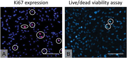
The pCECs of a native porcine cornea formed a uniform monolayer of tightly packed, hexagonal cells () with expression of the phenotypical markers ZO-1 and Na+/K+-ATPase (). During in vitro cell expansion, the pCECs also displayed a uniform layer of tightly packed cells and showed expression of ZO-1 and Na+/K+-ATPase (). Positive staining of Ki67 revealed that cultured pCECs retained the proliferative capacity (), while a low detection of nuclei of cells with compromised plasma membrane integrity (in green) indicated a high cell viability of the pCEC cultures (96 ± 5%) () which is also reflected by the low degree of Trypan Blue staining ().
Figure 1. Evaluation of cell morphology on porcine corneas and of pCEC cultures. (A) Porcine cornea stained with hypotonic Trypan Blue solution 0.04%. (B) Expression of ZO-1 detected via immunofluorescence on porcine corneal endothelium. (C) Expression of Na⁺/K⁺- ATPase detected via immunofluorescence on porcine corneal endothelium. (D) pCECs cultured at P2 upon FNC-coated glass coverslips and stained with hypotonic Trypan Blue solution 0.04%. (E) Expression of ZO-1 detected via immunofluorescence on pCECs cultured at P2 upon FNC-coated glass coverslips. (F) Expression of Na⁺/K⁺-ATPase detected via immunofluorescence on pCECs cultured at P2 upon FNC-coated glass coverslips. Scale bars: 100 µm.
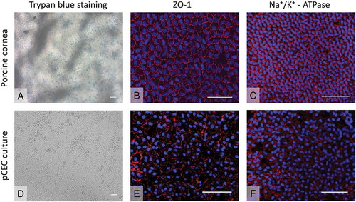
Figure 2. pCEC proliferation and viability at P2. (A) Evaluation of cell proliferation by expression of Ki67 (red, in white circles). Nuclei are stained by DAPI in blue. (B) Live-dead assay to determine cell viability (Blue: Live; Green, in circles: Dead). Scale bars: 100 µm.
pCEC morphology after seeding on different carriers
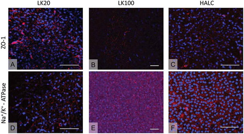
The pCECs were able to retain endothelial morphology and to form a monolayer composed by closely packed cells when seeded onto LK20, LK100, and HALC (). In addition, pCECs cultured on LK20, LK100, and HALC showed expression of the phenotypical markers ZO-1 ( A-C) and Na+/K+-ATPase (). Na+/K+ -ATPase expression had a more diffuse pattern all over the cell surfaces, whereas ZO-1 was mostly expressed on the cell borders.
Figure 3. pCEC morphology at P2 on different biocompatible carriers. Series of illustrative figures showing successful pCEC cultures at P2 on chosen biocompatible membranes. (A) LK20, (B) LK100, (C) HALC. Scale bars: 100 µm.
Figure 4. pCEC morphology check at P2 through immunofluorescence on different carriers. (A, B, C) ZO-1 expression detected in pCECs cultured on LK20, LK100, and HALC, respectively. (D, E, F) Na+/K+ expression detected in pCECs cultured on LK20, LK100, and HALC, respectively. Scale bars: 100 µm.
In vitro surgeries with pCEC-carrier constructs
DMEK-grafts were used as a positive control for in vitro surgeries () and served as a reference for the scoring (). The pCEC-HALC constructs behaved very similar to a DMEK-graft (, ) and were implanted using a modified DMEK technique. These constructs naturally rolled on themselves into a double roll, but with the endothelium on the internal surface, unlike a DMEK-graft where the endothelium is on the external surface. Staining of the pCEC-HALC constructs with 0.04% Trypan Blue for visualization compared well to a DMEK-graft (, ). The constructs could then be placed in a DMEK glass injector, with its correct orientation, i.e. curls of the double roll facing downwards, checked under a surgical microscope. The pCEC-HALC carrier was implanted in a DMEK-like normal fashion; combined manoeuvres with air, fluid, and corneal indentation could be used in order to successfully unfold the tissue and position it against the posterior surface of the corneal stroma. Although special caution was needed in order to position the endothelium in its correct orientation due to its “inverse endothelium inside” location when compared with the regular DMEK graft, this was successfully achieved in all cases. One hour post-in vitro surgery, after partial air bubble removal, all constructs showed at least partial attachment resulting in an average score of 3.5 (on a scale of 0–5 with 5 resembling a DMEK).
Table 1. In vitro surgeries with pCEC-carrier constructs: scoring scheme. Each parameter under evaluation was rated on a 5-point scale (i.e. 0–5) by two DMEK surgeons, with higher score indicating greater similarity to in vivo DMEK behaviour. A DMEK-graft was tested as a positive control and served as a reference point for the scoring. Values are reported as means.
Figure 5. In vitro surgeries with pCEC-carrier constructs. The upper three rows show each of the three tested pCEC-carriers constructs after staining with hypotonic Trypan Blue solution 0.04% (A,E,I), insertion into the artificial chamber (B,F,J), unfolding (C,G,K), and adherence to the bare stroma visualized by anterior segment OCT measurement (D,H,L). The lower row (M–P) shows the corresponding images of a DMEK-graft that served as a positive control and reference point for the scoring.
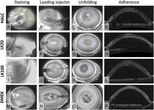
The pCEC-LK20 constructs showed a Trypan Blue staining behaviour similar to the pCEC-HALC constructs and DMEK-grafts (, ). These constructs had a tendency to form a large roll in BSS and could be loaded into the injector, but the “sticky” nature of the carrier prevented its easy insertion into the AC compared to a human DMEK-roll. Therefore, the pCEC-LK20 construct was folded over and inserted like a DSEK graft, a procedure in which the endothelial graft includes stromal tissue. Due to the increased thickness and rigidity, the DSEK-graft does not roll but is folded like a “taco” for insertion (). Implantation in the AC and unfolding were more technically demanding compared to a DMEK-graft or the pCEC-HALC construct, as the pCEC-LK20 construct tended to be sticky, fragile, and hard to centralize due to its biomechanical properties (, ). Adherence to the bare stroma was also not optimal (), resulting in score of 2.
Despite Trypan Blue staining (), pCEC-LK100 construct did not show any of the essential characteristics of a native DM (). Due to the thickness (100 μm) and increased rigidity of the carrier compared to normal DM, the construct was incapable to roll in BSS, and required a DSEK-like technique for implantation and unfolding (–K) with insufficient adherence (, ).
Discussion
In this study, we demonstrated that pCECs can be successfully cultured onto biocompatible, bioengineered collagen-based sheets, and onto HALC. Light microscopy and fluorescence microscopy analysis showed successful pCEC cultures up to P2 which retained their characteristic cell morphology, and distinctive expression of endothelial markers such as ZO-1 and Na+/K+-ATPase. However, upon in vitro transplantation, only the pCEC-HALC constructs behaved similar to DMEK-grafts in terms of staining, rolling in BSS, insertion in the anterior chamber, and attachment to the bare posterior stroma of a human donor corneo-scleral rim ().
Endothelial keratoplasty is now the preferred treatment for corneal endothelial disease, but scarcity of human donor tissue led to alternative approaches such as in vitro expansion of CEC on suitable cell carriers and injection of cultured human CEC.Citation9,Citation27–Citation29 In previous studies, it was shown that cultivated monkey CEC cultured on a collagen type I carriers for 4 weeks produced a confluent monolayer expressing ZO-1 and Na+/K+-ATPase, and at 6 months after transplantation into monkeys, the cornea was still clear with a normal cell density.Citation30 In addition, hCEC have been shown to grow into a confluent layer on collagen type I-coated culture plates,Citation31 and cultured hCEC on collagen sheets composed of cross-linked collagen type I transplanted in rabbits maintained 76–95% of pump function of human donor corneas.Citation11 Plastic compressed collagen type I, termed Real Architecture for 3D Tissues (RAFT), has been shown to support the growth of hCEC into a confluent monolayer expressing ZO-1 and Na+/K+-ATPase.Citation32 However, substrates like RAFT have been reported to give rise to inflammation in experimental animal models. In addition, cultivation of CEC on biological carriers such as DM, HALC, and amniotic membrane has been reported.Citation16,Citation17,Citation28,Citation33
Ideally, the carrier should mimic DM in its biological and biomechanical characteristics, since this may create a microenvironment required for cellular activity and mechanical support during transplantation using the DMEK technique.Citation28 To that end, we selected carriers that differed regarding their artificial extra-cellular matrix (ECM) composition, technological process, and biophysical (strength of cell-substrate adhesion or cell proliferation enhancement) and biomechanical properties (e.g. tensile strength, elasticity).
Our current study showed that pCECs cultured on the LinkCell™ collagen I-based sheets were able to retain endothelial morphology and to form a monolayer composed by closely packed cells with expression of ZO-1 and Na+/K+-ATPase. However, the behaviour of the pCEC-LK20 and pCEC-LK100 constructs during the in vitro DMEK surgery did not resemble that of a DMEK-graft since they had to be inserted in an artificial anterior chamber like a DSEK-graft (i.e. folded like a “taco”) instead of rolled-up like a DMEK-graft.
Our study suggests that HALC may be one approach for use as a biocompatible carrier for CEC, though the need for human-derived tissues is not eliminated through its use and further in vivo studies need to be performed. HALC has several benefits as it resembles DM in terms of composition; the major component of HALC is the basement membrane protein collagen IV and other matrix component include collagen types I and III, laminin, fibronectin, and heparin sulphate proteoglycans.Citation34 The use of HALC as a scaffold for the cultivation of different ocular cells,Citation35–Citation37 including hCEC,Citation17,Citation32 has already been reported. It was shown that hCEC seeded on de-epithelialized HALC grew to confluency and strongly expressed ZO-1 and Na+/K+-ATPase. We confirm these results with our pCEC-HALC constructs and showed that they can be used successfully in in vitro surgeries.
In conclusion, LK20 carrier showed good biocompatibility, but required a DSEK-adapted surgical protocol, while in vitro DMEK surgery showed HALC as the most suitable carrier for cultivated pCECs.
Ethical statement
The study was carried out following the tenets of the declaration of Helsinki.
Disclosure Statement
Dr. Melles is a consultant for DORC International/Dutch Ophthalmic USA and SurgiCube International. Dr. Dapena is a consultant for DORC International/Dutch Ophthalmic USA.
Dr. Rafat serves on the Board of Directors of the company LinkoCare Life Sciences AB, which is a spin-off firm developing products related to the research being reported, and holds relevant patents. Dr. Rafat’s terms of arrangements have been reviewed and approved by Linköping University in accordance with its policy on objectivity in research. The other authors have no conflicting relationship to disclose.
Additional information
Funding
References
- Joyce NC. Proliferative capacity of corneal endothelial cells. Exp Eye Res. 2012;95(1):16–23. doi:10.1016/j.exer.2011.08.014.
- Joyce NC, Meklir B, Joyce SJ, Zieske JD. Cell cycle protein expression and proliferative status in human corneal cells. Invest Ophthalmol Vis Sci. 1996;37:645–55.
- Joyce NC, Harris DL, Mello DM. Mechanisms of mitotic inhibition in corneal endothelium: contact inhibition and TGF-beta2. Invest Ophthalmol Vis Sci. 2002;43:2152–59.
- Murphy C, Alvarado J, Juster R, Maglio M. Prenatal and postnatal cellularity of the human corneal endothelium. A quantitative histologic study. Invest Ophthalmol Vis Sci. 1984;25:312–22.
- Tan DT, Dart JK, Holland EJ, Kinoshita S. Corneal transplantation. Lancet. 2012;379(9827):1749–61. doi:10.1016/S0140-6736(12)60437-1.
- Melles GR, Dapena I. How to get started with standardized ‘No-Touch’ descemet membrane endothelial keratoplasty (DMEK). 1st ed. Rotterdam: Netherlands Institute for Innovative Ocular Surgery; 2014.
- Melles GR, Ong TS, Ververs B, van der Wees J. Descemet membrane endothelial keratoplasty (DMEK). Cornea. 2006;25(2):987–90. doi:10.1097/01.ico.0000248385.16896.34.
- McCulley JP, Maurice DM, Schwartz BD. Corneal endothelial transplantation. Ophthalmology. 1980;87:194–201.
- Mimura T, Yamagami S, Amano S. Corneal endothelial regeneration and tissue engineering. Progr Retin Eye Res. 2013;35:1–17. doi:10.1016/j.preteyeres.2013.01.003.
- Ishino Y, Sano Y, Nakamura T, Connon CJ, Rigby H, Fullwood NJ, Kinoshita S. Amniotic membrane as a carrier for cultivated human corneal endothelial cell transplantation. Invest Ophthalmol Vis Sci. 2004;45:800–06.
- Mimura T, Yamagami S, Yokoo S, Usui T, Tanaka K, Hattori S, Irie S, Miyata K, Araie M, Amano S. Cultured human corneal endothelial cell transplantation with a collagen sheet in a rabbit model. Invest Ophthalmol Vis Sci. 2004;45(9):2992–97. doi:10.1167/iovs.03-1174.
- Watanabe R, Hayashi R, Kimura Y, Tanaka Y, Kageyama T, Hara S, Tabata Y, Nishida K. A novel gelatin hydrogel carrier sheet for corneal endothelial transplantation. Tissue Eng Part A. 2011;17(17–18):2213–19. doi:10.1089/ten.TEA.2010.0568.
- Liang Y, Liu W, Han B, Yang C, Ma Q, Zhao W, Rong M, Li H. Fabrication and characters of a corneal endothelial cells scaffold based on chitosan. J Mater Sci Mater Med. 2011;22(1):175–83. doi:10.1007/s10856-010-4190-6.
- Parikumar P, Haraguchi K, Ohbayashi A, Senthilkumar R, Abraham SJ. Successful transplantation of in vitro expanded human cadaver corneal endothelial precursor cells on to a cadaver bovine’s eye using a nanocomposite gel sheet. Curr Eye Res. 2014;39(5):522–26. doi:10.3109/02713683.2013.838633.
- Parikumar P, Haraguchi K, Senthilkumar R, Abraham SJ. Human corneal endothelial cell transplantation using nanocomposite gel sheet in bullous keratopathy. Am J Stem Cells. 2018;7:18–24.
- Kopsachilis N, Tsinopoulos I, Tourtas T, Kruse FE, Luessen UW. Descemet’s membrane substrate from human donor lens anterior capsule. Clin Exp Ophthalmol. 2012;40(2):187–94. doi:10.1111/j.1442-9071.2011.02678.x.
- Yoeruek E, Saygili O, Spitzer MS, Tatar O, Bartz-Schmidt KU, Szurman P. Human anterior lens capsule as carrier matrix for cultivated human corneal endothelial cells. Cornea. 2009;28(4):416–20. doi:10.1097/ICO.0b013e31818c2c36.
- Dapena I, Ham L, Melles GR. Endothelial keratoplasty: DSEK/DSAEK or DMEK–the thinner the better? Curr Opin Ophthalmol. 2009;20(4):299–307. doi:10.1097/ICU.0b013e32832b8d18.
- van Luijk CM, Bruinsma M, van der Wees J, Lie JT, Ham L, Melles GR. Combined chlorhexidine and PVP-I decontamination of human donor eyes prior to corneal preservation. Cell Tissue Bank. 2012;13(2):333–39. doi:10.1007/s10561-011-9260-6.
- Proulx S, Bourget JM, Gagnon N, Martel S, Deschambeault A, Carrier P, Giasson CJ, Auger FA, Brunette I, Germain L. Optimization of culture conditions for porcine corneal endothelial cells. Mol Vis. 2007;13:524–33.
- Brown DC, Gatter KC. Ki67 protein: the immaculate deception? Histopathology. 2002;40(1):2–11. doi:10.1046/j.1365-2559.2002.01343.x.
- Mikhailova A, Ilmarinen T, Ratnayake A, Petrovski G, Uusitalo H, Skottman H, Rafat M. Human pluripotent stem cell-derived limbal epithelial stem cells on bioengineered matrices for corneal reconstruction. Exp Eye Res. 2016;146:26–34. doi:10.1016/j.exer.2015.11.021.
- Barry PA, Petroll WM, Andrews PM, Cavanagh HD, Jester JV. The spatial organization of corneal endothelial cytoskeletal proteins and their relationship to the apical junctional complex. Invest Ophthalmol Vis Sci. 1995;36:1115–24.
- Yee RW, Geroski DH, Matsuda M, Champeau EJ, Meyer LA, Edelhauser HF. Correlation of corneal endothelial pump site density, barrier function, and morphology in wound repair. Invest Ophthalmol Vis Sci. 1985;26:1191–201.
- Vasquez Perez A, Liu C. Human ex vivo artificial anterior chamber model for practice DMEK surgery. Cornea. 2017;36(3):394–97. doi:10.1097/ICO.0000000000001112.
- Dapena I, Moutsouris K, Droutsas K, Ham L, van Dijk K, Melles GR. Standardized “no-touch” technique for Descemet membrane endothelial keratoplasty. Arch Ophthalmol. 2011;129(1):88–94. doi:10.1001/archophthalmol.2010.334.
- Bartakova A, Kunzevitzky NJ, Goldberg JL. Regenerative cell therapy for corneal endothelium. Curr Ophthalmol Rep. 2014;2(3):81–90. doi:10.1007/s40135-014-0043-7.
- Navaratnam J, Utheim TP, Rajasekhar VK, Shahdadfar A. Substrates for expansion of corneal endothelial cells towards bioengineering of human corneal endothelium. J Funct Biomater. 2015;6(3):917–45. doi:10.3390/jfb6030917.
- Kinoshita S, Koizumi N, Ueno M, Okumura N, Imai K, Tanaka H, Yamamoto Y, Nakamura T, Inatomi T, Bush J, et al. Injection of cultured cells with a ROCK inhibitor for bullous keratopathy. N Engl J Med. 2018;378(11):995–1003. doi:10.1056/NEJMoa1712770.
- Koizumi N, Sakamoto Y, Okumura N, Okahara N, Tsuchiya H, Torii R, Cooper LJ, Ban Y, Tanioka H, Kinoshita S. Cultivated corneal endothelial cell sheet transplantation in a primate model. Invest Ophthalmol Vis Sci. 2007;48(10):4519–26. doi:10.1167/iovs.07-0567.
- Choi JS, Kim EY, Kim MJ, Giegengack M, Khan FA, Khang G, Soker S. In vitro evaluation of the interactions between human corneal endothelial cells and extracellular matrix proteins. Biomed Mater. 2013;8(1):014108. doi:10.1088/1748-6041/8/1/014108.
- Levis HJ, Peh GS, Toh KP, Poh R, Shortt AJ, Drake RA, Mehta JS, Daniels JT. Plastic compressed collagen as a novel carrier for expanded human corneal endothelial cells for transplantation. PLoS One. 2012;7(11):e50993. doi:10.1371/journal.pone.0050993.
- Peh GSL, Ang HP, Lwin CN, Adnan K, George BL, Seah XY, Lin SJ, Bhogal M, Liu YC, Tan DT, et al. Regulatory compliant tissue-engineered human corneal endothelial grafts restore corneal function of rabbits with bullous keratopathy. Sci Rep. 2017;7(1):14149. doi:10.1038/s41598-017-14723-z.
- Danysh BP, Duncan MK. The lens capsule. Exp Eye Res. 2009;88(2):151–64. doi:10.1016/j.exer.2008.08.002.
- Hartmann U, Sistani F, Steinhorst UH. Human and porcine anterior lens capsule as support for growing and grafting retinal pigment epithelium and iris pigment epithelium. Graefes Arch Clin Exp Ophthalmol. 1999;237:940–45.
- Galal A, Perez-Santonja JJ, Rodriguez-Prats JL, Abad M, Alio J. Human anterior lens capsule as a biologic substrate for the ex vivo expansion of limbal stem cells in ocular surface reconstruction. Cornea. 2007;26(4):473–78. doi:10.1097/ICO.0b013e318033bd0f.
- Kozák I, Trbolová A, Zibrín M, Komorová T, Kolodzyeiski L, Juhás T. Electron microscopic study of anterior lens capsule allotransplants in chronic corneal ulcers. Cornea. 2004;23:797–803.