
Abstract
Purpose
The mechanism of action underlying prostaglandin analog (PGA) therapy involves changes in the expression of different metalloproteases to increase permeability of the sclera and allow increased aqueous humor outflow through this alternative drainage pathway. This alteration of structure impacts cornea/scleral biomechanics and may introduce artifact into the measurement of intraocular pressure (IOP) in the clinical setting.
Methods
A literature search reviewing the impact of PGA therapy on corneal and scleral biomechanics was conducted including basic studies, clinical studies with treatment naïve patients, and a clinical study examining the cessation of PGA therapy. Additional literature including engineering texts was added for greater clarity of the concepts underlying ocular biomechanics.
Results
One study with an animal model reported significant corneal stiffening with PGA treatment. Most longitudinal clinical studies examining the effects of initiation of PGA therapy in PGA naïve subjects failed to report biomechanical parameters associated with stiffness using the Corvis ST and only included those parameters strongly influenced by IOP. One study reported a significant reduction in scleral stiffness with IOP as a co-variate, highlighting the need to account for the effects of IOP lowering when assessing clinical biomechanics. The report of cessation of PGA therapy on corneal biomechanics showed no change in corneal compensated IOP after 6 weeks, raising the question of reversibility of the PGA-induced structural alteration.
Conclusions
Given that the findings in several clinical studies may merely reflect a reduction in IOP, further studies are warranted using Corvis ST parameters associated with corneal and scleral stiffness. The gold standard for IOP measurement in the clinical setting is Goldmann applanation tonometry, a technique previously shown to be affected by corneal stiffness. Since PGA therapy has been reported to alter not only scleral biomechanics, but also corneal biomechanics, it is essential to consider alternative tonometry technologies in the clinic.
Introduction
Over the last two decades, topical prostaglandin F2α analogs (PGA) have served as one of the first-line drugs for lowering intraocular pressure (IOP) in the treatment of glaucoma patients. The mechanism of action underlying this family of drugs has been shown to be largely dependent on the modulation of the uveoscleral outflow tract. Specifically, as demonstrated in both in vitroCitation1 and in vivoCitation2 models, prostaglandin analogs have been reported to upregulate the expression of matrix metalloproteinases (MMPs), proteases responsible for the degradation of the extracellular matrix in the eye.Citation1,Citation2 In addition, prostaglandin analogs have also been shown to decrease the expression of tissue inhibitors of metalloproteinases (TIMPs), protein regulators of MMP activity.Citation2,Citation3 The resulting shift in the ratio of MMPs to TIMPs thus causes an increased turnover of the collagen fibers that compose the extracellular matrix of the eye.Citation2,Citation3 This in turn, increases tissue permeability of the sclera and decreases the resistance to aqueous humor outflow.
More than simply increasing drainage of aqueous fluid, however, these changes to the extracellular matrix likely also affect the structural properties of the sclera. As the density of collagen fibrils in the extracellular matrix of the sclera decrease, more than just tissue permeability may be affected. Although no basic studies have been performed specifically examining the relationship between these PGA-induced changes and the mechanical properties of the sclera, it is likely that the decreased density of the collagen matrix would result in several changes including decreased stiffness and thus a lower resistance to deformation.
Given that studies have demonstrated the changes noted in MMP and TIMP expression to be pervasive throughout the conjunctival epithelium,Citation2,Citation3 it is also important to consider the effects of PGA treatment on other structures of the eye. The cornea, for example, has been indicated by recent studies as one of the structures potentially affected by concomitant changes secondary to PGA treatment.Citation4–6 Unfortunately, with only a few exceptions, there is once again a paucity of studies examining the specific effects of PGA on the cornea, as it is not directly involved in uveoscleral outflow. That being said, the effects of PGA on the structural properties of the cornea are of paramount importance as intraocular pressure (IOP) is traditionally measured through the cornea. As such, it is essential to closely examine the effects of PGA on the biomechanical properties of the cornea as these changes may affect standard IOP measurements.Citation7 To address these concerns, this review paper sets out to consolidate the current literature pertaining to PGAs and their effects on the biomechanical properties of specific structures of the eye.
Materials and methods
For this review paper, a literature review was performed primarily through the PubMed database using search terms to find studies pertaining to corneal biomechanics following prostaglandin treatment. Keywords included: prostaglandin analogs, cornea, corneal biomechanics, corneal hysteresis, Corvis ST, ocular response analyzer, and intraocular pressure. Due to the paucity of literature on the topic, most clinical studies pertaining to the topic were included based on the reporting of biomechanical parameters. Additional literature including basic science studies and engineering texts were included for greater clarity of the concepts underlying corneal biomechanics. Studies included were then further stratified into basic science studies, clinical studies with treatment naïve patients, and a clinical study examining the cessation of PGA therapy. The assessment systems used in the clinical studies, whether the Ocular Response Analyzer (ORA) or the Corneal Visualization Scheimpflug Technology (Corvis ST), were also included in our review.
Results
We identified articles pertaining to the effects of PGA therapy on corneal biomechanics, as well as scleral stiffness, and selected six core articles to discuss at length. The first was a basic science study demonstrating the effects of PGA therapy on strips of rabbit cornea. Four studies examining the effects of PGA therapy on treatment naïve patients and one report studying the effects of treatment cessation were also discussed.
The basics of biomechanics – stress and strain
Prior to delving into the intricacies of corneal and scleral biomechanics, it would be prudent to first introduce some of the core concepts underlying this discussion. This begins with the topic of material stiffness. Defined as the tendency for a material to return to its original form after being subjected to a force, the material stiffness of the cornea is difficult to characterize as the cornea is a composite material with regional variation in composition. As a result, studies examining the biomechanical properties of the cornea often focus on the tissue level relationship between stress and strain. Stress is defined as the amount of force distributed through a cross-sectional area and strain is the unitless deformation resulting from an applied stress relative to the original dimensions of the deforming structure.Citation8 For linear elastic materials, the relationship between stress and strain is linearly proportional and defined by a single mathematic constant or slope, known as the elastic modulus.Citation8 The cornea and other biologic materials, however, exhibit behavioral properties that are not so easily characterized.Citation9 Specifically, the cornea demonstrates a stress-strain relationship that is nonlinear, defined by a modulus that increases with higher IOP due to the crimping of collagen in the corneoscleral shell.Citation10 This nonlinear relationship has been well illustrated using ex vivo corneas.Citation11,Citation12 The result of a typical stress-strain plot of these experiments is shown in .
Figure 1. Schematic of the non-linear stress (load) vs strain (stretch) curve of the cornea. Some approaches for determining the tensile elastic modulus of the cornea include the secant and tangent modulus in which either the ratio of stress to strain or the derivative of the stress-strain curve is used for each point on the curve.
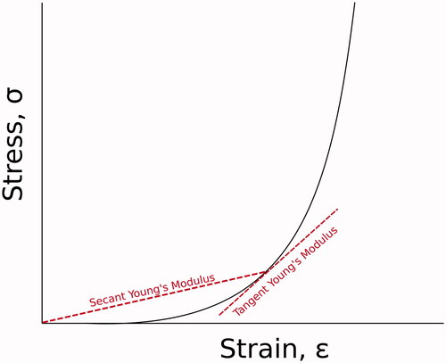
The non-linear nature of the cornea’s stress-strain relationship exemplifies the need to exercise caution when characterizing the mechanical properties of the cornea. The shape of the stress-strain curve illustrates that defining the cornea’s mechanical properties by a single elastic modulus is overly simplistic. In , two general approaches for determining the elastic modulus of the cornea for each point on the curve are shown: the secant and tangent modulus. The secant modulus is the slope of a line from the origin of the stress-strain diagram that intersects the stress-strain curve at the point of interest which can be calculated by the ratio of stress to strain (δ/ε). The tangent modulus on the other hand, is the slope of a line tangent to the stress-strain curve at the point of interest which requires calculating the slope or derivative (dδ/dε) of the stress-strain curve at the point of interest.Citation8
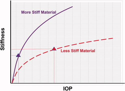
also demonstrates that the elastic modulus increases at greater levels of stress. It is important to note that an increased elastic modulus is associated with a stiffer response to applied forces.Citation8 This is clinically relevant as increased IOP results in greater applied stress to the inner surfaces of the eye and affects the biomechanical response to external forces applied to the cornea. Thus, when attempting to measure the biomechanical properties of the cornea, IOP has a confounding effect.Citation9 Specifically, as IOP increases, wall stress increases and provides increasing resistance to deforming forces. As such, it becomes more complicated to separate the innate biomechanical properties of the cornea from the effects brought about by changes in IOP. For example, at a higher IOP, a more compliant or softer cornea may exhibit stiffer behavior than that of a stiffer cornea at a lower IOP (). This highlights the need to contextualize the readings and findings we obtain from examining the biomechanics of the cornea, a consistent theme encountered as we delve further into the assessment of corneal and scleral biomechanical response.
Figure 2. Schematic illustrating the relationship of stiffness and IOP. This graph visually depicts how a more compliant cornea (dashed line) at a higher IOP (triangle on dashed line) may exhibit a stiffer response (greater resistance to deformation) than a cornea with stiffer properties (solid line) at lower IOP (triangle on solid line).Citation9
The basics of biomechanics – viscoelasticity
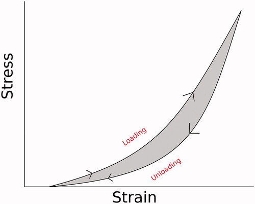
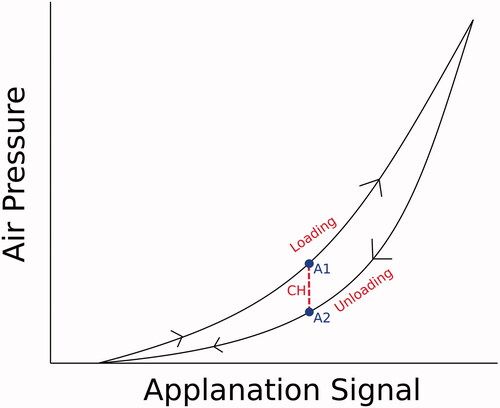
Adding even greater complexity to the discussion and examination of corneal biomechanics is the viscoelastic nature of the cornea. Unlike a purely elastic material that returns to its original shape and size via a mirrored path to that of its deformation or a viscous material in which force is entirely dissipated throughout the medium and lost as heat, the cornea exhibits both viscous and elastic properties when deforming. Thus, the tissue of the cornea is characterized as a viscoelastic material. As such, when stress is applied to the cornea, not all of the force applied is conserved as some of the energy is lost to internal friction, meaning the deformation brought about by the loading stress will be different from that of the stress during unloading. This difference between the resulting deformation associated with the loading and unloading stress is known as mechanical hysteresis and reflects the energy lost, as described above. This dissipation of energy can be visualized in comparing the stress-strain curve of the loading and unloading pathways for a stressor on the corneal tissue (). Compounded with the nonlinear elasticity of the cornea, the cornea’s viscoelastic nature adds an even greater challenge in measuring and characterizing the biomechanical properties of the cornea.
Figure 3. Schematic illustrating mechanical hysteresis. This curve demonstrates the different pathways taken during the loading and unloading phases of a nonlinearly elastic material. Mechanical hysteresis, the energy lost due to internal friction of the material, is the difference between these two pathways and is represented by the shaded area between the loading and unloading curves.
Due to its viscoelastic nature, the cornea deforms differently depending on the rate at which force is applied to it. Specifically, faster strain rates result in a stiffer corneal response, shifting the curve in to the left.Citation11 This means that both the amount of force and the time over which it is applied must be defined when attempting to examine the cornea’s biomechanical properties, as well as when comparing these properties between studies.
It is essential to interpret clinical IOP data gathered transcorneally in the context of the tonometry technique used to measure IOP. This is because different tonometers use different technologies representing the mechanism of force delivery which will influence the measurements gathered.Citation13,Citation14 It is well known that Goldmann Applanation Tonometry (GAT) is affected by central corneal thickness (CCT), resulting in overestimation of IOP in thick corneas and underestimation in thin corneas. However, it has also been theoretically shown to be affected by biomechanical properties, resulting in overestimation in a stiff cornea and underestimation in a compliant cornea.Citation7 As such, it is imperative to be aware of the potential measurement biases associated with each tonometer, especially when comparing studies using differing tonometry technologies.
The basics of biomechanics – properties related to corneal tissue
The stiffness response of the cornea is dependent on the structure of the corneal tissue and the cornea’s overall shape, which influences tonometry measurements.Citation7,Citation15,Citation16 Stiffness, defined as the resistance to deformation under an applied force, is influenced by a number of factors. In the cornea, both the thickness and collagen fibril density of the corneal tissue itself play a critical role. Specifically, a change in the thickness or collagen fibril density of the cornea represents a greater number of cross-linked collagen fibers, which in turn provides a greater resistance to deformation. This increased or decreased resistance to corneal deformation can lead to unaccounted overestimations or underestimations of IOP when measuring though the cornea.Citation17,Citation18 Although several algorithms have been proposed to account for this variance in corneal resistance, formulas based on a measured CCT are too simplistic as they only account for one aspect of this variance.Citation17 Indeed, studies have shown that numerous other factors can affect the collagen density of the cornea and by extension, the cornea’s tissue stiffness. These factors encompass a wide range of processes including age, hydration, and various common disease processes such as diabetes, keratoconus, and glaucoma.Citation18,Citation19
The basics of biomechanics – ocular rigidity
Ocular rigidity was described by Friedenwald as the measure of resistance that the eye exerts on distending forces of internal pressure, and serves as a parameter that defines the relationship between intraocular pressure and volume within the eye.Citation20 This relationship is nonlinear and is largely dependent not only on the material and tissue properties of the ocular shell, but also on the eye’s size and architecture.Citation21 Under the assumption that the properties governing ocular rigidity are largely unchanged by treatment, this relationship between pressure and volume is the governing rationale behind most current glaucoma therapies: the idea that a reduction in volume within the eye will follow Friedenwald’s curve and result in a corresponding decrease in IOP.
Reduction of corneal stiffness in an animal model after PGA treatment
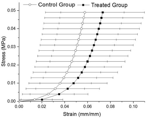
As mentioned previously, the core mechanisms of action for the Prostaglandin F2α analog (PGA) family of drugs are changes in MMP and TIMP activity that ultimately result in the increased degradation of the collagen fiber matrix in the eye and increased uveoscleral outflow.Citation1,Citation3 As the collagen fibril density is a contributor to the biomechanical properties of the cornea, changes brought about by PGAs could decrease corneal resistance to deformation and therefore introduce important inaccuracies when measuring IOP through the cornea.Citation22 Zheng et al. sought to explore this possibility through a uniaxial tensile testing model using strips of rabbit cornea treated with travoprost. Visualized in the figure (), the results from Zheng’s study demonstrated a rightward shift of the stress-strain curve of the lapin corneal tissue treated with travoprost when compared to the untreated tissue. This shift indicated less resistance to deformation which aligns well with the theory of that PGA treatment may generate underestimation of IOP clinically by reducing the stiffness of the tissue. Given the difficulty of clinically delineating the effects of PGA in reducing IOP versus the potential reduction brought about by the reduction in tissue stiffness, this study also notably removes the confounding variable of IOP to examine the effects of PGA on the tissue properties of the cornea. While the reduction of IOP following PGA treatment would normally result in a decrease in measured tissue stiffness, the rightward shift in , indicating a greater strain for a given stress, suggests that PGA treatment directly affects tissue stiffness.Citation23 The results of this study raise interesting points regarding the need for accuracy in IOP measurement, especially when used clinically as a guidepost for therapy in glaucoma management.
Figure 4. The mean stress-strain behavior of the corneas in Zheng’s study. Note that the treated group (black) exhibited a shift to the right from control (white), indicating a reduced resistance to deformation and greater strain for a given stress, compared to the control groups.Citation36
Clinical assessment of corneal biomechanics
The assessment of corneal biomechanics in a clinical setting poses yet another challenge as loads placed on the eye must be nondestructive. Additionally, the inability to precisely control the intraocular pressure during observation means that IOP must be taken into account when assessing parameters influenced by IOP. As illustrated in , failure to account for IOP would lead to inaccurate interpretation. As such, the biomechanical response of the cornea is assessed by examining parameters related to characteristics of the cornea’s deformation and the relationship of these parameters to stiffness. This is done using one of two commercial systems for clinically assessing corneal biomechanical deformation response: the Ocular Response Analyzer (ORA) or the Corneal Visualization Scheimpflug Technology (Corvis ST). Similar to traditional air-puff tonometers, both systems use an air jet to deform the cornea and assess response. However, the detection schemes and the air puff strategies for these two systems are starkly different. Since biomechanical response is a function of the applied load, these devices can be considered complimentary as they provide different biomechanical information.
The Ocular Response Analyzer
The first of the two systems, the Riechert ORA, uses a variable air puff to deform the cornea, such that an eye with higher IOP receives a greater magnitude air puff than an eye with lower IOP. As seen in , the detection scheme includes an infrared (IR) emitter aligned with the cornea and an IR detector to generate a spike on the detector when applanation (flattening) produces a mirror-like reflection. This occurs twice, once in the inward direction at the first applanation during the loading phase (A1), and once in the outward direction at the second applanation (A2), during the unloading phase.Citation24 Following A1, the cornea becomes slightly concave as the air pressure magnitude continues to rise to a maximum based on the timing of A1. The earlier A1 occurs, the lower is the maximum air pressure magnitude. As the cornea then returns toward its natural convex shape, it passes through A2. The air pressures measured at A1 and A2 are different as a result of the viscoelastic properties of the cornea. The difference between these two air pressures is defined as Corneal Hysteresis (CH), a measure of the cornea’s ability to dissipate energy. It is always a positive value in a valid measurement.
Figure 5. Example of an ORA measurement. As the cornea deforms under an air puff, the infrared (IR) applanation signal (red) represents the light collected by the detector with the corresponding geometry of the cornea shown at the top. Also plotted is the air-pressure signal (blue). As the cornea applanates, the light generates the first peak on the detector. The applanation signal then decreases as the cornea enters a state of slight concavity and peaks again in the outgoing direction during unloading, as the cornea goes through applanation a second time as it recovers its convex shape. P1 and P2 are the corresponding applanation pressures and Pmax is the maximum pressure reached.Citation9 Reprinted with permission by Wolters Kluwer Health, Inc.
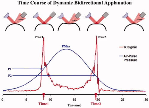
Analogous to the stress-strain curve demonstrated in , also visually demonstrates what CH represents with stress represented as a function of air pressure and strain represented by the deforming shape of the cornea captured by the Applanation signal of the ORA. A1 and A2 are at the same point on the Applanation signal since they are both the same flattened shape.
Figure 6. Schematic illustrating corneal hysteresis (CH). Specifically, this term is defined as the difference between inward and outward applanation pressures using the Ocular Response Analyzer (ORA). Pressures at applanation points, A1 and A2, are captured by the ORA during the loading and unloading pathways and used to calculate CH.
Over the years, the relationship between CH and several ocular disease processes have been examined. Perhaps most promisingly, CH has been reported as a potential indicator of progression in glaucoma. Specifically, eyes with low corneal hysteresis have been associated with visual field loss, potentially as a result of the diminished capacity to dissipate the additional stress caused by a high IOP.Citation25 As studies continue to examine the applications of CH, however, it is essential to remember that corneal hysteresis is not a static property. Corneal hysteresis is affected by several factors including IOP and tissue structure. As such, CH must always be interpreted in the context of other measurements, most importantly, IOP. The ORA also provides Corneal Compensated IOP (IOPcc), which was empirically developed to account for biomechanical affects on estimation of IOP and therefore provides a more accurate assessment than GAT.Citation9
Corvis ST studies and IOP-independent parameters
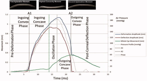
In contrast to the ORA, the Corvis ST uses an ultra-high-speed camera to image the corneal deformation in response to a consistent, precisely metered air puff. Every eye receives the same delivered air puff. Once the cornea reaches its limit of deformation and the air puff magnitude continues to increase, the whole eye moves in the backward direction. This allows capture of parameters related to whole eye motion in addition to those associated with corneal deformation. This system thus provides more measurable parameters to examine when assessing corneal and ocular biomechanics ().
Figure 7. Example of a Corvis ST measurement. As the cornea deforms under an air puff, the high-speed camera captures the changing geometry of the cornea shown at the top. Applanation events are noted as A1 and A2. The oscillation phase begins at the time point when the cornea has reached its limit of maximum deflection and the whole eye begins to move backwards as the air pressure continues to increase.Citation16 Reprinted with permission from SLACK Incorporated.
In expanding upon studies that utilize Corvis ST to examine and measure the biomechanical response of the eye, it is essential to analyze and report parameters that are less influenced by changes in IOP. Examples of these parameters focus on measurements that are dependent on the shape of deformation, rather than the magnitude of deformation. These include the integrated inverse radius (IIR) and the deformation amplitude ratio (DAR). Other metrics include those that focus on stiffness, including the Stress-Strain Index (SSI), the stiffness parameter at first applanation (SP-A1), and the stiffness parameter at highest concavity (SP-HC) which has been linked to scleral stiffness.Citation26 Collectively, these parameters could be used to examine the changes in corneal and scleral stiffness seen with prostaglandin therapy. We encourage authors who have already gathered data on the current population using Corvis ST to revisit their data in order to investigate these values and urge those designing studies in the future to ensure that these parameters are included.
Clinical studies on PGA treatment naïve patients
Although it remains relatively sparse, literature regarding the long-term effects of PGA treatment on the biomechanical properties of the cornea has expanded over the years. Despite this growth, however, many of the existing studies report findings that are confounded by differing experimental parameters as well as misconceptions caused by the complex interplay of factors affecting the measurement of the cornea’s biomechanical response.
In one investigation of the effects of PGA therapy in treatment naïve eyes with primary open angle glaucoma (POAG), Wu et al. suggests that long-term PGA treatment results in significant changes to corneal deformation parameters including a reduced time to first applanation (AT1), an increased first applanation velocity (AV1), a longer time until second applanation (AT2), a slower second applanation velocity (AV2) and an increased deformation amplitude (DA) as measured using Corvis ST.Citation4 Notably, all of the aforementioned parameters have been shown to be significantly associated with IOP and therefore correspond with changes that would be seen with a decrease in IOP, as expected from PGA treatment.Citation27,Citation28 AT1 is the time denoted when the magnitude of the air puff applied to the cornea causes first applanation of the cornea in the inward direction. AV1 describes the speed of the cornea at that time. The lower the IOP, the sooner the applanation occurs and the higher the speed of deformation. At the point of maximum concavity, DA is measured; this measurement is also primarily a function of IOP, as greater deformation is associated with lower IOP. This is addressed within the article as a possible explanation for the changes seen in corneal deformation, even alluding to other studies citing IOP as the strongest predictor of DA.Citation29 Unfortunately, these parameters alone are insufficient to suggest changes to the biomechanical properties of the cornea. The only conclusion that can be drawn from these parameters is that IOP was reduced. Other parameters that are less affected by changes in IOP, as discussed in , are available for analysis but were not reported in this study.
Table 1. Corvis ST parameters are separated into those that are primarily dependent on IOP versus those that are more predominantly influenced by corneal stiffness.Citation16,Citation28,Citation41
A study conducted by Bolivar et al. suggests a PGA-induced increase of CH unrelated to the IOP decrease induced by PGA treatment. In Bolivar’s study, however, this conjecture is premised on the notion that a regression analysis demonstrated a lack of correlation between the magnitudes of the increase seen in CH and the decrease in IOP following PGA treatment.Citation30 It should be noted that as previously described, corneal hysteresis is a measure of energy dissipation described by the difference between applanation pressures at loading and unloading. As such, the lack of a linear correlation does not indicate CH is unrelated to IOP.
In another study, Sanchez-Barahona et al. examined IOP readings following the initiation of topical latanoprost therapy in treatment naïve POAG patients using three tonometry methods (GAT, ORA and Corvis ST). Following a three month treatment period, Sanchez-Barahona reported a statistically significant difference between the measured average IOP reduction reported by Corvis ST compared to those measured using GAT and ORA.Citation31 Corneal compensated IOP (IOPcc), an ORA-specific measurement that attempts to account for the biomechanics of the cornea, was used to measure IOP with the ORA. In a similar fashion to Wu, they also reported significant changes in corneal applanation time (AT1 and AT2) and deformation amplitude (DA). As mentioned above in the discussion of Wu et al.’s study, AT and DA are parameters that are strongly influenced by IOP and thus, the changes seen can be explained by the reduction in IOP secondary to PGA therapy. Similarly, the only conclusion that can be drawn from the changes in these parameters is that IOP was reduced.Citation32 Also, the variance in IOP reduction noted between the tonometers should also be interpreted with caution as the differences may be explained by the differing tonometry technologies, both static (GAT) and dynamic (ORA and Corvis ST). In both Sanchez-Barahona’s and Wu’s case, Corvis ST parameters that have been shown to be less dependent on IOP, as previously described in , were not utilized in examining the potential changes to the biomechanical response of the cornea secondary to PGA therapy.
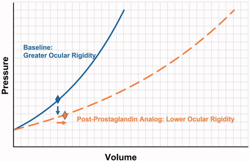
In a more recent study by Scott et al., the effects of PGA therapy on anterior chamber volume (ACV) and IOP were investigated. In this study, a decrease in IOP following PGA therapy was paradoxically shown to not be contingent upon reduction of the anterior chamber volume, in contrast to Friedenwald’s definition of ocular rigidity. From the findings of the study, 36% of the study eyes demonstrated an increase in anterior chamber volume (ACV) despite a measured mean reduction in IOP.Citation33 A reduction of volume within the eye is the primary goal when attempting to reduce IOP, thus these results provide compelling evidence that PGA therapy also alters ocular rigidity, possibly through changes in the properties of the cornea and/or sclera. Significant decreases in scleral stiffness, measured by SP-HC, was shown with IOP as a co-variate at 4 months following initiating of PGA treatment, offering a rationale for the reduction in ocular rigidity. visually demonstrates how an increase in anterior chamber volume can be accompanied by a decrease in IOP if there is also a change in ocular rigidity.
Figure 8. Visual representation demonstrating how a reduction in IOP (vertical arrow) from baseline value (diamond on solid line) to new value (diamond on dashed line) after Prostaglandin Analog (PGA) treatment can be accompanied by an increase in anterior chamber volume (horizontal arrow) if the ocular rigidity drops from a greater value at baseline (solid curve) to a lower value (dashed curve) caused by the reduction in scleral stiffness with PGA treatment.Citation33
Clinical studies on the cessation of PGA therapy
In examining patients that had been receiving prostaglandin therapy for at least one year, Meda et al. reported significant increases in CH, CRF, CCT and GAT in POAG patients after the cessation of PGA therapy in the study eye for 6 weeks, compared to the control eye which continued to receive PGA therapy.Citation5 Although an increase was reported in both CH and GAT in the study eye, this is incongruent with the well-established inverse relationship of CH and IOP. Investigating potential explanations for this discrepancy, it is important to note that Meda’s study reported a concomitant increase in CCT with the cessation of PGA therapy. This increased thickness would affect the stiffness response of the cornea and in turn, overestimate IOP when using Goldmann Applanation tonometry.Citation34,Citation35 Thus, it is likely that the true IOP value in this study was unchanged or changed very little, reinforced by the lack of significant change in IOPcc. This suggests that a washout period of only six weeks was insufficient for PGA therapy and that the measured increase in CH, CRF and GAT in this study could be explained by measurement artifact caused by an increase in CCT. It is likely that PGA-induced changes to the structure of the cornea and sclera have already occurred in these patients and as reflected by the lack of change in IOPcc, were likely not reversed within the six-week study period. As such, conclusions drawn from this study should be taken with caution, especially when comparing these results to studies of PGA therapy in treatment naïve patients.
Future studies – the reversibility of prostaglandin therapy?
Looking to the future, there are still many areas to be further studied in examining the potential effects of prostaglandin therapy on the biomechanics of the cornea and sclera. Unlike other glaucoma treatments, PGA therapy has been shown to cause changes to the properties of the cornea and sclera distinct from the confounding biomechanical effects brought about by alterations in IOP.Citation36 Scott et al.’s findings further reinforce the possibility of structural changes brought about by PGA therapy that may influence IOP measurements apart from the reduction of intraocular volume.Citation33 Based on the changes in MMP and TIMP expression following PGA induction, as described in studies conducted by Zheng et al. and Park et al., future studies could possibly examine the changes seen in MMP and TIMP expression following the cessation of PGA therapy. Currently, no studies exist regarding the effects of PGA cessation on MMP and TIMP expression. Studies could also be done to examine the specific structural changes possibly occurring in both the induction and cessation of PGA therapy. In doing so, studies could examine the potential reversibility of the modifications seen in the extracellular matrix and perhaps better characterize the tissue changes observed in the cornea and sclera following PGA therapy. As with Meda’s study, more research could also be done on the specific effects of PGA therapy washout and the potential reversibility of the structural changes secondary to prostaglandin analog use.Citation37
Clinical ramifications of PGA-induced biomechanical changes
Lastly, given that the gold standard for IOP measurement in the clinical setting is Goldmann applanation tonometry, a technique which has been shown to be affected by changes to corneal stiffness, it is imperative to consider the use of different tonometry technologies when studying the effects of PGA on IOP. It is likely that topical PGAs alter tissue properties of the cornea, thus, other technologies in addition to GAT should be used when measuring IOP, since these changes in tissue properties would result in artifact when measuring IOP. Even when assessing individual patients, it is likely that the decreased tissue stiffness would result in an overestimation of PGA efficacy with an underestimation of the post-PGA treatment IOP value. Although previous studies have suggested that PGA therapy is equal to or possibly superior to non-PGA therapies, such as beta-blocker therapy, it is essential to consider that if efficacy is defined by the magnitude of IOP reduction, PGA therapies will potentially have an innate advantage brought about by this IOP measurement artifact which would exaggerate IOP lowering effect.Citation38–40 In other words, a portion of the IOP lowering effect may be due to underestimation artifact related to a reduction of stiffness and a portion due to true reduction in pressure. As such, in future studies comparing PGA therapies to non-PGA therapies, it will be essential to consider the possibility of measurement artifact brought about by potential changes to corneal and scleral biomechanics.
Limitations of this review include the paucity of papers on the topic and the use of parameters that are influenced by IOP in several of the published studies to evaluate the potential biomechanical changes introduced by PGA therapy in the setting of a reduction in IOP. Given that many of the findings in these clinical studies may merely reflect lowering of IOP, further studies using Corvis ST parameters that are relatively independent of IOP, such as SP-A1, SP-HC, SSI, IIR and DAR are warranted.
There is still much to be explored with regard to the effects of PGA therapy on corneal and scleral biomechanics. We hope this literature review provides the foundation for future studies as there is still many unknown and important findings to learn.
Acknowledgements
Dr. Cynthia Roberts is a consultant to Oculus Optikgeräte GmbH and Ziemer Ophthalmic Systems AG.
Disclosure statement
The authors declare that they have no known competing financial interests or personal relationships that could have appeared to influence the work reported in this paper.
References
- Kim JW, Lindsey JD, Wang N, Weinreb RN. Increased human scleral permeability with prostaglandin exposure. Invest Ophthalmol Vis Sci. 2001; 42(7):1514–1521.
- Honda N, Miyai T, Nejima R, Miyata K, Mimura T, Usui T, Aihara M, Araie M, Amano S. Effect of latanoprost on the expression of matrix metalloproteinases and tissue inhibitor of metalloproteinase 1 on the ocular surface. Arch Ophthalmol. 2010; 128(4):466–471. doi:10.1001/archophthalmol.2010.40.
- Lopilly Park H-Y, Kim JH, Lee KM, Park CK. Effect of prostaglandin analogues on tear proteomics and expression of cytokines and matrix metalloproteinases in the conjunctiva and cornea. Exp Eye Res. 2012; 94(1):13–21. doi:10.1016/j.exer.2011.10.017.
- Wu N, Chen Y, Yu X, Li M, Wen W, Sun X. Changes in corneal biomechanical properties after long-term topical prostaglandin therapy. PloS One. 2016; 11(5):e0155527. doi:10.1371/journal.pone.0155527.
- Meda R, Wang Q, Paoloni D, Harasymowycz P, Brunette I. The impact of chronic use of prostaglandin analogues on the biomechanical properties of the cornea in patients with primary open-angle glaucoma. Br J Ophthalmol. 2017; 101(2):120–125. doi:10.1136/bjophthalmol-2016-308432.
- Amano S, Nejima R, Inoue K, Miyata K. Effect of topical prostaglandins on the biomechanics and shape of the cornea. Graefes Arch Clin Exp Ophthalmol. 2019; 257(10):2213–2219. doi:10.1007/s00417-019-04435-7.
- Liu J, Roberts CJ. Influence of corneal biomechanical properties on intraocular pressure measurement: quantitative analysis. J Cataract Refract Surg. 2005; 31(1):146–155. doi:10.1016/j.jcrs.2004.09.031.
- Palko JR, Liu J. Definitions and concepts. In Roberts CJ, Liu J (Ed). Corneal biomechanics: from theory to practice. 2016.
- Roberts CJ. Concepts and misconceptions in corneal biomechanics. J Cataract Refract Surg. 2014; 40(6):862–869. doi:10.1016/j.jcrs.2014.04.019.
- Gogola A, Jan N-J, Brazile B, Lam P, Lathrop KL, Chan KC, Sigal IA. Spatial patterns and age-related changes of the collagen crimp in the human cornea and sclera. Invest Ophthalmol Vis Sci. 2018; 59(7):2987–2998. doi:10.1167/iovs.17-23474.
- Elsheikh A, Wang D, Brown M, Rama P, Campanelli M, Pye D. Assessment of corneal biomechanical properties and their variation with age. Curr Eye Res. 2007; 32(1):11–19. doi:10.1080/02713680601077145.
- Elsheikh A, Alhasso D, Rama P. Biomechanical properties of human and porcine corneas. Exp Eye Res. 2008; 86(5):783–790. doi:10.1016/j.exer.2008.02.006.
- Salouti R, Alishiri AA, Gharebaghi R, Naderi M, Jadidi K, Shojaei-Baghini A, Talebnejad M, Nasiri Z, Hosseini S, Heidary F. Comparison among Ocular Response Analyzer, Corvis ST and Goldmann Applanation Tonometry in healthy children. Int J Ophthalmol. 2018; 11(8):1330–1336. doi:10.18240/ijo.2018.08.13.
- Hong J, Xu J, Wei A, Deng SX, Cui X, Yu X, Sun X. A new tonometer—the Corvis ST Tonometer: clinical comparison with noncontact and Goldmann Applanation Tonometers. Invest Ophthalmol Vis Sci. 2013; 54(1):659. doi:10.1167/iovs.12-10984.
- Kwon TH, Ghaboussi J, Pecknold DA, Hashash YMA. Effect of cornea material stiffness on measured intraocular pressure. J Biomech. 2008; 41(8):1707–1713. doi:10.1016/j.jbiomech.2008.03.004.
- Roberts CJ, Mahmoud AM, Bons JP, Hossain A, Elsheikh A, Vinciguerra R, Vinciguerra P, Ambrósio R. Introduction of two novel stiffness parameters and interpretation of air puff-induced biomechanical deformation parameters with a dynamic Scheimpflug Analyzer. J Refract Surg. 2017; 33(4):266–273. doi:10.3928/1081597X-20161221-03.
- Shih CY, Graff Zivin JS, Trokel SL, Tsai JC. Clinical significance of central corneal thickness in the management of glaucoma. Arch Ophthalmol. 2004; 122(9):1270–1275. doi:10.1001/archopht.122.9.1270.
- Krueger RR, Ramos-Esteban JC. How might corneal elasticity help us understand diabetes and intraocular pressure? J Refract Surg. 2007; 23(1):85–88. doi:10.3928/1081-597X-20070101-13.
- Blackburn BJ, Jenkins MW, Rollins AM, Dupps WJ. A review of structural and biomechanical changes in the cornea in aging, disease, and photochemical crosslinking. Front Bioeng Biotechnol. 2019; 7:66. doi:10.3389/fbioe.2019.00066.
- Friedenwald JS. Contribution to the theory and practice of tonometry*. Am J Ophthalmol. 1937; 20(10):985–1024. doi:10.1016/S0002-9394(37)90425-2.
- Kotliar K, Maier M, Bauer S, Feucht N, Lohmann C, Lanzl I. Effect of intravitreal injections and volume changes on intraocular pressure: clinical results and biomechanical model. Acta Ophthalmol Scand. 2007; 85(7):777–781. doi:10.1111/j.1600-0420.2007.00939.x.
- Meek KM, Blamires T, Elliott GF, Gyi TJ, Nave C. The organisation of collagen fibrils in the human corneal stroma: a synchrotron X-ray diffraction study. Curr Eye Res. 1987; 6(7):841–846. doi:10.3109/02713688709034853.
- Roberts CJ. Corneal effects of prostaglandins (Basic Science). Int Glaucoma Rev. 2019;20–1:22–24.
- Luce DA. Determining in vivo biomechanical properties of the cornea with an ocular response analyzer. J Cataract Refract Surg. 2005; 31(1):156–162. doi:10.1016/j.jcrs.2004.10.044.
- Zimprich L, Diedrich J, Bleeker A, Schweitzer JA. Corneal hysteresis as a biomarker of glaucoma: current insights. Clin Ophthalmol. 2020; 14:2255–2264. doi:10.2147/OPTH.S236114.
- Nguyen BA, Reilly MA, Roberts CJ. Biomechanical contribution of the sclera to dynamic corneal response in air-puff induced deformation in human donor eyes. Exp Eye Res. 2020;191:107904. doi:10.1016/j.exer.2019.107904.
- Miki A, Yasukura Y, Weinreb RN, Maeda N, Yamada T, Koh S, Asai T, Ikuno Y, Nishida K. Dynamic Scheimpflug ocular biomechanical parameters in untreated primary open angle glaucoma eyes. Invest Ophthalmol Vis Sci. 2020; 61(4):19. doi:10.1167/iovs.61.4.19.
- Vinciguerra R, Elsheikh A, Roberts CJ, Ambrósio R, Kang DSY, Lopes BT, Morenghi E, Azzolini C, Vinciguerra P. Influence of pachymetry and intraocular pressure on dynamic corneal response parameters in healthy patients. J Refract Surg. 2016; 32(8):550–561. doi:10.3928/1081597X-20160524-01.
- Huseynova T, Waring GO, Roberts C, Krueger RR, Tomita M. Corneal biomechanics as a function of intraocular pressure and pachymetry by dynamic infrared signal and Scheimpflug imaging analysis in normal eyes. Am J Ophthalmol. 2014; 157(4):885–893. doi:10.1016/j.ajo.2013.12.024.
- Bolívar G, Sánchez-Barahona C, Teus M, Castejón MA, Paz-Moreno-Arrones J, Gutiérrez-Ortiz C, Mikropoulos DG. Effect of topical prostaglandin analogues on corneal hysteresis. Acta Ophthalmol. 2015; 93(6):e495–e498. doi:10.1111/aos.12689.
- Sánchez-Barahona C, Bolívar G, Katsanos A, Teus MA. Latanoprost treatment differentially affects intraocular pressure readings obtained with three different tonometers. Acta Ophthalmol. 2019; 97(8):e1112–e1115. doi:10.1111/aos.14170.
- Roberts CJ. Assessing PG effects on IOP (Medical Treatment). Int Glaucoma Rev. 2020;20(4):61–62.
- Scott JA, Roberts CJ, Mahmoud AM, Jain SG. Evaluating the relationship of intraocular pressure and anterior chamber volume with use of prostaglandin analogues. J Glaucoma. 2021;30(5):421–427. doi:10.1097/IJG.0000000000001736.
- Agarwal DR, Ehrlich JR, Shimmyo M, Radcliffe NM. The relationship between corneal hysteresis and the magnitude of intraocular pressure reduction with topical prostaglandin therapy. Br J Ophthalmol. 2012; 96(2):254–257. doi:10.1136/bjo.2010.196899.
- Tsikripis P, Papaconstantinou D, Koutsandrea C, Apostolopoulos M, Georgalas I. The effect of prostaglandin analogs on the biomechanical properties and central thickness of the cornea of patients with open-angle glaucoma: a 3-year study on 108 eyes. Drug Des Devel Ther. 2013; 7:1149–1156. doi:10.2147/DDDT.S50622.
- Zheng XB, Wang Y, Zhao Y, Cao S, Zhu R, Huang W, Yu A, Huang JH, Wang QM, Wang JJ, et al. Experimental evaluation of travoprost-induced changes in biomechanical behavior of ex-vivo rabbit corneas. Curr Eye Res. 2019; 44(1):19–24. doi:10.1080/02713683.2018.1516781.
- Diaconita V, Quinn M, Jamal D, Dishan B, Malvankar-Mehta MS, Hutnik C. Washout duration of prostaglandin analogues: a systematic review and meta-analysis. J Ophthalmol. 2018;2018:3190684–3190688. doi:10.1155/2018/3190684.
- Sherwood M, Brandt J. Six-month comparison of bimatoprost once-daily and twice-daily with timolol twice-daily in patients with elevated intraocular pressure. Surv Ophthalmol. 2001; 45:S361–S368. doi:10.1016/S0039-6257(01)00219-3.
- Alm A, Stjernschantz J. Effects on intraocular pressure and side effects of 0.005% latanoprost applied once daily, evening or morning. Ophthalmology. 1995; 102(12):1743–1752. doi:10.1016/S0161-6420(95)30798-1.
- Fellman RL, Sullivan EK, Ratliff M, Silver LH, Whitson JT, Turner FD, Weiner AL, Davis AA. Comparison of travoprost 0.0015% and 0.004% with timolol 0.5% in patients with elevated intraocular pressure. Ophthalmology. 2002;109(5):998–1008. doi:10.1016/S0161-6420(02)01010-2.
- Eliasy A, Chen K-J, Vinciguerra R, Lopes BT, Abass A, Vinciguerra P, Ambrósio R, Jr. Roberts CJ, Elsheikh A. Determination of corneal biomechanical behavior in-vivo for healthy eyes using CorVis ST tonometry: stress-strain index. Front Bioeng Biotechnol. 2019;7:105. doi:10.3389/fbioe.2019.00105.