Abstract
The aim of this study was to evaluate radiation-induced pulmonary abnormalities of breast cancer patients. Altogether 202 consecutive patients receiving postoperative radiotherapy entered the study. Plain chest radiographs taken at entry and 3, 6 and 12 months after radiotherapy were evaluated according to modified Arriagada classification. In addition, pulmonary symptoms were recorded. Supplementary high-resolution computed tomography (HRCT) was employed in a subgroup of patients (n = 15). Plain radiographs were interpreted by a radiologist, and uncertain findings were re-evaluated by a radiologist together with a radiation oncologist. Grade 2 pneumonitis was the most common abnormality. The proportion of patients yielding a grade 2 finding was 22.5%, 28.1%, and 16.0% at three, six, and twelve months, respectively. There were 2 normal findings in HRCTscans, and 8 in plain radiographs of the same patients. Radiological lung abnormalities are common after radiotherapy, but they are usually reversible, and their significance for the patient's well-being is minor. No correlation between symptoms and lung or pleural reactions was seen.
Postoperative radiotherapy is widely used in breast cancer treatment and its value in reducing the risk of local and loco-regional recurrence is well recognized Citation[1–5]. Postoperative radiotherapy after breast cancer can however cause lung injuries as a complication Citation[6], Citation[7]. The pulmonary radioreaction in the irradiated area is an interstitial pulmonary inflammation, pneumonitis, with a certain degree of an intra-alveolar exudative component. If the threshold of potential complete restitution is crossed, the pneumonitis will develop into pulmonary fibrosis. Lung injury from radiation is divided into three sequential pathologic phases: an exudative phase, an organizing or proliferative phase, and a chronic fibrotic phase Citation[8]. In radiographs, these injuries are categorized as pneumonitis or fibrosis. Pneumonitis generally appears 3–6 months and fibrosis 6–12 months after radiotherapy. These side effects have usually been monitored by observing the patient's symptoms and plain chest radiographs. Although radiological changes in the lung after irradiation have been known for many decades, there are few prospective studies on the subject Citation[9–13]. Numbers of patients have been small in these studies Citation[9], Citation[10], Citation[13]. The information obtainable by computed tomography (CT) is more detailed than that from plain radiographs Citation[9], Citation[14]. High-resolution computed tomography (HRCT) has proved to be sensitive in detecting diffuse pulmonary changes after radiotherapy Citation[15], Citation[16]. CT has been used to compare different radiotherapy techniques and the lung injuries they cause Citation[17], Citation[18], but not for routine follow-up; plain radiographs have been the routine in follow-up studies. They have also been used in the comparison of lung abnormalities caused by different radiotherapic techniques Citation[19], Citation[20].
In the present study we evaluated the prevalence and grade of radiotherapy-induced lung injuries as seen in plain radiographs. Since interpretation of plain films is difficult we used consensus reading in cases of uncertain findings. For more detailed information we carried out HRCT on 15 consecutive patients to compare the information seen in the plain radiographs and HRCT.
Material and methods
During the period from October 10, 1997 to December 31, 1998 all (202) consecutive breast cancer patients who started postoperative radiotherapy were entered into a prospective study at the Department of Oncology in Tampere University Hospital in Finland. Patients were operated either by mastectomy (n = 42) or lumpectomy (n = 127) and all had axillary clearance for histologically confirmed carcinoma of the breast. Altogether 16 patients were excluded, eight because chest radiographs were not taken at entry as planned, six because the patient refused to fill in the questionnaire concerning symptoms and two because they did not receive the planned dose of radiotherapy. The number of patients eligible for final analysis was 186. All patients received either local (N-0) or locoregional (N-1) radiotherapy.
Computed tomography images were used in the planning of the target volume and in the dose calculation. The imaging process is described more detailed in our previous publication Citation[21]. PTV delineation was carried out in a 3-D radioptherapy planning system (CADPLAN®, Varian Associates Inc.). In node-negative patients the clinical target volume comprised the entire breast, including fatty tissue and skin. In node positive patients the thoracic wall and ipsilateral internal mammary and axillary and supraclavicular lymph nodes were also included. There are detailed instructions for the PTV delineation Citation[21]. The dose specification was according to ICRU Report 50 Citation[22]. The total dose of 50 Gy was given in 2 Gy fractions during 5 weeks, starting 4–8 weeks after surgery.
After lumpectomy the radiotherapy technique consisted of two opposite or nearly opposite isocentric tangential photon fields (5 or 6 MV). After mastectomy the radiotherapy comprised 6 MV photons for anterior and for regional lymph node areas (axillary, supraclavicular) and an extra posterior field of 25 MV for the axillary area. Furthermore an electron field was given for chest wall and internal mammary lymph node areas. The radiotherapy tecnique used is described in more detail in our previous publication Citation[21]. Adjuvant chemotherapy was given concomitantly with radiotherapy only for node-positive patients (n = 80). The chemotherapy regimen was intravenous cyclophosphamide, methotrexate, fluorouracil (CMF) Citation[21]. Patients reported all respiratory symptoms (dyspnea, cough, fever and chest pain) experienced before radiotherapy, during the first 3 months and the following 3 months' period after radiotherapy, using a standardized questionnaire Citation[21]. The questionnaire did not include smoking as a separate item.
Posterior-anterior and lateral chest radiographs were obtained before radiotherapy and at 3, 6 and 12 months after the end of radiotherapy. Altogether 17 chest radiographs were not taken as planned at three months and 8 at six months after radiotherapy. The same radiologist evaluated all radiographs retrospectively, having only the patients’ chest X-ray films with date available, but no other radiographs or other information such as clinical findings, symptoms, or which breast had been irradiated. The status of both lungs was evaluated in radiographs taken before commencement of radiotherapy. Pleura was similarly evaluated on both sides. Three months after the end of radiotherapy a new set of chest X-rays were taken and evaluated according to the modified Arriagada classification Citation[23], i.e. grade 0 = normal, 1 = uncertain, 2 = linear streaky, 3 = dense diffuse opacification, 4 = dense uniform opacification. The thickness of opacification in centimeters was measured in lateral chest radiographs. Also pleural reactions were evaluated and graded on both sides: 0 = normal, 1 = uncertain, 2 = certain new pleural reaction. The same evaluation was made at 6 and 12 months after radiotherapy. In this study pneumonitis is a radiological diagnosis.
Findings considered uncertain by the radiologist were re-evaluated by the same radiologist together with an experienced radiation oncologist and reclassified either as normal or grade 2, but still without additional information on the patient. The first fifteen patients also underwent CT and high-resolution computed tomography (HRCT) of the lungs at six months after the end of radiotherapy. CT scans were made on a Somatom CR scanner (Siemens, Erlangen, Germany) without intravenous contrast agent. Images were taken during maximal inspiration with 8 mm collimation over the entire lung area. Treated areas were imaged separately with 2 mm collimation for reconstruction, using a high spatial frequency (bone) algorithm and a 512 x 512 matrix.
HRCT images were evaluated by the same radiologist as the plain radiographs, without information regarding patient's symptoms and other findings. The interstitial abnormalities seen on HRCT scans were coded using criteria according to Schratter-Sehn and associates Citation[15]. The criteria were modified by quantitative analysis (mild, moderate, strong) (). The thickness of the pleural reaction adjacent to the reaction area of the lung was also evaluated. It was considered mild if the width was less than 0.5 cm, moderate between 0.5 and 1.0 cm, and strong over 1.0 cm. Grading was performed on the HRCT scan showing the strongest reactions. HRCT findings were compared to plain radiograph findings obtained on the same day. The study protocols were approved by the Ethical Committee of Tampere University Hospital.
Table I. Lung abnormalities by score in HRCT scans.
Results
Plain chest X-ray films were available in 169 cases at three months, 178 at six and 156 at one year after radiotherapy. Of these patients 127 (75.1%) had been irradiated after lumpectomy and 42 (24.9%) after mastectomy. The mean age of all patients was 58.9 years (range 31–80 years). There were 167 (98.8%) women and 2 men in the whole study.
Interpretation of plain radiographs
The radiographs taken before the radiation therapy showed solitary fibrotic scars in 41 patients (23.0% of all) on the side to be treated. No diffuse lung disease was observed. Of these 41, 20 patients (49.0%) did not develop any post-radiotherapy reactions. Lung reactions detected in plain radiographs at different intervals after radiotherapy are presented in . There were 23 (13.6%) uncertain findings (grade 1) among all radiographs viewed by the radiologist at three months after radiotherapy. In re-evaluation, fourteen (60.9%) of these uncertain findings were reclassified as normal and nine (39.1%) as grade 2 (linear streaky opacification). At six months after radiotherapy there were also 23 (12.9%) uncertain findings, of which 21 (91.3%) were reclassified as normal and two as grade 2 after consensus reading. In one case old scars and opacification in the plain radiograph misled the radiologist to grade the untreated side in error. All 3 uncertain findings at one year were reclassified as normal after consensus reading.
Table II. Lung reactions by grade detected in plain radiographs after radiotherapy.
Chest X-ray findings
The chest radiographs were normal in 54.4% (n = 92) at three months, in 61.2% (n = 109) at six months, and in 84.0% (n = 131) at one year after radiotherapy. Pneumonitis, grade 2 (linear, streaky) was the most common abnormality noted in 28.1% (n = 50) of all findings at the end of six months. The abnormalities in the pleura consisted of an elevated diaphragm, a diffuse contour or adhesions of the diaphragm. Pleural reaction was observed in 17.2% at three months and in 18.8% at six months after radiotherapy. Pleural fluid associated with radiation reactions was not detected in any plain radiographs during the whole study. Grade 3 (dense, diffuse opacification) radiation pulmonary reaction was noted in 13.6% (n = 23) of all cases and two patients evinced abnormalities in the pleura at three months. In 35.7% a pleural reaction was detected at six months after radiotherapy. Grade 4 (dense uniform opacification) abnormalities were found in 16 patients (9.5%) at three months and in 5 (2.8%) at six months after radiotherapy. In both of these groups there were two patients with pleural reaction. One patient had no lung abnormalities but a pleural reaction after the end of three months, and two at one year. Of all patients with grade 4 lung reaction two had received concomitant chemotherapy.
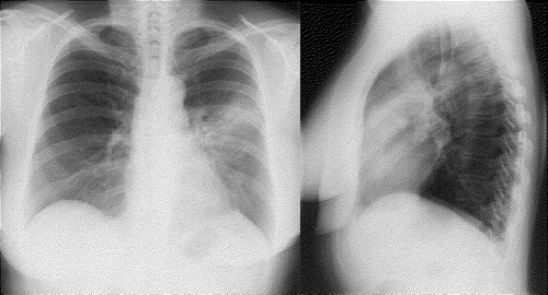
The opacification was thicker and therefore better measurable in grade 3 and 4 in the lateral plain radiograph. At the end of one year 25 patients (16.0%) showed mild streaky opacities (grade 2), and of these 61.5% evinced a pleural reaction (). Four of the 25 patients had received concomitant chemotherapy. Of these pleural abnormalities 77.8% were found at the end of one year, but not earlier. The distribution of reactions seen in chest radiographs at 3, 6 and 12 months after radiotherapy among patients having some reaction at 12 months, is presented in . Nine patients of the 25 with grade 2 reaction at one year had received loco-regional radiotherapy (no statistical difference between local and loco-regional groups; χ2=0.247), and four had received concomitant chemotherapy. Altogether 21 patients showed a grade 4 lung reaction. The mean dose volume histogram for these 21 patients was only slightly higher compared to that of all patients (a). Of these 21, only 4 had pulmonary symptoms. The individual DVHs of these patients are presented in b. Two of them were treated with corticosteroids. Chest radiographs of one of these two patients (patient 4 in b) are presented in . There was no correlation between symptoms reported by patients and lung reactions observed by the radiologist (χ2=0.313). Of those patients that had pre-therapy radiological changes on the treated side and had also post-therapy lung reactions in radiographs, 5 patients had subjective pulmonary symptoms (cough, dyspnea). Concomitant adjuvant chemotherapy did not seem to increase the likelihood of lung reactions Citation[21]. The thickness of opacification in chest radiographs was measurable in 34 cases at three months; mean thickness was 2.82 cm (range 1.0–6.0 cm) and in 17 cases at six months 3.2 cm (range 0.8–8.0 cm).
Figure 1. Radiological pulmonary findings (grade 0–4) at 3, 6 and 12 months (mo) after radiotherapy for the patients evincing some reaction at 12 months.
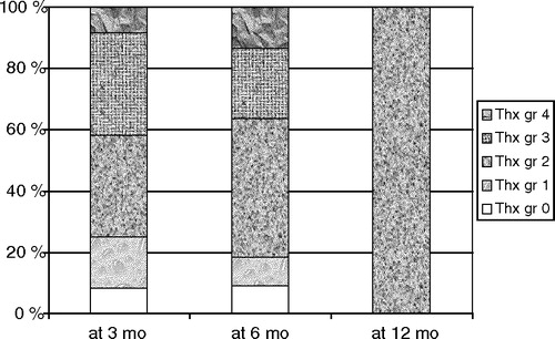
Figure 2. (a) Mean dose volume histograms (DVHs) for all patients and patients with grade 4 pulmonary findings in plain radiographs at either 3 or 6 months after radiation therapy according to type of operation (BCS = breast conserving surgery), M = mastectomy), and (b) DVHs of four individual patients with grade 4 findings also showing pulmonary symptoms.
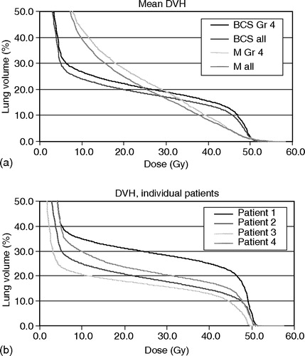
Figure 3. A 57-year old woman (Patient 4) 3 months after radiotherapy of the left breast. Grade-4 lesions = dense uniform opacification in upper lobe and lingula.
Findings in HRCT scans
The abnormalities seen in plain radiographs and in HRCT-scans in 15 patients at six months after the end of radiotherapy are summarized in . Two patients had received also chemotherapy. Two of these 15 patients (13.3%) yielded normal findings in the lungs and pleura in HRCT-scans, although plain radiographs were normal in 8 (53.3%) cases. Streaky linear opacities (grade 2) were found in plain radiographs of 7 patients (46.7%), but of these three (42.9%) had score 1 (thickened septal lines) with a quantitative analysis result strong (>10 lines) in HRCTscans, and in one patient also ground glass appearance, alveolar filling indicating an acute radiologic change. If the patient had other lung findings than score 1, they were all mild. Pleural reactions were found with lung abnormalities in HRCT scans, but of those three patients who had strong score 1 findings, only one evinced a moderate pleural reaction and the other two mild.
Table III. Lung abnormalities in plain radiographs and in HRCTscans.
Discussion
Radiographs are an essential part of the diagnosis and follow-up of pulmonary radiation damage in breast cancer patients. Acute and late lung reactions after radiotherapy for breast cancer have been evaluated mainly in small studies Citation[9], Citation[13], Citation[24–26]. The aim of this prospective study was to evaluate the prevalence of pulmonary lesions after radiotherapy and correlations to symptoms among breast cancer patients. The plain radiograph was normal in 54% of patients at three and in 61% at six months after radiotherapy, while earlier reports have reported some 80% as normal Citation[9], Citation[11–13], Citation[19], Citation[25]. The retrospective interpretation of radiographs may increase the number of positive findings. The great number of uncertain findings, 13.6% at three months and 12.9% at six months after radiotherapy, indicates the difficulty of interpretation of plain radiographs. The radiologist had no clinical data on the patient when interpreting the radiographs, which added to the uncertainty. We used consensus reading to resolve uncertain findings, as has been the approach in reading mammograms Citation[27]. There are only a few reports of irritative pleural reactions Citation[9], Citation[28]. In general pleural and lung reactions are interconnected. In our study there were three patients who evinced only pleural abnormalities without lung changes. We found no correlation between pleural reactions and grade of lung abnormalities. More than half of the patients with mild lung reactions had also pleural abnormalities. Of these almost all were found in plain radiographs only at one year after radiotherapy. Svane and colleagues reported delayed lung reactions at 4 years without preceding detection of radiologic changes Citation[7]. According to our data pleural reactions may likewise be delayed. Evaluation of lung changes was difficult, because the abnormalities were mainly linear streaky and diffuse opacities in plain radiographs.
In our study on HRCT, 13 (86.7%) of 15 patients evinced discrete parenchymal abnormalities after radiotherapy. None of them had symptoms, i.e. cough, dyspnea, fever or chest pain. The incidence of symptomatic radiation pneumonitis varies from less than 1% to 23%, but in most studies it is uncommon Citation[7], Citation[18], Citation[20], Citation[21], Citation[25], Citation[26], Citation[29–31]. This discrepancy is explained by the small lung volume irradiated for breast cancer. Kimsey and associates have shown that symptomatic pneumonitis occurs in patients who have more than 10% of the total lung volume irradiated Citation[24]; and a group under Lind reported an association between increasing chest radiograph scores and irradiated lung volumes Citation[19]. In our data symptomatic therapy (corticosteroid) was needed by only two patients (10%) with grade-4 findings in radiographs. Dense uniform opacification in radiograph usually corresponds to pathologically diagnosed excudative phase Citation[8]. Hernberg and colleagues reported on 10 symptomatic patients, only one of them needing symptomatic therapy Citation[26]. There was no correlation between symptoms and lung reactions seen in radiographs, even concomitant adjuvant chemotherapy did not increase lung reactions Citation[21]. Earlier lung changes on the treated side did not increase radiation reactions or symptoms. All different scores were seen here on HRCT, in contrast to Schratter-Sehn and associates, who reported no score-3 lesions in their seventeen-patient material Citation[15]. In our study, 8 of 15 patients had normal chest radiographs, although 6 of them showed lesions with scores 1–5 on HRCT, albeit none strong in quantitative analysis. Schratter-Sehn and group reported no normal finding in plain radiographs although all patients evinced changes in HRCT Citation[15]. Ooi and associates reported that all their 30 patients yielded HRCT findings at three months after radiotherapy and the situation was the same up to one year Citation[31]. Score-4 lesions, honeycombing areas, were seen in 3 patients, all mild reactions in our study. Since we had only a small subgroup of the total material in HRCT analysis and no follow-up with this method, we cannot evaluate the progression or regression of the lesions. Linear opacities at the margins of the radiation port, reticular opacities similar to those seen in patients with UIP (usual interstitial pneumonia), or discrete consolidation conforming to the shape of the radiation portals but not uniformly involving the irradiated lung, can be seen in some patients. The importance of HRCT for irradiated breast cancer patients emerges in differential diagnostics, especially when the patients have pulmonary symptoms. Irradiation induced changes in the lung tissue are not specific and in most cases radiographic findings are confined to the field of radiation. It is important to differentiate them from infection and metastatic spread.
As the symptoms do not correlate to radiographic findings, we do not recommend plain radiographs as a routine follow-up method after radiotherapy.
The trial was supported by the Medical Research Fund of Tampere University Hospital.
References
- Veronesi U, Saccozi R, Del Vecchio M, Banfi A, Clemente C, De Lena M, et al. Comparing radical mastectomy with quadrantectomy, axillary dissection, and radiotherapy in patients with small cancers of the breast. N Engl J Med 1981; 306: 6–11
- Høst H, Brennhovd IO, Loeb M. Postoperative radiotherapy in breast cancer: long-term results from the Oslo study. Int J Radiat Oncol Biol Phys 1986; 12: 727–32
- Overgaard M, Juul Christensen J, Johansen H, Nybo-Rasmussen A, Brincker H, van der Kooy P, et al. Postmastectomy irradiation in high-risk breast cancer patients. Present status of the Danish Breast Cancer Cooperative Group trials. Acta Oncol 1988; 27: 707–14
- Fisher B, Redmond C, Poisson R, Margolese R, Wolmark N, Wickerham L, et al. Eight-year results of a randomised clinical trial comparing total mastectomy and lumpectomy with or without irradiation in the treatment of breast cancer. N Engl J Med 1989; 320: 822–8
- Rutqvist LE, Cedermark B, Glas U, Johansson H, Rotstein S, Skoog L, et al. Radiotherapy, chemotherapy and tamoxifen as adjuvants to surgery in early breast cancer: a summary of three randomized trials. Int J Radiat Oncol Biol Phys 1989; 16: 629–39
- Sigmund G, Slanina J, Hinkelbein W. Diagnosis of radiation-pneumonitis. Recent Results Cancer Res 1993; 130: 123–31
- Svane G, Rotstein S, Lax I. Influence of radiation therapy on lung tissue in breast cancer patients. Acta Oncol 1995; 34: 845–9
- Davis SD, Yankelevitz DF, Henschke CI. Radiation effects on the lung: clinical features, pathology,and imaging findings. AJR Am J Roentgenol 1992; 159: 1157–64
- Bell J, McGivern D, Bullimore J, Hill J, Davies ER, Goddard P. Diagnostic imaging of post-irradiation changes in the chest. Clin Radiol 1988; 39: 109–19
- Roberts MC, Foulcher E, Zaunders JJ, Bryant DH, Freund J, Cairns D, et al. Radiation pneumonitis: A possible lymphocyte-mediated hypersensitive reaction. Ann Intern Med 1993; 118: 696–700
- Holli K, Pitkänen M. Tangential breast irradiation with or without internal mammary chain irradiation: results of a randomized trial. Radiother Oncol 1995; 36: 172–6
- Fernando IN, Powles TJ, Ashley S, Grafton D, Harmer CL, Ford HT. An acute toxicity study on the effects of synchronous chemotherapy and radiotherapy in early stage breast cancer after conservative surgery. Clin Oncol 1996; 8: 234–8
- Ooi GC, Kwong DL, Ho JC, Lock DT, Chan FL, Lam WK, et al. Pulmonary sequelae of treatment for breast cancer: a prospective study. Int J Radiat Oncol Biol Phys 2001; 50: 411–9
- Coscina WF, Arger PH, Mintz MC, Coleman BG. CT demonstration of pulmonary effects of tangential beam radiation. J Comput Assist Tomogr 1986; 10: 600–2
- Schratter-Sehn AU, Schurawitzki M, Zach M, Schratter M. High-resolution computed tomography of the lungs in irradiated breast cancer patients. Radiother Oncol 1993; 27: 198–202
- Majurin M-L, Valavaara R, Varpula M, Kurki T, Kulmala J. Low-dose and conventional-dose high resolution CT of pulmonary changes in breast cancer patients treated by tangential field radiotherapy. Eur J Radiol 1995; 20: 114–9
- Newman G, Bell J, Goddard P, Bullimore JA. Pulmonary changes in breast cancer patients treated by three different radiotherapy techniques. Clin Oncol 1989; 1: 91–6
- Wennberg B, Gagliardi G, Sundbom L, Svane G, Lind P. Early response of lung in breast cancer irradiation: radiologic density changes measured by CT and symptomatic radiation pneumonitis. Int J Radiat Oncol Biol Phys 2002; 52: 1196–206
- Lind PARM, Bylund H, Wennberg B, Svensson C, Svane G. Abnormalities on chest radiographs following radiation therapy for breast cancer. Eur Radiol 2000; 10: 484–9
- Lind PARM, Marks LB, Hardenbergh PH, Clough R, Fan M, Hollis D, et al. Technical factors associated with radiation pneumonitis after local±regional radiation therapy for breast cancer. Int J Radiat Oncol Biol Phys 2002; 52: 137–43
- Holli K, Pitkänen M, Järvenpää R, Rajala J, Lahtela S, Hyödynmaa S, et al. Early skin and lung reactions in breast cancer patients after radiotherapy: prospective study. Radiother Oncol 2002; 64: 163–9
- International Commission on Radiation Units and Measurements. Prescribing, recording, and reporting photon beam therapy. ICRU Report 50. Bethesda, MD:ICRU;1994.
- Arriagada R, Ladron de Guevara JC, Mouriesse H, Hanzen C, Couanet D, Ruffie P, et al. Limited small cell lung cancer treated by combined radiotherapy and chemotherapy: evaluation of a grading system of lung fibrosis. Radiother Oncol 1989; 14: 1–8
- Kimsey FC, Mendenhall NP, Ewald LM, Coons TS, Layon AJ. Is radiation treatment volume a predictor for acute or late effect on pulmonary function?. Cancer 1994; 73: 2549–55
- Cazzaniga LF, Bossi A, Cosentino D, Frigerio M, Martinelli A, Monti A, et al. Radiological findings when very small lung volumes are irradiated in breast and chest wall treatment. Radiat Oncol Investig 1998; 6: 58–62
- Hernberg M, Virkkunen P, Maasilta P, Keyriläinen J, Blomqvist C, Bergh J, et al. Pulmonary toxicity after radiotherapy in primary breast cancer patients: results from a randomized chemotherapy study. Int J Radiat Oncol Biol Phys 2002; 52: 128–36
- Anttinen I, Pamilo M, Soiva M, Roiha M. Double reading of mammography screening films-One radiologist or two?. Clin Radiol 1993; 48: 414–21
- Fennessy JJ. Irradiation damage to the lung. J Thorac Imaging 1987; 2: 68–79
- Rotstein S, Lax I, Svane G. Influence of radiation therapy on the lung-tissue in breast cancer patients: CT-assessed density changes and associated symptoms. Int J Radiat Oncol Biol Phys 1990; 18: 173–80
- Shapiro CL, Recht A. Side effects of adjuvant treatment of breast cancer. N Engl J Med 2001; 344: 1997–2008
- Ooi GC, Kwong DLW, Chan KN, Ngan H, Lock DTW, Lam WK, et al. Serial HRCT lung changes after 3-field radiation treatment of breast cancer. Clin Radiol 2000; 55: 817–24