Abstract
Female sex steroids are implied in breast cancer development. The estrogen (ER) and progesterone (PR) receptor subtypes may have different roles to modulate the cellular response. Paired samples of cancer and adjacent normal tissue were collected from postmenopausal women at surgery for ductal breast cancer. The expression of ERα, ERß, PRA and PRB was quantified by immunostaining and digitized image analysis. We found ERß to be significantly reduced in breast cancer tissue (35% vs 50%; p = 0.001) and there was also a decrease of the ERß/ERα ratio. Among women using hormones at the time of diagnosis tumor tissue showed higher values for both PRB and PRA, as compared to women without such treatment. The results extend previous animal data to be valid also in women. There is evidence that loss of ERß expression may relate to estrogen dependent tumor progression. Increased PR expression could possibly relate to breast cancer risk during combined estrogen/progestogen treatment.
The expression of sex steroid receptors in breast cancer tissue has been established to predict the clinical response to endocrine treatment. Thus assessment of breast cancer receptor status in individual patients is mandatory for the choice of treatment both in recurrent disease and in the adjuvant setting.
The sex steroid receptors ER and PR are nuclear proteins which function as transcription factors to regulate the expression of specific genes. The first estrogen receptor, ERα was sequenced in 1986 and the second called ERß was discovered and sequenced ten years later Citation[1]. Although derived from different genes they are virtually similar in the DNA binding domain but to a lesser extent (about 58%) in the ligand binding domain. The two receptor variants seem to elicit a different response to compounds with estrogen agonist and/or antagonist properties Citation[2].
ERß has been reported to be more abundant in normal breast tissue and there are indications that ERß may modulate and even counteract the effects of ERα Citation[3]. Data suggest that ERß is poorly activated at low estradiol concentrations and functions when activated as an inhibitor of ERα Citation[4]. ERß expression is high in resting breast epithelium, while low values have been found in proliferating breast tissue. A change in the mRNA ratio for ERß /ERα expression has been suggested to occur during breast carcinogenesis Citation[3], Citation[5].
Studies in cell cultures and in animal models on the two progesterone receptor isoforms PRA and PRB indicate that PRA may have a repressive function on PRB action Citation[6]. It is suggested that the regulated expression of PRA and PRB is critical to the mammary gland response to progesterone Citation[7].
Numerous women are currently treated with estrogen and progestogen for the alleviation of menopausal symptoms. Epidemiological and clinical data show an increased risk of breast cancer during such hormonal therapy (HT) Citation[8]. There is substantial evidence that progestogens are mitogenic on the human breast when given long term to postmenopausal women Citation[9].
Animal experiments and observations in women suggest that treatment with estrogen/progestogen may have diverging effects on the expression of ER and PR isoforms in the breast epithelium. Previously in preinvasive mammary tumors, decreased levels of ERß protein were found to be associated with increased cell proliferation Citation[3]. A suppression of the proliferation inhibiting isoforms ERß and PRA could be one mechanism for increased cell proliferation during combined estrogen/progestogen treatment Citation[2], Citation[10], Citation[11].
At present it is unclear how receptor expression is regulated and how it may change between normal and cancerous tissue and during exogenous hormonal treatment Citation[11]. In particular there is a lack of human data. Most of our knowledge so far comes from cell culture experiments and studies in rodents and other animal models Citation[12]. Therefore, we performed a pilot-study in postmenopausal women with primary breast cancer. Pair-wise samples of normal and malignant breast tissue were collected during surgery and the expression of ER and PR subtypes was quantified by immunohistochemical image analysis.
Material and methods
Patient characteristics
Consecutive breast tissue samples were collected from postmenopausal women undergoing breast cancer surgery at St Görans Hospital, Stockholm, Sweden. From this material a total of 37 women with invasive primary ductal breast cancer were identified. Among these women 14 were on hormonal treatment and 23 did not receive such treatment at the time of diagnosis. Women were defined as hormone users if they had been on systemic treatment with estrogen only (estradiol or conjugated estrogens) or estrogen in combination with progestogen (levonorgestrel, norethisterone or medroxyprogesterone acetate) until at least one month before diagnosis. The study was approved by the local ethics committee of the Karolinska Institute (98–173) and all women gave their informed consent to participate. Data on patient characteristics e.g. age, tumor size, ER status and type of surgery are given in . There were no significant differences between groups.
Table I. Patient characteristics in 37 postmenopausal women with and without hormone therapy at diagnosis, undergoing surgery for invasive ductal breast cancer.
Breast tissue sampling
All tissues were collected at breast surgery for therapeutic purpose. For each patient, tissue blocks containing the tumor and also adjacent histologically normal glandular structures at the periphery of lesions were collected and frozen at −70°C until analysis. In addition to routine pathological analysis all samples were subject to a central reanalysis by an experienced pathologist to define invasive ductal carcinoma and normal tissue. Tissue was primarily used for immunohistochemistry (IHC) and when large enough also for mRNA analyses of ERα and ERß.
Immunohistochemistry
Paraffin sections (5 µm) were used and a standard immunohistochemical technique (avidin-biotin-peroxidase) was carried out to analyze ER and PR immunostaining intensity and distribution. Monoclonal mouse anti-human antibodies were used for detection of ERα (08-1149, Zymed Laboratories, Inc., South San Francisco, CA), ERβ (MCA 1974, Serotec Ltd, Oxford, UK), PRA (Novacastra, Newcastle, UK) and PRB (MAI-411, Affinity Bioreagents Inc San Francisco, CA). Negative controls were prepared by replacing the primary antibodies with mouse IgG.
After the tissue sections were dewaxed and rehydrated, an antigen retrieval procedure was performed. Sections were pretreated in a microwave oven at high power, in 0.01M sodium citrate buffer (pH 6.0) for 10 min and then allowed to cool for a further 20 min at room temperature (RT). Following washing in buffer (0.1M PBS, pH 7.4, for ERα, PRA, PRB and 0.1M Tris-buffered saline (TBS), pH 7.4, for ERβ), non-specific endogenous peroxidase activity was blocked by treatment with 3% hydrogen peroxide (Merck, Darmstadt, Germany) in methanol for 10 min at RT. Following 10 min wash in buffer, sections were exposed for 30 (ERα, PRA, PRB) and 45 (ERβ) min respectively to normal horse serum (Vector Laboratories, Burlingame, CA) diluted in PBS (ERα, PRA, PRB) or TBS containing 5% (w/v) BSA (ERβ) in a humidified chamber at RT. The tissue sections were thereafter incubated with the primary antibody. The following antibody dilutions were used: ERα 1:5 in PBS, ERβ 1:20 in TBS with 5% BSA, PRA 1:500, and PRB 1:100. All incubations with monoclonal antibodies were carried out at 4°C overnight (ERα, ERβ) or 60 min RT (PRA, PRB). The sections were then incubated for 30 (ERα), 45 (PRA, PRB) or 60 (ERβ) min with the second antibody: a biotinylated horse anti-mouse IgG (Vector Laboratories), diluted in normal horse serum. After incubation for 30 (ERα, ERβ, PRA) or 60 (PRB) min with horseradish peroxidase-avidin biotin complex (Vectastain Elite, Vector, CA), the bound enzyme was visualized by the application of 3,3′-diaminobenzidine (DAKO Cytomation, Carpinteria, CA, USA). The sections were counterstained with haematoxylin and dehydrated before mounted with Pertex®.
Image analysis
A Leica microscope connected to a computer, using Colorvision software (Leica Imaging System Ltd. Cambridge, UK), was used to assess immunostaining quantitatively by a computer image analysis system. Quantification of immunostaining was performed on the digitized images of systematic randomly selected fields of mammary glands, from which the stroma was interactively excluded. Ten fields of glandular cells were measured separately in each tissue section except for a few cases with less material, where all glands in the section were measured. Using color discrimination software the total area of all positively stained nuclei irrespective of staining intensity was measured and expressed as the ratio of the total area of cell nuclei. Staining intensity was not included in the analysis. The reproducibility and diagnostic relevance of this parameter has been questioned Citation[13], Citation[14]. The results from image analysis were compared with manual scoring of cells with a positive or negative staining (two different observers blinded to treatment). There was a strong positive correlation between the two methods (rs 0.80; p < 0.001).
mRNA determinations
The fresh frozen tissue specimens were homogenized in a SDS-containing buffer, digested with Proteinase K and subsequently extracted with phenol:chloroform for preparation of total nucleic acids (TNA). The concentration of DNA in the TNA samples was measured fluorometrically at the wavelength 458 nm. For measurements of specific mRNA, probes were synthesized in vitro (using reagents supplied from Promega Biotech., Madison, Wisconsin) and radio labelled with 35S-UTP (Amersham, Bucks., UK). Solution hybridization assays were then performed. In short, 35S-UTP labelled cRNA was hybridized (20.000 cpm/incubation) at +70°C to the TNA samples. Incubations were performed in duplicate in microcentrifuge tubes (Treff AG, Switzerland) in a total volume of 40 µl containing 0.6 mol/l NaCl, 20 mmol/l Tris-HCl pH 7.5, 4 mmol/l EDTA, 0.1% SDS, 0.75 mmol/l dithiothreitol, and 25% formamide. After incubation overnight, each sample was treated for 45 min at 37°C in 1 ml of a solution containing 40 µg RNase A, 0.3 µg RNase T1 (Boehringer-Mannheim, Mannheim, Germany) and 100 µg calf thymus DNA, to digest non-hybridized RNA. Labelled hybrids protected from RNase digestion were precipitated by addition of 100 µl 100% trichloroacetic acid (TCA) and collected on filters (Whatman GF/C). The radioactivity on the filters was measured and the results were compared with a standard curve of known amounts of in vitro synthesized mRNA complementary to the probe used. Results were expressed as a mol (10−18) mRNA/µg DNA in the TNA samples.
Hybridization probes
The probe used for ERα mRNA determinations was descended from bcpe1, a full length cDNA of 4838 bases containing the whole open reading frame of the human estrogen receptor. The cDNA was inserted in a pGEM7zf vector. Restriction of this vector with BglII allows the synthesis of a probe corresponding to nucleotides 4262-4838 which encode the C-terminal half of the steroid binding domain (E) and all of domain F.
The probe used for ERβ mRNA determinations was derived from a pBS plasmid with an insert of a 187 bp PvuII/EcoRI fragment corresponding to nucleotides 774-979 in the N-terminal part of the human ERβ gene.
Statistical analysis
Differences between groups were assessed by the Mann-Whitney U-test and differences between paired normal and malignant tissues by the Wilcoxon signed rank test. Correlations were calculated using the Spearman's rank correlation test. A p-value of < 0.05 was considered as significant.
Results
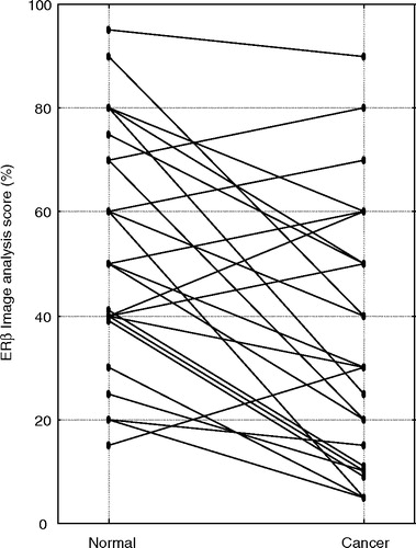
Samples of apparently normal and malignant tissue were collected during primary cancer surgery. In some cases pair wise assessment could not be performed due to low amounts of relevant epithelial tissue. ERα, ERβ, PRA and PRB were all expressed in the nuclei of both normal and malignant breast tissue (). Overall the immunostaining for the ER subtypes was more abundant than that of PRA and PRB. The expression of ERα and ERβ isoforms as assessed by image analysis (median and range) in primary ductal carcinoma and normal tissue for the whole material is illustrated in . Breast cancer tissue showed numerically higher values for ERα as compared to normal tissue (81% vs 52%; p = 0.06). In contrast there was a decreased expression of ERß in cancer tissue. Median values as compared to normal tissue were for the total material 35% vs 50% (p = 0.001), in women without prior HT 40% vs 55% (p < 0.01) and in women with HT at diagnosis 30% vs 50% (p = 0.07) respectively. Individual values for the total material of paired samples are displayed in . In breast cancer tissue there was also a significant decrease of the ERß/ERα ratio (p < 0.02).
Figure 1. Representative pictures of immunostaining for ERα (a: normal; b: cancer) and ERß (c: normal; d: cancer) in paired breast tissue samples. Pictures e–h denote immunostaining for PRA (e: no HT; f: HT) and PRB (g: no HT; h: HT) in tumor tissue from two individual women without and with hormone therapy. Pictures i–l represent negative controls (i: ERα normal; j: ERß normal; k: PRA cancer and l: PRB cancer).
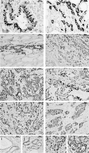
Figure 2. Image analysis score for the whole material. Percentage area of cells positive for ERα (normal n = 29; cancer n = 33) and for ERß (normal n = 29; cancer n = 32). Box-and-whisker plots representing the median value with 50% of all data falling within the box. The “whiskers” represent the range.
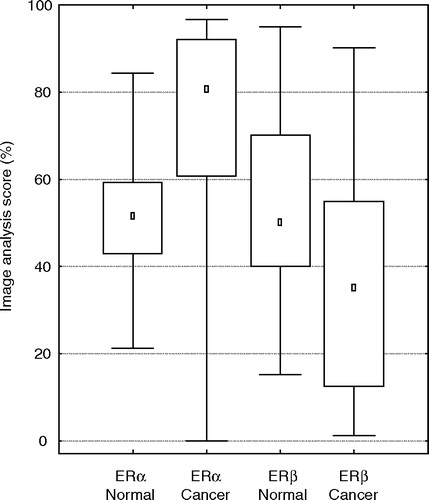
Figure 3. Image analysis score for the expression of ERß in paired samples of normal and cancer tissue obtained from 25 postmenopausal women.
In the total material the mRNA levels for ERα were significantly increased in tumorous (n = 24) as compared to normal (n = 21) tissue (mean 11.6 vs 6.3 amol/µg DNA; p < 0.03). A similar increase was found also for ERß (mean 34.5 vs 18.7 amol/µg DNA; p < 0.01). These differences were apparent among women not using HT (mean mRNA ERα: 12.8 vs 6.2; mean mRNA ERß: 37.0 vs 17.7) but in the subgroup of 14 women on hormone therapy at the time of diagnosis there were no significant differences between the mRNA levels (mean mRNA ERα: 8.9 vs 6.5; mean mRNA ERß: 28.5 vs 20.7).
As regards PR expression at image analysis there was an overall strong positive correlation between the percentage of PRA and PRB stained cells (rs 0.89; p < 0.001). In the total material values for PRA, PRB and also the ratio PRA/PRB, did not differ between normal (PRA n = 30; PRB n = 28) and tumorous (PRA n = 32; PRB n = 33) tissue. However tumor tissue from the subgroup of women using hormones at the time of diagnosis showed more positively stained cells for both PRB and PRA than those without such treatment (). Median values were for PRB 6% vs 0.03% (p < 0.03) and for PRA 25% vs 12% (p < 0.03) respectively.
Discussion
In the present study using digitized image analysis for quantification, we found the percentage of ERß positive cells to be significantly reduced in breast cancer as compared to normal tissue. These results from postmenopausal women with ductal breast cancer add to previous findings in cell cultures and animal experiments Citation[2], Citation[15], Citation[16]. Data are also in agreement with reports that ERß expression may decrease in precancerous lesions during enhanced proliferation Citation[3], Citation[16]. This was also indicated in a small study on eleven ER positive breast cancer specimens Citation[5]. Expression of ERα has been found to display a strong positive correlation with tumor grade and it was suggested that ERß would reduce mitogenic activity Citation[15]. A decreased level of ERß may be associated with breast carcinogenesis and DNA methylation could be an important mechanism for ERß gene silencing in breast cancer. There is accumulating evidence that loss of ERß expression may be an important step in estrogen dependent tumor progression not only in the breast but also in e.g. the prostate, ovary and colon Citation[17].
Whereas both ERα and ERß can activate transcription, ERß can moderate ERα activity. The response to estrogens may depend on the relative amounts of ER subtypes Citation[18], Citation[19]. Accordingly in the present study we found the ERß/ERα ratio to be reduced in breast cancer tissue. However there was still an increase in mRNA level for both receptor subtypes. The apparent discrepancy between increased mRNA levels and a decreased protein staining for ERß in tumor tissue, could tentatively reflect an increased turnover or a deficient/reduced translation of this receptor subtype
The mechanism for a putative protective influence of ERß in breast carcinogenesis is unknown. Functionally the heterodimerization of ERß with ERα has a dominant regulator effect on ligand-dependent ERα reporter gene transactivation Citation[2]. Studies in cancer cell lines have shown differences in stimulating the transcriptional activity function (AF 1) of the receptor and activator protein (AP 1) cross talk Citation[20], Citation[21]. ERß in stromal and vascular cells close to malignant epithelial cells might also play a paracrine role in controlling cell proliferation Citation[22].
Estrogen treatment is known to induce PR expression. Studies in breast cancer cells, have suggested this effect to reflect mainly an increase of the PRB isoform. Also variations in the PRA/PRB ratio have been reported in human breast cancers Citation[23]. Here we found levels for both PRA and PRB to be twofold increased in cancer tissue from women who were using hormone therapy at diagnosis. However, in contrast to the apparent differences in ER expression between normal and malignant tissue we found no evidence for a similar change in the PRB/PRA balance and there was a strong positive correlation between the two receptor isoforms.
There are diverging results about the mitogenic effects of progestogens on breast epithelial cells Citation[9]. In transgenic mice an excess of PRA results in a disproportionate ductal branching during mammary gland development while an over expression of PRB is associated with inappropriate lobuloalveolar growth. In MCF-7 breast cancer cells PRA mediates antiestrogenic effects on endogenous ER activity Citation[24]. A different effect of the two PR isoforms on estradiol-dependent transcription has been reported and PRB initiated ER activation. Available data indicate that PRA and PRB are functionally different and PRA has been shown to act as a dominant repressor of PRB function Citation[6].
Female sex steroids are implied in breast cancer development and the balance between the effects of estrogen and progesterone seems to be of particular importance. Our finding of an increased PR expression in breast tumor tissue among women with ongoing hormone therapy could possibly be relevant in this respect. The different receptor subtypes may have different roles to modulate the cellular response. While combined estrogen/progestogen treatment clearly seems to increase the risk of breast cancer Citation[8], the effects of estrogen alone is much more uncertain. In fact within the large prospective WHI trial estrogen alone for a mean duration of seven years showed no risk increase but rather a tendency towards a protective effect Citation[25].
In the present pilot-study, we have demonstrated significant differences in receptor isoform expression between normal and malignant human breast tissue and also a possible influence of hormonal treatment. Clearly the results need further confirmation in larger materials. In the future better knowledge of the ERα and ERß content in tumor tissue might help to improve the use of endocrine treatment in individual patients.
According to clinical experience some women with breast cancer initially respond well to treatment with tamoxifen or oophorectomy but later become resistant to endocrine therapy. There are observations to suggest that human breast tumors can adapt to endocrine therapy by developing hypersensitivity to estradiol. Long-term estrogen deprivation of breast cells up-regulates both the MAP kinase and phosphatidyl-inositol 3-kinase pathways and during this process ERα is markedly increased Citation[26]. There are also indications that ERß positive tumors are generally of low biological aggressiveness and are likely to respond to hormonal therapy Citation[27]. ERß protein expression has recently been associated with increased disease free survival in women with breast cancer Citation[28], Citation[29].
The ERα cDNA was a generous gift from Donald McDonnell, Department of Pharmacology, Duke University Medical Centre, Durham, North Carolina, USA. We are grateful to Eva Enmark, Department of Medical Nutrition, Karolinska Institute, Novum, Huddinge, Sweden, for providing the human ERβ cDNA. We thank Britt Masironi (immunohistochemistry assays and image analyses), Monica Lindberg (ER mRNA determinations) and Catharina Karlsson for skilful technical assistance.
This work was supported by grants from the Swedish Cancer Society, Swedish Research Council (Project no. 5982), Stockholm County Council (Project no. 7391), Cancer Society of Stockholm, Gustaf V Jubilee Fund, Swedish Society of Medicine and Karolinska Institute. The sources of funding had no influence on any parts of this work.
References
- Kuiper GG, Enmark E, Pelto-Huikko M, Nilsson S, Gustafsson JA. Cloning of a novel receptor expressed in rat prostate and ovary. Proc Natl Acad Sci 1996; 93: 5925–30
- Gustafsson JA, Warner M. Estrogen receptor beta in the breast, role in estrogen responsiveness and development of breast cancer. J Steroid Biochem Mol Biol 2000; 74: 245–8
- Roger P, Sahla ME, Makela S, Gustafsson JA, Baldet P, Rochefort H. Decreased expression of estrogen receptor beta protein in proliferative preinvasive mammary tumors. Cancer Res 2001; 61: 2537–41
- Hall JM, McDonnell DP. The estrogen receptor beta-isoform (ERbeta) of the human estrogen receptor modulates ERalpha transcriptional activity and is a key regulator of the cellular response to estrogens and antiestrogens. Endocrinology 1999; 140: 5566–78
- Leygue E, Dotzlaw H, Watson PH, Murphy LC. Altered estrogen receptor alpha and beta messenger RNA expression during human breast tumorigenesis. Cancer Res 1998; 58: 3197–201
- Conneely OM, Lydon JP, De Mayo F, O'Malley BW. Reproductive functions of the progesterone receptor. J Soc Gynecol Investig 2000; 7: S25–32
- Hopp TA, Weiss HL, Hilsenbeck SG, Cui Y, Allred DC, Horwitz KB, et al. Breast cancer patients with progesterone receptor PR-A-rich tumors have poorer disease-free survival rates. Clin Cancer Res 2004; 10: 2751–60
- Chlebowski RT, Hendrix SL, Langer RD, Stefanick ML, Gass M, Lane D, et al. Influence of estrogen plus progestin on breast cancer and mammography in healthy postmenopausal women, the Women's Health Initiative Randomized Trial. JAMA 2003; 289: 3243–53
- Santen RJ. Risk of breast cancer with progestins, critical assessment of current data. Steroids 2003; 68: 953–64
- Isaksson E, Wang H, Sahlin L, von Schoultz B, Cline JM, von Schoultz E. Effects of long-term HRT and tamoxifen on the expression of progesterone receptors A and B in breast tissue from surgically postmenopausal cynomolgus macaques. Breast Cancer Res Treat 2003; 79: 233–9
- Althuis MD, Fergenbaum JH, Garcia-Closas M, Brinton LA, Madigan MP, Sherman ME. Etiology of hormone receptor-defined breast cancer, a systematic review of the literature. Cancer Epidemiol Biomarkers Prev 2004; 13: 1558–68
- Cheng G, Li Y, Omoto Y, Wang Y, Berg T, Nord M, et al. Differential regulation of estrogen receptor (ER)alpha and ERbeta in primate mammary gland. J Clin Endocrinol Metab 2005; 90: 435–44
- Löfgren L, Skoog L, von Schoultz E, Tani E, Isaksson E, Fernstad R, et al. Hormone receptor status in breast cancer–a comparison between surgical specimens and fine needle aspiration biopsies. Cytopathology 2003; 14: 136–42
- Harvey JM, Clark GM, Osborne CK, Allred DC. Estrogen receptor status by immunohistochemistry is superior to the ligand-binding assay for predicting response to adjuvant endocrine therapy in breast cancer. J Clin Oncol 1999; 17: 1474–81
- Shaw JA, Udokang K, Mosquera JM, Chauhan H, Jones JL, Walker RA. Oestrogen receptors alpha and beta differ in normal human breast and breast carcinomas. J Pathol 2002; 198: 450–7
- Skliris GP, Munot K, Bell SM, Carder PJ, Lane S, Horgan K, et al. Reduced expression of oestrogen receptor beta in invasive breast cancer and its re-expression using DNA methyl transferase inhibitors in a cell line model. J Pathol 2003; 201: 213–20
- Bardin A, Boulle N, Lazennec G, Vignon F, Pujol P. Loss of ERbeta expression as a common step in estrogen-dependent tumor progression. Endocr Relat Cancer 2004; 11: 537–51
- Turgeon JL, McDonnell DP, Martin KA, Wise PM. Hormone therapy, physiological complexity belies therapeutic simplicity. Science 2004; 304: 1269–73
- Hall JM, Couse JF, Korach KS. The multifaceted mechanisms of estradiol and estrogen receptor signaling. J Biol Chem 2001; 276: 36869–72
- Cowley SM, Parker MG. A comparison of transcriptional activation by ER alpha and ER beta. J Steroid Biochem Mol Biol 1999; 69: 165–75
- Paech K, Webb P, Kuiper GG, Nilsson S, Gustafsson J, Kushner PJ, et al. Differential ligand activation of estrogen receptors ERalpha and ERbeta at AP1 sites. Science 1997; 277: 1508–10
- Cunha GR, Young P, Hom YK, Cooke PS, Taylor JA, Lubahn DB. Elucidation of a role for stromal steroid hormone receptors in mammary gland growth and development using tissue recombinants. J Mammary Gland Biol Neoplasia 1997; 2: 393–402
- Richer JK, Jacobsen BM, Manning NG, Abel MG, Wolf DM, Horwitz KB. Differential gene regulation by the two progesterone receptor isoforms in human breast cancer cells. J Biol Chem 2002; 277: 5209–18
- Shyamala G, Yang X, Cardiff RD, Dale E. Impact of progesterone receptor on cell-fate decisions during mammary gland development. Proc Natl Acad Sci 2000; 97: 3044–9
- Anderson GL, Limacher M, Assaf AR, Bassford T, Beresford SA, Black H, et al. Effects of conjugated equine estrogen in postmenopausal women with hysterectomy, the Women's Health Initiative randomized controlled trial. JAMA 2004; 291: 1701–12
- Santen RJ, Song RX, Zhang Z, Kumar R, Jeng MH, Masamura S, et al. Adaptive hypersensitivity to estrogen, mechanism for superiority of aromatase inhibitors over selective estrogen receptor modulators for breast cancer treatment and prevention. Endocr Relat Cancer 2003; 10: 111–30
- Jarvinen TA, Pelto-Huikko M, Holli K, Isola J. Estrogen receptor beta is coexpressed with ERalpha and PR and associated with nodal status, grade, and proliferation rate in breast cancer. Am J Pathol 2000; 156: 29–35
- Myers E, Fleming FJ, Crotty TB, Kelly G, McDermott EW, O'Higgins NJ, et al. Inverse relationship between ER-beta and SRC-1 predicts outcome in endocrine-resistant breast cancer. Br J Cancer 2004; 91: 1687–93
- Omoto Y, Inoue S, Ogawa S. Clinical value of the wild-type estrogen receptor beta expression in breast cancer. Cancer Lett 2001; 163: 207–12