Abstract
We prospectively evaluated the safety, local tumor control, and impact on survival parameters of percutaneous laser ablation (PLA) in patients with colorectal liver metastases not amenable to surgical resection. The study included 44 individuals with 75 unresectable liver metastases and no known extrahepatic disease. The median number of metastases treated for each patient was one, with a range of 1–4. Metastases had a median diameter of 3.4 cm (range 0.5–9 cm), and a median volume of 16.8 cm3 (range 0.4–176.4 cm3). All patients also received systemic chemotherapy with modalities that differed according to the type of response to PLA. After treatment, 61% (46/75) of the tumors were ablated completely. The likelihood of achieving a complete ablation was significantly higher when metastases had a diameter <3.0 cm (p = 0.004). Overall survival was 30.0±12.7 months in patients with a complete ablation, and 20.2±10.2 months in those with a partial ablation (p = 0.002). There were no major complications during or after PLA, the most frequent side effect being abdominal pain that required analgesics. These findings indicate that PLA can be safely used as an adjunct to chemotherapy in unresectable colorectal liver metastases, and may have a positive impact on survival.
Liver metastases are the main cause of death in patients with colorectal cancer. They are already present in 15–25% of the patients at the time of detection of the primary tumor, and will develop in another 20% of patients following treatment of the colorectal primary Citation[1]. Cases presenting with isolated liver metastases may undergo several local regional therapeutic procedures. Surgical resection is the accepted first line treatment for patients with resectable disease, with 5-year survival figures ranging from 25% to 39% Citation[2]. Unfortunately, only approximately 15% of patients are candidates for surgical resection, due to extent or distribution of disease or concurrent medical disability Citation[3]. Interest has recently focused on interstitial ablative techniques to increase the number of patients who may benefit from tumor eradication and to decrease the morbidity of therapy. Several new techniques have been developed such as cryoablation, radiofrequency, and microwave Citation[1], Citation[4]. Among hyperthermic methods, currently the most widely used techniques, percutaneous laser ablation (PLA) is emerging as a minimally invasive, palliative, and potentially curative option Citation[5–7]. In this study we evaluated the local tumor control and side effects of PLA in patients with isolated unresectable liver metastases secondary to colorectal cancer.
Patients and methods
Patients
The time for this study was from September 1994 to June 2003. Individuals with unresectable liver metastases from colorectal cancer underwent treatment after informed consent had been obtained. Patients’ pretreatment characteristics are shown in . Many patients were referred to our institution by oncologists from other hospitals in central and southern Italy.
Table I. Demographic and clinical data of patients.
Eligibility criteria were the following: primary colorectal adenocarcinoma with unresectable metastases to the liver, irrespective of their location (more specifically, we also included lesions located less than 1 cm away from large vessels, gallbladder, main bile ducts, or Glisson's capsule); a number of metastases ≤5 (as documented by imaging); maximum size of any single metastasis 10 cm in case of a single metastasis, no more than 6 cm in case of multiple lesions; radical resection of primary colon or rectal tumor required; no evidence of metastases outside of the liver; no prior chemotherapy with the exception of 5-Fluorouracil plus leucovorin (5-FU + LV) completed six months or more prior to registration; no prior procedure of interstitial tumor ablation; age ≥18 years; performance status WHO 0–2; renal, hepatic, cardiac and pulmonary functions grade 0–1 (WHO).
Unresectability of liver metastases was established for all patients by a team that included a surgeon from the hepatobiliary unit, an oncologist and a radiologist, all from our institution. Criteria for unresectability, according to our institution's policy, were: unfavorable location of the metastases (contiguity with at least two hepatic veins, the inferior vena cava or the liver hilum); disease extent (number and/or size of metastases) requiring a major hepatectomy; insufficient liver reserve (>70% of the liver was involved); prior metastasectomy; age and/or comorbidities for which patients were considered at high risk of developing severe perisurgical complications; or refusal to consent for surgery.
Exclusion criteria included other concurrent progressive malignancies except for basal cell or squamous cell skin carcinoma, pregnant or lactating women, concomitant diseases with severe organ dysfunction, psychiatric, addictive, or any disorder which could compromise ability to give truly informed consent for partecipation in this study, and patients receiving other investigational agent(s).
The choice of a maximum of five metastases and a maximum size of 10 cm in case of a single metastasis was dictated by technical reasons, because in our experience these were the cases that could be managed with a limited number of PLA sessions without incurring in major complications Citation[8], Citation[9]. In this regard, it should be noted that in an attempt to create a tumor-free zone, we tried to extend ablation beyond the tumor margin identified by imaging.
On study entry, patients had signed an institutional review board-approved informed consent.
Interventions
PLA sessions were performed before chemotherapy, following the technique previously described Citation[8]. In brief, a Neodymium:Yttrium Aluminium Garnet (Nd:YAG) laser operating at the wavelength of 1064 nm and plane-cut tip optical fibers with a quartz core of 300 µm were used (D.E.K.A.-M.E.L.A.; Firenze, Italia). With ultrasound guidance, four fibers were inserted within the lesion to be treated through 21-gauge access needles. Fibers were positioned in a square configuration; with interneedle spacing of 1.5–2.0 cm. Simultaneous activation of the fibers was obtained with an optical beam-splitting device (SMART 1064 HCC; DEKA-M.E.L.A., Florence, Italy). Power was set at 5 W and each ablation was continued for 6–12–18 minutes depending on the size of the lesion. One laser illumination lasting 6 minutes was considered a single tumor treatment. Up to three illuminations were performed during a single session. Our standard technique was performed during conscious sedation. Just before needles insertion, local anesthesia was achieved by using 5 mL of 2% xylocaine, then once needles were correctly positioned, both 3–7 mg of midazolam maleate and 4 ml of fentanyl citrate were administered intravenously with continuous monitoring of the cardiovascular and respiratory systems. The goal of PLA was to destroy the entire tumor plus a 0.5–1 cm cuff of adjacent normal liver. The cuff of normal liver is destroyed in an attempt to create a tumor-free margin.
Chemotherapy consisted of the Douillard regimen (irinotecan [Campto®; Pfizer Italia, Rome, Italy] 180 mg/m2 on day 1, 5-FU [Fluorouracile Teva®; Teva Pharma Italia, Milan, Italy] 400 mg/m2 bolus and 600 mg/m2 by 22 h infusion, plus Leucovorin [Lederfolin®; Wyeth Lederle, Aprilia, Italy] 200 mg/m2 on days 1 and 2, every 2 weeks) Citation[10]. Patients with a complete ablation of the metastases after PLA received 12 cycles of this regimen as adjuvant treatment. Those with a partial ablation after PLA received treatment until disease progressed, developed unacceptable toxic effects, or consent was withdrawn. At disease progression, irinotecan was replaced by oxaliplatin [Eloxatin®; Sanofi-Aventis, Milan, Italy] at the dose of 100 mg/m2 (FOLFOX regimen) Citation[11]. Additional lines of chemotherapy were administered to fit patients with tumor progression. At the end of the treatment period, patients who had received adjuvant chemotherapy were followed up for progression every 3 months.
Study parameters and monitoring of patients
Patients’ evaluation before entry included a complete history and physical examinations, helical CT of chest and complete abdomen, bone scan, and an electrocardiogram. Baseline laboratory evaluation included a complete blood cell count (CBC) with reticulocytes, routine serum chemistry, coagulation tests and urinalysis, and the serum tumor markers CEA, TPA, and Ca 19-9. Vital signs and CBC were monitored once weekly. The same pathologist team reviewed all the histologic slides.
Evaluation of patients after PLA
An abdominal CT scan was performed 24–48 hours after each PLA session to assess the extent of treatment-induced coagulation necrosis. Areas of hypoattenuation that did not enhance with contrast medium were considered to represent necrotic tissue Citation[6], Citation[8], Citation[12], Citation[13]. In dubious cases, liver biopsies were obtained to prove absence of disease. The World Health Organization response criteria were adopted Citation[14]. A complete response was defined as disappearance of the tumor, confirmed by the absence of contrast-enhancing tissue. A partial response was defined in patients with residual disease but a reduction of at least 50% in the area of all measurable lesions on CT scanning. Follow-up and outcome were documented by subsequent clinical visits with liver tests, tumor markers, chest and abdominal CT every 6–8 courses of chemotherapy. Additional tests were performed as required by the clinical evaluation of the patients. For patients with residual disease undergoing chemotherapy, stable disease was defined as a reduction of less than 50% in the area of all measurable lesions, or an increase of less than 25%. Tumor progression was defined as an increase of at least 25% in the overall area of the tumor or appearance of new lesions.
Statistical analysis
Statistical evaluation was performed with the STATISTICA for Windows (StatSoft, Inc.; Tulsa, OK) software package on an IBM-compatible computer. Mann-Whitney U-test was used to compare continuous variables between responders and nonresponders. Fisher's exact test was used to compare categorical variables. Actuarial curves were computed using the Kaplan-Meier technique. Overall survival was recorded from the time of first laser ablation to the time of last follow-up. Time to progression was defined as the time from first laser ablation to the time of local recurrence or volume increase of partially ablated liver metastases, appearance of new liver metastases, or appearance of metastases in other organs. A “p” value of 0.05 or less was designated as statistically significant. All p values are two-tailed. The cut-off date for the statistical analyses was 15th December 2004.
Results
Local tumor control
The median number of laser sessions per metastasis was 1 (range 1–6), while the median number of sessions for each patient was 2 (range 1–7). Fifteen patients underwent a single PLA session. After PLA, 61% (46/75) of metastases were ablated completely and 35% (26/75) partially. The magnitude of response was associated with tumor size (). A complete ablation was achieved in 39/45 (87%) tumors with a diameter ≤3.0 cm, and only in 7/30 (23%) tumors with a diameter > 3.0 cm (p < 0.001).
Table II. LA results by size of liver metastases.
A complete response was observed in 20/44 (45%) patients, and a partial response in 21/44 (48%). Unfavorable tumor location in the proximity of great vessels or other critical structures was the reason for not achieving a complete ablation in all these cases. and show representative cases.
Figure 1. (a) Recurrent colorectal metastasis of the liver in a patient who had already undegone a partial resection of the right lobe. A helical CT scan shows a hypoattenuating lesion 2.7 cm in diameter in segment 3 of the left lobe.
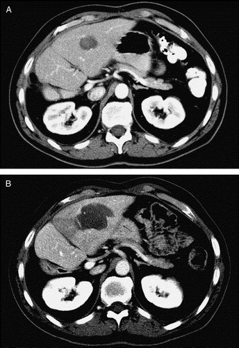
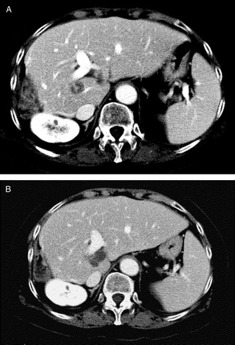
Figure 2. (b) 24 hours after percutaneous laser ablation a contrast-enhanced CT scan shows complete ablation of the lesion. The necrotic area appears as a hypoattenuating area 4.5 cm in diameter. During follow-up (40 months) new lesions appeared in segment 2, which were successfully treated with percutaneous laser ablation.
The median time from the end of PLA sessions to start of chemotherapy was 12 days (range 7–21 days). The median number of chemotherapy courses was 16 (range 1–28), with a relative dose intensity of 0.875. Local tumor control data for patients with a complete response are detailed in . Disease relapse around the sites of previously ablated metastases occurred in 10 (50%) of these patients. In patients with a partial response, chemotherapy resulted in maintenance of response for at least 3 months in 10 cases. In no case chemotherapy transformed a partial response to PLA into a complete response.
Figure 3. Kaplan-Meier estimate of overall survival in patients with complete and partial necrosis of metastases.
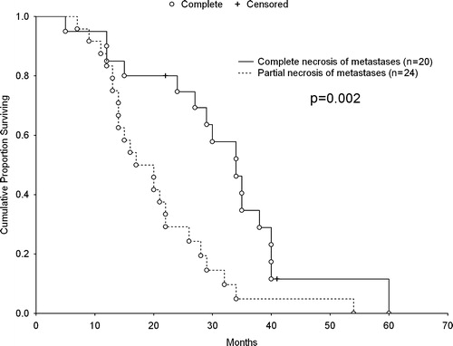
Table III. Local tumor control data, in cases where a complete necrosis was achieved, at 3 and 6 months after LA.
Safety
No major complications were encountered during or after PLA sessions. The most frequent side effect was moderate abdominal pain that required analgesics. Pain at the right shoulder of a few days’ duration occurred in all patients with lesions in close proximity to the liver capsule. Pleural effusions were observed in patients in whom lesions were located at the dome of the liver (i.e. segments 3, 4, 7 and 8). None of these patients required intensive care or interventional treatment, and all effusions cleared up after a few weeks. A transient and mild body temperature increase (37.5°–38.5° C) due to the necrotic tissue was observed in all cases. As a matter of fact, most patients had fully recovered and were capable of starting chemotherapy a few days after the last PLA session. There was no treatment-related death. Seeding of metastases along the insertion tract occurred in one patient.
Survival parameters
Time to progression was 11.2±9.0 months in patients in whom a complete ablation was obtained, and 6.3±3.9 months in those with a partial ablation (p = 0.024). Reasons of progression were local recurrence or volume increase of partially ablated liver metastases in 21 (48%) patients, appearance of new liver metastases in 13 (30%), and metastases in other organs in 10 patients (22%). Overall survival was 24.7±13.0 months (median 22 months). Survival was 30.0±12.7 months in patients with a complete ablation, and 20.2±10.2 months in those with a partial or minor response (p = 0.002; ). The 3-year survival for patients with a complete response was 35%. In these patients, there was no difference in the duration of survival or time to progression between those who had a size of metastasis ≤3.0 cm, and those who had a size of metastasis > 3.0 cm.
Discussion
The PLA technique used in this study proved to be safe, with a few and quite easily manageable minor treatment-related adverse events. In particular, we did not observe abscess formation, intraabdominal bleeding, or visceral injury, which have been described with radiofrequency ablation Citation[15], Citation[16] and, occasionally, with other laser ablation techniques Citation[6], Citation[12]. Ultrasound-guidance was preferred to CT-guidance because in our experience the former is easier to use, even for lesions under the diaphragm or near the bowels. In fact, US-guidance allows real-time monitoring of the fiber tips in the target area and is less time-consuming than CT-guidance. The extent of coagulation necrosis cannot be predicted from mathematical models because of the complex interactions between laser light and tumor tissue Citation[17], highlighting the importance of accurate real-time monitoring of necrosis. In addition, the use of fine needles and US-guidance allows a more flexible and safe approach even through the intercostal route.
A complete ablation of metastases was achieved in less than 50% of patients. This figure appears lower than in other recently reported series, in which complete tumor ablation was achieved in almost all cases Citation[6], Citation[12], Citation[13], Citation[16], Citation[18]. In this regard, we have to underline that the clinical success of PLA and of all hyperthermic techniques is based on a number of factors, the most important of which is tumor location. As a matter of fact, many of our cases presented with a difficult technical approach due to the location of metastases near great vessels or main bile ducts. This also partly explains why small lesions (≤3.0 cm) were more easily eradicated than larger ones.
Regarding local tumor control, we observed recurrences in 24% of cases. This compares favorably with the results of Solbiati et al. Citation[13] and Gillams et al. Citation[16], while Vogl et al. reported a surprisingly low recurrence rate of less than 5% Citation[6].
Our patients had a median overall survival of 22 months; with a 1-year survival was 95%. However, while cases who did not achieve a complete response after PLA had survival figures comparable with those of chemotherapy alone Citation[10], patients who had their tumors completely ablated had survival rates approaching those of surgery Citation[2]. However, direct comparisons with general groups of patients with unresectable tumors should be interpreted cautiously. In fact, randomized controlled trials with ablative therapies are lacking. Other thermal ablation groups have reported encouraging data on survival in phase II trials. Gillams et al. carried out PLA in 69 patients, including 20 patients with extrahepatic disease. The average number of liver metastases was 2.9 (range, 1–16), and the mean maximal diameter was 3.9 (range, 1–8) cm. The 3-year survival rate and median survival time from liver metastasis diagnosis was 34% and 27 months, respectively Citation[12]. Subsequently the same group treated 167 patients with radiofrequency ablation Citation[16]. The mean number of metastases at the time of first radiofrequency ablation was 4.1 (range 1–27), and the mean maximum diameter of the largest lesion in each patient was 3.9 cm (range 1–12). Median overall survival for the whole group was 32 months, with a 3-year survival rate of 40%. Solbiati et al. reported their results of radiofrequency ablation in 117 patients with up to four metastases, mean diameter 2.8 cm. The majority (88%) of their patients had either one or two tumors. The median survival in this cohort was 36 months, and their 3-year survival was 46% Citation[13]. Vogl et al. have reported a median survival of 35 months and a 3-year survival rate of 56% in patients with five tumors or less, maximum diameter 5 cm. This group uses a different Nd:YAG laser ablation technique and a combination of CT guidance for applicator placement and high field MR monitoring of the thermal effect Citation[6].
In conclusion, our data confirm previous reports about the safety and feasibility of PLA in patients with liver metastases from colorectal cancer. Our series is relatively small and inhomogeneous to give definitive results in term of local tumor control and survival. However, there is an indication that PLA probably has a positive impact on survival parameters in those individuals in whom the tumors can be completely ablated. These findings warrant randomized trials to test the efficacy of the combination PLA plus chemotherapy compared to chemotherapy alone. Currently it is reasonable to offer this therapeutic option to patients with limited size liver disease who are not suitable candidates for resection.
References
- Ruers T, Bleichrodt RP. Treatment of liver metastases, an update on the possibilities and results. Eur J Cancer 2002; 38: 1023–33
- Fong Y, Cohen AM, Fortner JG, Enker WE, Turnbull AD, Coit DG, et al. Liver resection for colorectal metastases. J Clin Oncol 1997; 15: 938–46
- Schlag PM, Benhidjeb T, Stroszczynski C. Resection and local therapy for liver metastases. Best Pract Res Clin Gastroenterol 2002; 16: 299–317
- Tranberg KG. Percutaneous ablation of liver tumours. Best Pract Res Clin Gastroenterol 2004; 18: 125–45
- Wacker FK, Reither K, Ritz JP, Roggan A, Germer CT, Wolf KJ. MR-guided interstitial laser-induced thermotherapy of hepatic metastasis combined with arterial blood flow reduction: Technique and first clinical results in an open MR system. J Magn Reson Imaging 2001; 13: 31–6
- Vogl TJ, Straub R, Eichler K, Sollner O, Mack MG. Colorectal carcinoma metastases in liver: laser-induced interstitial thermotherapy–local tumor control rate and survival data. Radiology 2004; 230: 450–8
- Wietzke-Braun P, Schindler C, Raddatz D, Braun F, Armbrust T, Nolte W, et al. Quality of life and outcome of ultrasound-guided laser interstitial thermo-therapy for non-resectable liver metastases of colorectal cancer. Eur J Gastroenterol Hepatol 2004; 16: 389–95
- Pacella CM, Bizzarri G, Cecconi P, Caspani B, Magnolfi F, Bianchini A, et al. Hepatocellular carcinoma: Long-term results of combined treatment with laser thermal ablation and transcatheter arterial chemoembolization. Radiology 2001; 219: 669–78
- Pacella CM, Bizzarri G, Magnolfi F, Cecconi P, Caspani B, Anelli V, et al. Laser thermal ablation in the treatment of small hepatocellular carcinoma: Results in 74 patients. Radiology 2001; 221: 712–20
- Douillard JY, Cunningham D, Roth AD, Navarro M, James RD, Karasek P, et al. Irinotecan combined with fluorouracil compared with fluorouracil alone as first-line treatment for metastatic colorectal cancer: A multicentre randomised trial. Lancet 2000; 355: 1041–7
- Tournigand C, Andre T, Achille E, Lledo G, Flesh M, Mery-Mignard D, et al. FOLFIRI followed by FOLFOX6 or the reverse sequence in advanced colorectal cancer: A randomized GERCOR study. J Clin Oncol 2004; 22: 229–37
- Gillams AR, Lees WR. Survival after percutaneous, image-guided, thermal ablation of hepatic metastases from colorectal cancer. Dis Colon Rectum 2000; 43: 656–61
- Solbiati L, Livraghi T, Goldberg SN, Ierace T, Meloni F, Dellanoce M, et al. Percutaneous radio-frequency ablation of hepatic metastases from colorectal cancer: Long-term results in 117 patients. Radiology 2001; 221: 159–66
- Miller AB, Hoogstraten B, Staquet M, Winkler A. Reporting results of cancer treatment. Cancer 1981; 47: 207–14
- de Baere T, Risse O, Kuoch V, Dromain C, Sengel C, Smayra T, et al. Adverse events during radiofrequency treatment of 582 hepatic tumors. AJR Am J Roentgenol 2003; 181: 695–700
- Gillams AR, Lees WR. Radio-frequency ablation of colorectal liver metastases in 167 patients. Eur Radiol 2004; 14: 2261–7
- Jacques SL. Laser-tissue interactions. Photochemical, photothermal, and photomechanical. Surg Clin North Am 1992; 72: 531–58
- Gillams AR. Thermal ablation of liver metastases. Abdom Imaging 2001; 26: 361–8