Abstract
The study was undertaken in order to compare dose plans for intensity-modulated radiotherapy (IMRT) with 3D conformal radiotherapy (3D-CRT) dose plans in patients with nasopharyngeal carcinoma (NPC). Clinical data from 20 consecutive patients treated with IMRT are presented. For 11 patients 3D-CRT plans were made and compared to the IMRT plans with respect to doses to the planning target volumes (PTVs) and to organs at risk (OARs). For comparison of the conformation of dose to defined target volumes the conformity index (CI) was used. Target volume coverage and critical organ protection were significantly improved with IMRT compared to 3D-CRT. One-year loco-regional control, distant metastasis-free survival, and overall survival were 79%, 72%, and 80%. Two patients have had recurrence in the clinical target volume (CTV) only and seven patients have relapsed in distant organs and/or in head-and-neck areas outside the target areas. The study confirms that IMRT is superior to 3D-CRT in the treatment of NPC. As locoregional control of NPC improves we are facing an increasing number of recurrences outside the irradiated area.
The incidence of NPC varies significantly from less than 1 per 100 000 person years in most western countries and up to 25 per 100 000 person years in Southeast Asia, Greenland and North Africa Citation[1]. This variation seems to be partly due to early infection with Epstein-Barr virus in genetically predisposed individuals, but chemical carcinogens have also been associated with the development of NPC Citation[2]. Radiotherapy is the most important treatment modality, and the prognosis for patients with tumor localized in the nasopharynx only (AJCC/UICC stage I) is excellent Citation[3], but unfortunately most patients (∼90%) have lymph node metastases and/or invasion in adjacent structures at the time of diagnosis. The superiority of combined chemo-radiotherapy over radiotherapy alone has been demonstrated in patients with stage III and IV disease and is now standard treatment for these patients in most radiotherapy centres Citation[4].
Nasopharyngeal tumors are located at the base of the skull in close proximity to several important organs (e.g. eyes, optic nerves/chiasm, brain stem, inner ear, and salivary glands) which may be impossible to protect with 3D conformal radiotherapy. CT/MR-guided intensity-modulated radiotherapy (IMRT) seems to improve target coverage and protect critical structures when applied to patients with cancer of the nasopharynx Citation[5], Citation[6]. At the Department of Oncology, The Finsen Centre, Rigshospitalet, Copenhagen all patients with NPC have been treated with IMRT since 2001. All treatment plans are based on fusions of planning CT-scans with MR-scans and in some cases also PET-scans. The present study was undertaken in order to demonstrate the superiority of IMRT compared with 3D conformal radiotherapy regarding target volume coverage as well as critical organ protection and to present survival and recurrence data for 20 consecutive patients treated with IMRT between January 2001 and March 2004.
Material and methods
Patients
Data from 20 consecutive patients (six female, 14 male), aged 14–78 years (median 47) treated between January 2001 and March 2004 were analyzed. Thirteen patients had undifferentiated carcinoma, five patients had poorly differentiated squamous cell carcinoma, one had intermediary differentiated squamous cell carcinoma, and one had adenoid cystic carcinoma. The distribution of patients in T- and N-stages is given in . Median follow-up was 23 months (range 13–51). All patients were treated with radiotherapy (described in detail below) and the radiosensitizer Nimorazol (Naxogin) 1200 mg/m2 orally 90 minutes before each irradiation Citation[7]. Weekly concomitant cisplatin 40 mg/m2 (max. 70 mg) i.v. was introduced in our institution in 2003, and only two of the 20 patients received concomitant cisplatin.
Table I. Number of patients in the different T- and N-stages. The distribution of patients in AJCC/UICC stages was: Stage I: 0 patients, stage II: 2 patients, stage III: 5 patients, and stage IV: 13 patients.
Treatment planning
The patients were all treated on a carbon couch positioned by an individual mask covering the head and shoulders. The energy used for head and neck treatments is 6 MV. In all patients, treatment planning was based on fusion of MRI performed routinely at the department of Radiology and CT-scans performed as routine therapy-planning CT-scans at the Department of Oncology.
The lymph node levels were outlined according to the guidelines of Gregoire et al. Citation[8]. Target definitions and dose specification were in accordance with the recently developed guidelines for IMRT of head and neck cancer developed by the Danish Head & Neck Cancer Group (DAHANCA). Briefly, these guidelines include the following:
Total radiation dose to the gross tumor volume (GTV) (tumor and lymph node metastases): 68 Gy/34 fractions, 5 fractions per week.
Elective clinical target volume – high-risk (CTVE-h) defined as areas close to the GTV at high risk for microscopic disease (e.g. margins around the primary, the whole lymph node level in which lymph node metastases have been found, possibly also the next lymph node level below the level with metastases): Minimum dose of 60 Gy.
Elective clinical target volume – low-risk (CTVE-l) defined as volumes judged to be at lower but still significant risk of microscopic disease (uninvolved lymph node levels II, III, IV, V, retropharyngeal): Minimum dose of 50 Gy.
Simultaneous integrated boost, minimum fraction size for subclinical disease 1.5 Gy.
Maximum doses to specific organs at risk and to the corresponding planning volumes (PRV) are defined in .
Dose limitations to other OARs: Parotid glands: 50% of the volume should be kept below 26 Gy. Larynx: 67% of the volume should be kept below 50 Gy.
Table II. Maximum prescribed doses to organs at risk (OARs) and planning risk volumes (PRVs).
For 11 patients conventional 3D-CRT plans were made retrospectively on CadPlan (owing to the lack of electron calculation on our previous version of Eclipse). The plans were made according to our practice before the introduction of IMRT without using the CTVs and PTVs. The traditional anterior-posterior and cranial-caudal boundaries were used. Primary fields to 46 Gy using 4-6 MV photons included the spinal cord using two lateral opposed fields with a half-beam technique and one anterior field inferiorly for the supra-clavicular area. Compensation for inhomogeneity was used to obtain a sagital midline dose of 2 Gy daily for the opposing fields. From 46–68 Gy the fields were moved off the spinal cord and if necessary, electron fields were added to cover the involved lymph nodes close to the spinal cord. Between 46 and 50 Gy was allowed to the spinal cord and orbits were to be protected as much as possible. No other OARs were taken into consideration. The tumor + lymph nodes were to receive 68 Gy (±5%). The 3D-CRT plans were created by a highly experienced dose planner, in order to compare the best possible conventional plans with the IMRT plans.
Dose-volume histograms (DVHs) were calculated for IMRT and 3D-CRT plans and maximum and minimum doses extracted. Maximum dose was defined as the dose received by 1% of the volume in question and minimum dose was defined as the dose received by 99% of the volume.
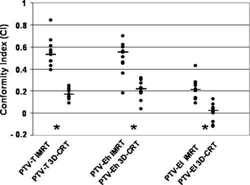
The ability to cover target volumes can be assessed by the conformity index (CI), which estimates how the 95% isodose level conforms to the PTV Citation[6]:where VPTV=volume of PTV, V95%=volume enclosed by the 95% isodose level, V∼95%=volume of PTV receiving dose < 95% isodose level. The conformity index should ideally be 1. In situations where the 95% isodose level does not exactly cover the PTV, the CI decreases and may even become negative.
Statistics
The Mann-Whitney U test was used for comparison of minimum and maximum doses to target volumes and organs at risk and for comparison of CIs. For comparison of fractions of patients receiving doses above/below the doses prescribed or allowed, Fisher's exact test was applied. A two-sided p-value less than 0.05 was considered significant.
Results
The distribution of maximum and minimum doses to target volumes are shown in . There were no significant differences between IMRT and 3D-CRT in maximum and minimum doses to PTV-T, but the maximum doses to PTV-Eh and PTV-El were significantly lower with IMRT than with 3D-CRT. In contrast, the minimum doses to PTV-Eh and PTV-El were significantly higher and varied less with IMRT than with 3D-CRT. The IMRT doses were generally more uniformly distributed around and closer to the defined target dose of 60 Gy for PTV-Eh and 50 Gy for PTV-El. A minimum dose of 95% (57 Gy) to the PTV-Eh was reached in 2/11 IMRT-plans vs. 0/11 3D-CRT plans (not significant), but the volume of PTV-Eh receiving less than 57 Gy (5% less than the prescribed dose to CTV) was significantly lower with IMRT (median 2%) than with 3D-CRT (median 12%) (p < 0.001). Five of 10 patients received the 95% dose limit (47.5 Gy) to all of the PTV-El with IMRT compared to 0/10 with 3D-CRT (p = 0.03) and the median volume of PTV-El receiving less than 47.5 Gy was 0.25% and 14.5% for IMRT and 3D-CRT, respectively (p = 0.0002). These results translate into a significantly improved conformity index of all PTVs by IMRT compared to 3D-CRT dose planning ().
Figure 1. Doses to the planning target volumes. Maximum (received by 1% of the volume in question) and minimum (received by 99%) target doses for PTV-T, PTV-Eh, and PTV-El with IMRT and 3D-CRT, respectively. Each dot represents the value obtained from one patient; the small horizontal lines are medians. The large horizontal lines indicate 95% of the prescribed dose to the target volume. *p < 0.05.
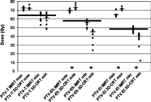
Figure 2. Comparison of IMRT and 3D-CRT conformity index for PTV-T, PTV-Eh, and PTV-El. Each dot represents the value obtained from one patient, the horizontal lines are medians. *p < 0.05.
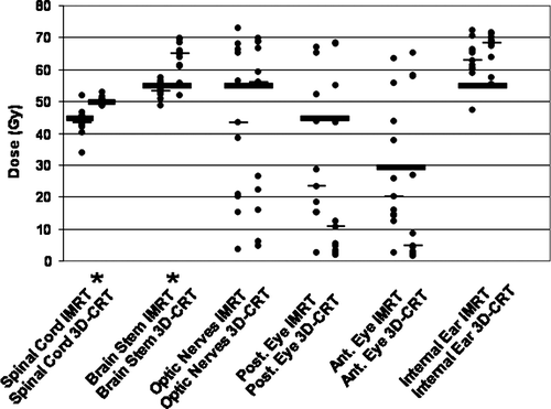
Maximum doses to organs at risk (OARs) with primary priority are shown in . The maximum doses to the spinal cord and brain stem were significantly lower with IMRT than with 3D-CRT. Since the spinal cord was allowed to receive different doses with IMRT (45 Gy) and 3D-CRT (46–50 Gy) it was not possible to compare the maximum doses to the spinal cord directly. In 9/11 IMRT plans the dose to the spinal cord was kept at or below 45 Gy, whereas the dose limit of 50 Gy was exceeded in 5/11 3D-CRT plans. The brain stem dose was kept below 54 Gy in 8/11 IMRT plans but only in 1/11 3D-CRT plans. There were no differences in organ protection or maximum doses to the optic nerves, posterior/anterior eyes, or inner ears with IMRT and 3D-CRT plans, respectively, although the maximum dose to one or both inner ears could be kept below the allowed 54 Gy in 8/11 patients with IMRT compared to 3/11 patients with 3D-CRT (p = 0.08).
Figure 3. Doses to risk organs of primary priority. Maximum doses received by 1% of the organ in question. Each dot represents the value obtained from one patient; the small horizontal lines are medians. The large horizontal lines indicate the prescribed maximum dose. *p < 0.05.
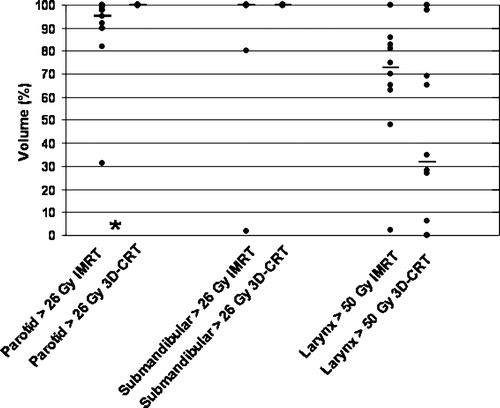
The entire volume of parotid glands received more than 26 Gy in all 3D-CRT plans, whereas IMRT allowed for protection of small areas of the parotid gland in 9/11 cases (p < 0.001), but only to a minor extent (). The median volume receiving less than 26 Gy was 5% by IMRT (p = 0.001). IMRT was not superior to 3D-CRT with respect to larynx or submandibular gland protection.
Figure 4. Irradiated volumes in risk organs of secondary priority. Each dot represents the relative organ volume receiving more than the prescribed maximum mean dose. The horizontal lines are medians. *p < 0.05
Of the 20 treated patients 11 are alive, eight without recurrence. Nine patients have died, three died without evidence of recurrent disease.
In total, nine patients of 20 treated had recurrences, seven of these with distant metastases. In these seven patients, ten distant metastastic sites were identified (four hepatic, two subcutaneous, two pulmonary, one osseous, and one inguinal). Two of nine patients with recurrence had inoperable local recurrence without evidence of distant metastases.
Four patients had distant metastases without evidence of disease in the head and neck area. Until now, five patients had recurrence in the GTV and CTV. Median survival has not been reached at a median follow-up of 23 months. One-year loco-regional control, distant metastasis-free survival, and overall survival were 79%, 72%, and 80%, respectively.
Discussion
NPC is curable with combined chemo-radiotherapy even in advanced stages without distant metastases. Sufficiently high dose delivery to the PTV is crucial for local tumor control and patient survival but with conventional 3D-radiotherapy even treatment of GTV to 66–68 Gy occurs at the cost of considerable morbidity Citation[9]. The risk of severe side-effects (e.g. radiation-induced myelopathy) has limited the obtainable doses in 3D-CRT target volumes and led to the introduction of IMRT in order to increase the target dose with improved conformity and to reduce the dose to organs at risk. There is a continuum between conventional 3D-planning and IMRT; forward 3D-planning can be sophisticated by attempting to cover the PTV-Eh and PTV-El to the appropriate doses and multiple fields can be added by forward planning or inverse 3D-CRT to approach the dose distribution obtainable with IMRT Citation[6], Citation[10]. We chose to compare the IMRT plans with our previous practice of conventional 3D-CRT in order to demonstrate what could be achieved by moving directly to IMRT.
In the present study, the minimum dose to the high- and low-risk elective target volumes was significantly higher for the IMRT plans than for 3D-CRT (). Furthermore, the CI was significantly higher (closer to 1) for PTV-T, PTV-Eh and PTV-El with IMRT than with 3D-CRT (). CI was originally introduced by Knöös et al. Citation[11] as the degree to which the 95% isodose curve covers the entire PTV (CI = PTV/V95%). Wu et al. introduced an additional component to the equation in order to lower CI in the situation where PTV and V95% are of the same size but not enclosing exactly the same areas. In this case, the fraction of PTV receiving less than 95% of the prescribed dose should be subtracted Citation[6]. The results of our study clearly demonstrate the superiority of IMRT in conforming doses to given target volumes, which should also be reflected in significant risk organ protection. In fact, it was possible to decrease the maximum dose to the spinal cord and brain stem and to induce significant parotid protection with IMRT compared to 3D-CRT ( and , ). Corresponding results were reported in a study of one patient with NPC by Xia et al. Citation[12], and Wu et al. found a significantly improved CI with IMRT compared to 3D-CRT in 40 patients, but did not report any data regarding doses to OARs Citation[6]. Their mean CI of 0.59±0.07 with IMRT is very close to our median value of 0.53 (range: 0.39–0.84) even though our patients seem to have more advanced disease stages, since the mean volume and standard deviation of PTV-T in our series was 186±93 cm3 compared to 69.0±34.7 cm3 in the study by Wu et al. Citation[6].
Table III. Number of patients (n = 11) in which the dose constraint was achieved with IMRT and 3D-CRT, respectively. Maximum dose allowed to the spinal cord was 45 Gy for IMRT but in the 3D-CRT plans the spinal cord was treated to 46 Gy before field reduction. Consequently, the dose limit was 45 Gy for IMRT and 50 Gy for 3D-CRT.
The parotid glands could be protected to a minor extent (median 5% of the parotid gland volume) with IMRT, but not at all with 3D-CRT. However, parotid gland volume receiving ≤26 Gy varied considerably and was as high as 68.7% in one patient (). IMRT has been shown to preserve the function of the salivary glands Citation[13] and a recent randomised trial has demonstrated a significant preservation of parotid saliva flow rate and a reduction of severe xerostomia from 86% to 46% already at six weeks after radiotherapy with IMRT compared to 2D-CRT of 50 patients with NPC Citation[14].
Only two of our patients (10%) had recurrences in the primary target volume without distant metastases and seven patients (35%) developed distant metastases. It is not possible to draw firm conclusions regarding tumor control or patient survival from the present study, but experience with IMRT to NPCs was recently published from the Memorial-Sloan Kettering Cancer Center, New York Citation[15] and from Prince of Wales Hospital, Hong Kong Citation[16]. Both centres had excellent treatment results with a 3-year locoregional control rate of 93% Citation[15] and 3-year overall survival of 83% Citation[15] and 90% Citation[16] with improved normal-tissue protection. These results seem better than previous results with 3D-CRT, but as the loco-regional control rate increases, the data presented here and the results from both centers demonstrate that distant metastases have become the predominant form of failure with 3-year metastasis free survival rates of 78% Citation[15] and 79% Citation[16], respectively. Concomitant chemotherapy was given to 93% of patients treated at Memorial Sloan-Kettering and 25% treated in Hong Kong. It is now part of the standard treatment for most patients with stage III–IV NPC, but the high incidence of distant metastases in spite of concomitant chemotherapy may suggest a role for neoadjuvant chemotherapy in eradication of microscopic metastases Citation[4], Citation[17].
A multivariate analysis of the data from Hong Kong showed dose escalation above 66 Gy to be a favourable prognosticator, whereas the volume of GTV was a significant adverse factor Citation[16]. In our study there was no obvious correlation between the volume of PTV-T and CI, indicating that tumor location (in close proximity to high risk organs) may be as important for PTV coverage as tumor size.
Whether the demonstrated improved dose distribution with IMRT leads to improved survival remains to be proven. The excellent tumor control and patient survival data from several other centres using IMRT as standard treatment for patients with NPC seem to diminish the need for randomized trials, which would require the inclusion of several hundred patients with this relatively rare type of cancer. Even with multicenter collaborations it will be almost impossible to recruit a sufficient number of patients and the routine use of IMRT in most large radiotherapy centres as standard treatment to patients with NPC has made it difficult (and unethical?) to convince patients to enrol in a trial with a 50% chance of receiving a treatment with at significantly increased toxicity and possibly reduced effect compared to the standard IMRT treatment. There are, however, several unresolved aspects of IMRT. The distribution of lower doses of irradiation to larger volumes of the patient may result in a significant increase in the incidence of secondary cancers and close follow-up and registration is essential.
In conclusion, IMRT increased the conformity index and the minimum dose to elective high- and low-risk target volumes compared to 3D-CRT in patients with NPC. IMRT also led to a decrease in maximum dose to the spinal cord and brain stem and enabled protection of a part of the parotid glands. As concomitant chemo/radiotherapy improves the loco-regional control, distant metastases seem to emerge more frequently and neoadjuvant chemotherapy may play an important role in early eradication of metastatic cells.
References
- Parkin, DM, Whelan, SL, Ferlay, J, Raymond, L, Young, J, editors. Cancer incidence in five continents. Volume VII. IARC Sci Publ no. 143; 1997.
- Chan AT, Theo PM, Huang DP. Pathogenesis and treatment of nasopharyngeal carcinoma. Semin Oncol 2004; 31: 794–801
- Chua DT, Sham JS, Kwong DL, Au GK. Treatment outcome after radiotherapy alone for patients with Stage I–II nasopharyngeal carcinoma. Cancer 2003; 98: 74–80
- Langendijk JA, Leemans ChR, Buter J, Berghof J, Slotman BJ. The additional value of chemotherapy to radiotherapy in locally advanced nasopharyngeal carcinoma: A meta-analysis of the published literature. J Clin Oncol 2004; 22: 4604–12
- Emami B, Sethi A, Petruzzelli GJ. Influence of MRI on target volume delineation and IMRT planning in nasopharyngeal carcinoma. Int J Radiat Oncol Biol Phys 2003; 57: 481–8
- Wu VWC, Kwong DLW, Sham JST. Target dose conformity in 3-dimensional conformal radiotherapy and intensity modulated radiotherapy. Radiother Oncol 2004; 71: 201–6
- Overgaard J, Hansen HS, Overgaard M, Bastholt L, Berthelsen A, Specht L, et al. A randomized double-blind phase III study of nimorazole as a hypoxic radiosensitizer of primary radiotherapy in supraglottic larynx and pharynx carcinoma. Results of the Danish Head and Neck Cancer Study (DAHANCA) Protocol 5–85. Radiother Oncol 1998; 46: 135–46
- Gregoire V, Levendag P, Ang KK, Bernier J, Braaksma M, Budach V, et al. CT-based delineation of lymph node levels and related CTVs in the node-negative neck: DAHANCA, EORTC, GORTEC, NCIC, RTOG consensus guidelines. Radiother Oncol 2003; 69: 227–36
- Wang CC. Radiation therapy for head and neck neoplasms. John Wright, Boston 1983
- Kam MKM, Chau RMC, Suen J, Choi PHK, Teo PML. Intensity-modulated radiotherapy in nasopharyngeal carcinoma. Dosimetric advantage over conventional plans and feasibility of dose escalation. Int J Radiat Oncol Biol Phys 2003; 56: 145–57
- Knoos T, Kristensen I, Nilsson P. Volumetric and dosimetric evaluation of radiation treatment plans: Radiation conformity index. Int J Radiat Oncol Biol Phys 1998; 42: 1169–76
- Xia P, Fu KK, Wong GW, Akazawa C, Verhey LJ. Comparison of treatment plans involving intensity-modulated radiotherapy for nasopharyngeal carcinoma. Int J Radiat Oncol Biol Phys 2000; 48: 329–37
- Saarilahti K, Kouri M, Collan J, Hamalainen T, Atula T, Joensuu H, et al. Intensity modulated radiotherapy for head and neck cancer: Evidence for preserved salivary gland function. Radiother Oncol 2005; 74: 251–8
- Kam, MK, Leung, SF, Zee, B, Choi, PH, Chau, RM, Cheung, KY, et al. Impact of intensity-modulated radiotherapy (IMRT) on salivary function in early-stage nasopharyngeal carcinoma (NPC) patients: A prospective randomized study. Proceedings of ASCO Annual Meeting. 2005; #5501.
- Wolden SL, Chen WC, Pfister DG, Kraus DH, Berry SL, Zelefsky MJ. Intensity-modulated radiation therapy (IMRT) for nasopharynx cancer: Update of the Memorial Sloan-Kettering experience. Int J Radiat Oncol Biol Phys 2006; 64: 57–62
- Kam MK, Teo PML, Chau RM, Cheung KY, Choi PH, Kwan WH, et al. Treatment of nasopharyngeal carcinoma with intensity-modulated radiotherapy: The Hong Kong experience. Int J Radiat Oncol Biol Phys 2004; 60: 1440–50
- Lin JC, Liang WM, Jan JS, Jiang RS, Lin AC. Another way to estimate outcome of advanced nasopharyngeal carcinoma – is concurrent chemoradiotherapy adequate?. Int J Radiat Oncol Biol Phys 2004; 60: 156–64