To the Editor
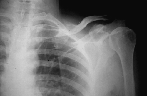
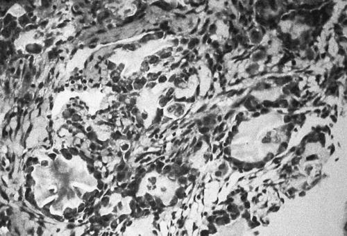
A 42 year old female presented to the OPD with complaints of swelling and pain for past 4 months on the left clavicular region. It progressed in size rapidly with early rise in pain. Pain was continuous and increased with movements, partially remitting with analgesics. There was no history of trauma or fever. On examination a bony hard swelling arising from medial two-thirds of clavicle measuring 8 cm×6 cm in size, free from overlying skin was found. There was no fluctuation or bruit and cross-illumination was negative. Passive range of movements at shoulder was full. Distal pulses were well felt and neurologically the limb was unremarkable. On x-ray, large erosive lesion of clavicle with irregular margins involving medial two-thirds of clavicle with surrounding soft tissue swelling was noted (). Comparing the previous available radiographs, a rapid increase in size of the lesion with bone destruction was seen. Non-contrast CT-scan () showed a destructive lesion of left clavicle in the middle thirds of clavicle extending into medial thirds with a pathological fracture. Open biopsy was done and histopathological analysis showed lesion compatible with aggressive osteoblastoma (). On clinico-radiological correlation a diagnosis of aggressive osteoblastoma was made. The lesion was resected and acromio-clavicular joint was preserved along with the coraco-clavicular ligaments. The tumor was found to invade deltoid and pectoralis muscle attachments to the clavicle. The involved muscles with a 2 cm wide margin were resected along with the tumor. The curettings from the distal stump of the bone and tissue from left out margins were free from tumor. Patient was followed regularly and was asymptomatic four years after the surgery, without any recurrence.
Figure 1. The radiograph demonstrates an ill defined lytic lesion involving medial two thirds of the clavicle with a soft tissue shadow extending beyond the confines of the bone.

Figure 2. The computed tomographic images of the same patient reveals a ballooned out lesion (1) involving the medial two thirds of the clavicle with a thin shell of bone around. There is extension of the lesion into soft tissue (3) and cortical breach is well demonstrated (2).

Figure 3. Histopathology: Microscopic examination showed areas with abundant osteoid formation with low degree of cellularity with no atypia. Undecalcified sections depicted partial calcification of osteoid, elsewhere highly cellular areas were present comprising oval osteoblast like cells (epitheloid osteoblasts) and moderate nuclear pleomorphs with occasional mitosis and intermingled tumor giant cells.

Osteoblastoma is a very rare benign bone tumor akin to osteoid osteoma. By definition, all osteoblastomas are larger than 1.5 cm Citation[1]. Majority (>90%) of patients are diagnosed before the age of 30 years with male predominance Citation[1], Citation[2]. Generally osteoblastomas are expansile lesions, but in majority of cases the cortex remains intact. The behavior of occasional aggressive lesions, often referred to as “aggressive osteoblastomas” or “malignant osteoblastomas,” may simulate that of malignant neoplasms, thereby complicating differentiation of osteoblastoma from osteosarcoma in certain situations. The term “aggressive osteoblastoma” is applied to large, locally destructive lesions that mimic a low-grade osteosarcoma on microscopic examination Citation[1–3]. The presence of epithelioid osteoblasts, lace- or sheet-like osteoid production, and a permeative pattern of tumor growth in osteoblastomas is thought to be associated with an aggressive clinical behaviour Citation[4]. These tumors may represent a continuum between typical benign osteoblastomas and low-grade osteoblastic osteosarcomas or somehow represent the previously misdiagnosed or unrecognized cases of osteosarcoma. Also there are rare reports of spontaneous malignant transformation of osteoblastoma with distant metastases Citation[1], Citation[3]. This tumor occurs without any regard to age being more common in young adults Citation[1–3]. The primary region is either a long or the flat bones of body, but recently involvement of short and flat bones is increasingly recognised. Only three cases of osteoblastoma of clavicle are reported till date, none of them being “aggressive” osteoblastoma. Radiographic differential diagnosis of this lesion includes low-grade osteosarcoma, aneurysmal bone cyst, eosinophilic granuloma (Langerhans cell histiocytosis), chondrosarcoma, giant cell tumor, contiguous pulmonary metastasis, distant metastasis and tuberculosis. Differentiating aggressive osteoblastoma from low-grade osteosarcoma relies on the presence of osteoblasts, epithelioid osteoblasts, characteristic histopathology, absent mitosis especially absence of bizarre ones, circumscribed nature of lesion with no metastasis. It is of utmost importance to distinguish this lesion from osteosarcoma with a close histopathological examination as the treatment and prognosis of the two conditions differ considerably. Osteosarcoma typically tends to be more invasive. Although osteoblastoma and osteosarcoma can produce osteoid, osteoblastoma does not show the malignant histologic features. Also, osteosarcoma exhibits sheets of osteoblasts, while in osteoblastoma only a single layer of these cells lines the bony trabeculae.
Computed tomography scanning is an important imaging modality that very well shows intralesional bone production and the bony shell that surrounds the lesion. In addition, CT-scans delineate well, areas of cortical destruction and soft tissue extension. Osteoblastoma presents a challenge to the clinician in diagnosis and treatment. The variable presentations of osteoblastoma emphasize the need to individualize the treatment. According to Enneking Citation[5], the minority of osteoblastomas are “pseudomalignant” stage 3 lesions, with atypical aggressive clinical, radiographic, and histologic patterns which “have a propensity for pervasive recurrence and have been fatal in vital anatomic locations” as in the presented case. Good quality imaging, meticulous biopsy providing sufficient material to the pathologist and careful analysis are required to make an accurate diagnosis. Osteoblastomas needs surgical treatment when detected. Aggressive osteoblastomas in particular should be properly diagnosed before embarking upon treatment keeping in mind the striking resemblance to osteosarcoma, the treatment of which is radical resection. Patient should be kept under close follow-up for early detection of recurrence considering the tendency of this particular lesion to recur.
References
- Dorfman, HD, Czerniak, B. Benign osteoblastic tumors. In:. Bone Tumors. 1st ed. St Louis, Mo: Mosby; 1998. p 85–127.
- Dorfman HD, Weiss SW. Borderline osteoblastic tumors: Problems in differential diagnosis of aggressive osteoblastoma and low-grade osteosarcoma. Semin Diagn Pathol 1984; 1: 215–34
- Kunze E, Enderle A, Radig K, Schneider-Stock R. Aggressive osteoblastoma with focal malignant transformation and development of pulmonary metastases. A case report with a review of literature. Gen Diagn Pathol 1996; 141: 377–92
- Cheung FM, Wu WC, Lam CK, Fu YK. Diagnostic criteria for pseudomalignant osteoblastoma. Histopathology 1997; 31: 196–200
- Enneking, WF. Osteoblastoma (37 cases). In: WF Enneking, editor. Musculoskeletal tumor surgery. Volume 2. New York: Churchill Livingstone Inc.; 1983. p 1043–54.
