To the Editor
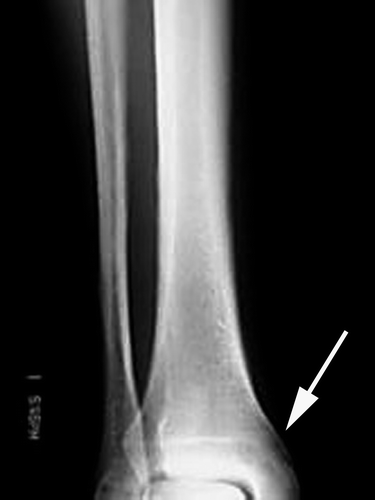
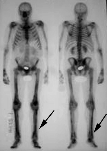
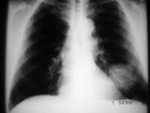
A 66-year-old man was being followed up by the urology services for a borderline elevation of his PSA. A further slight rise in his PSA, in addition to an isolated elevation of serum alkaline phosphatase, led to an isotope bone scan being requested. This showed lower limb changes typical of hypertrophic osteoarthropathy (). Bilateral plain films of tibia and fibula confirmed this diagnosis (). The reporting radiologist suggested imaging the chest. A chest x-ray showed a 6 cm solid lesion in the left lower zone (). Computed tomography (CT) of thorax showed a well-defined, mainly solid pleural-based mass in the left lower zone, measuring approximately 5.5×5.6×5.6 cm in size. CT-guided biopsy revealed a spindle-cell neoplasm, with immunohistochemistry findings consistent with a primitive neuroectodermal tumour (PNET, or soft tissue Ewing's). CT staging of thorax, abdomen and pelvis showed no evidence of metastatic disease.
Figure 2. Plain x-ray of tibia and fibula showing cortical elevation typical of hypertrophic osteoarthropathy.

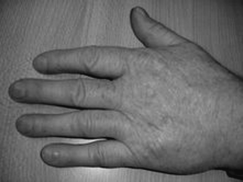
The patient was referred to the medical oncology services. On clinical examination there was Grade 4 clubbing of the upper limb digits (). He commenced treatment with chemotherapy as per the P6 protocol (vincristine, cyclophosphamide and doxorubicin alternating with ifosfamide and etoposide). The doses of chemotherapy were modified due to concerns regarding his ability to tolerate this regimen, which is more commonly used in children. Restaging CT scans performed following cycle 3 and cycle 5 of chemotherapy showed unchanged appearances of the mass. In view of the poor radiological response to chemotherapy, the patient did not proceed to surgery. He instead received radical chest irradiation, 60 Gy administered over 30 fractions. This was completed on 18/05/2006. Clinically, the fingertip clubbing decreased substantially during his treatment. A repeat chest x-ray performed in June 2006 showed a residual mass measuring at least 5×6 cm, with surrounding interstitial changes, likely reflecting radiation pneumonitis. He has remained clinically well in the intervening period, with no symptoms. His most recent CT of thorax, abdomen and pelvis in April 2007 showed significant reduction in the size of the mass to 5.3×3 cm, with interval development of local scarring and traction bronchiectasis, consistent with post radiotherapy change. A chest x-ray in September 2007 shows no evidence of disease progression.
Hypertrophic osteoarthropathy is characterized by finger clubbing, periosteal new bone formation and polyarthritis Citation[1]. Patients may complain of pain and swelling in the metacarpophalangeal joints, knees, ankles, wrists and even shoulders or elbows. Physical examination shows finger clubbing and tenderness over the adjacent long bones. Radionuclide bone scans with radioactive 99mTc typically shows increased binding of isotopes at sites of development of periosteal reaction in the long bones and elsewhere when the disease is active Citation[2]. This may be confirmed by evidence of periosteal new bone formation on plain film. It may be primary or secondary to a variety of malignant and non-maligant causes. When secondary to malignancy, hypertrophic osteoarthropathy is most commonly associated with bronchogenic neoplasms, particularly adenocarcinoma. However HPOA has been reported in association with many other solid malignancies, including melanoma Citation[3], cancers of the nasopharynx, oesophagus, breast and thyroid, Hodgkin lymphoma and sarcomas Citation[4]. It has not previously been reported in the setting of PNET. Reversibility of clubbing and/or hypertrophic osteoarthropathy after therapy (including surgery, radiotherapy and chemotherapy) has previously been described Citation[5].
PNETs belong to the Ewing's family of tumours (EFTs) of bone and soft tissue tumours which primarily affect children and young adults. They are defined by specific chromosomal abnormalities, most commonly the t(11,22)(q24,12) translocation. PNETs of thoraco-pulmonary origin were originally described by Askin et al., and are sometimes referred to as Askin tumours. Historically these tumours have had a very poor prognosis. More recently, locoregional EFTs have been treated with more success with intensive chemotherapy. A phase II, single-arm non-randomised evaluation of the MSKCC P6 protocol achieved an event-free survival rate in patients with locoregional disease of 82% at 4 years Citation[6], with most patients proceeding to an extensive surgical resection after their third treatment cycle. The majority of patients proceeding to surgical resection had a complete response or very good partial response after three cycles. A review from the Royal Marsden sarcoma unit of 59 patients with Ewing's sarcoma/PNET treated with heterogenous treatment regimens demonstrated an estimated 5-year progression free survival rate of 34% in non-metastatic patients Citation[7]. It appears that the response to initial chemotherapy is predictive of the likelihood of achieving cure with surgery. In the Royal Marsden review, disease bulk and response to primary therapy were strongly associated with PFS and OS, although these two parameters were closely correlated with each other. Age and tissue of origin of the tumour did not influence outcome.
References
- Utine EG, Yalçin B, Karnak İ, Kale G, Yalçin E, Dogru D, et al. Childhood intrathoracic Hodgkin lymphoma with hypertrophic pulmonary osteoarthropathy: A case report and review of the literature. Eur J Pediatr 2007; Jun 29. [Epub ahead of print].
- Jajic Z, Jacic I, Nemcic T. Primary hypertrophic osteoarthropathy: Clinical, radiologic and scintigraphic characteristics. Arch Med Res. 2001; 32: 136–42
- Thompson MA, Warner NB, Hwu WJ. Hypertrophic osteoarthropathy associated with metastatic melanoma. Melanoma Res 2005; 15: 559–61
- Burstein HJ, Janicek MJ, Skarin AT. Hypertrophic osteoarthropathy. J Clin Oncol 1997; 15: 2759–60
- Albrecht S, Keller A. Postchemotherapeutic reversibility of hypertrophic osteoarthropathy in a patient with bronchogenic adenocarcinoma. Clin Nucl Med 2003; 28: 463–6
- Kolb EA, Kushner BH, Gorlick R, Laverdiere C, Healey JH, LaQuaglia MP, et al. Long-term event-free survival after intensive chemotherapy for Ewing's family of tumours in children and young adults. J Clin Oncol 2003; 21: 3423–30
- Verrill MW, Judson IR, Harmer CL, Fisher C, Thomas JM, Wiltshaw E. Ewing's sarcoma and primitive neuroectodermal tumor in adults: Are they different from Ewing's sarcoma and primitive neuroectodermal tumor in children?. J Clin Oncol 1997; 15: 2611–21


