To the Editor
We previously reported on a patient in this journal (Acta Oncol 2006;45:750–2) who had a gastric mucosa-associated lymphoid tissue (MALT) lymphoma (MALToma) which showed intense F-18-fluoro-deoxyglucose (FDG) uptake into the tumor Citation[1]. FDG positron emission tomography (PET) has been considered the first-line modality for staging, restaging, and monitoring the therapeutic response for lymphomas. However, variable FDG avidity in MALTomas has been reported in the literature, and the usefulness of this modality for MALTomas is controversial Citation[2–4]. A recent report suggested that a MALToma with plasmacytic differentiation may be related to FDG uptake Citation[5]. We recently had a patient with an invasive thyroid MALToma which showed intense FDG uptake in the postoperative residual tumor.
A 77-year-old female received a total thyroidectomy for a large, rapidly growing mass in her left thyroid. The operative findings revealed that the mass was elastic and firm with an ill-defined margin and local invasion to the surrounding muscle and vessels. Incomplete removal of the tumor was noted during the operation. Gross pathological findings showed that the specimen was diffusely fibrotic and there was an ill-defined grayish-white tumor in the upper left thyroid. Histopathology revealed an invasive MALT lymphoma in the tumor associated with chronic thyroiditis and fibrosis. Immunohistochemical stains were positive for CD 20, negative for CD5, cyclin D1, CD10 and CD3, which excluded the possibility of other small B-cell lymphomas and follicular cell lymphoma. The cytokeratin stain enhanced the presence of lymphoepithelial lesion. The origin of the neoplastic lymphoid tissue turned out to be marginal zone B cell lymphoma (MALToma). In addition, the tumor had invaded the perithyroidial soft tissue and nerve (). Two months after the operation, a whole-body FDG-PET study showed focally intense FDG uptake in the anterior aspect of the left side of the neck, suggestive of a viable residual tissue ().
Figure 1. Tumor in the left thyroid gland showing a picture of a malignant lymphoma with diffuse lymphocyte infiltration and focal lymphoid nodular proliferation on a background of chronic thyroiditis. The nucleus of the tumor cells is small to intermediate sized and irregularly shaped, as shown in the inset (lower right).
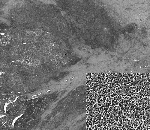
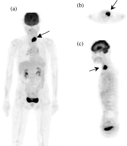
Figure 2. Whole-body PET performed 45 min after an intravenous injection of 370 MBq FDG using a Siemens ACCEL PET scanner. (A) Maximal intensity projection (MIP) view, (B) transverse section, and (C) sagittal section revealing focally intense FDG uptake in the anterior left side of the neck. The maximal standard uptake value (SUVm) of the lesion was 12.2. No abnormal uptake was demonstrated on the right side of the neck or elsewhere.
A thyroid lymphoma is a rare, heterogeneous disease comprising approximately 1∼5% of all thyroid malignancies and 1∼2.5% of all lymphomas Citation[6]. Pathogenically, the acquired lymphoid tissue from autoimmune thyroiditis might evolve to MALT and even transform to an aggressive lymphoma in the thyroid Citation[7–10]. Derringer et al. reported that among 108 cases of primary thyroid lymphomas, MALT was identified in 66 cases (61%), including mixed diffuse large B cell lymphoma (DLBCL) with MALT in 36 cases. In addition, lymphocytic thyroiditis was found in 94% of the cases, and 69% of the patients had perithyroidial soft-tissue infiltration Citation[9]. Thieblemont et al. reported that among 26 cases, 23% were MALTomas, 30% were DLBCLs, and 20% were mixed DLBCLs with MALTs. All patients with a thyroid MALToma were followed-up for various periods of time under a diagnosis of Hashimoto's thyroiditis Citation[6]. Sasal et al. reported that MALTomas comprised 77% of all thyroid lymphomas Citation[11].
FDG uptake in a benign lesion of the thyroid is not uncommon Citation[12–14]. Our previous study revealed that the diffuse and intense uptake in the thyroid glands was a clue to a diagnosis of chronic thyroiditis with hypothyroidism Citation[12]. FDG uptake into the DLBCL of the thyroid has been reported Citation[15–17]. However, few reports about thyroid MALTomas with FDG uptake are available Citation[18]. Because of the coexistence of chronic thyroiditis and MALTomas, utilization of histopathology and/or FDG-PET for initial evaluation and for follow-up to detect a residual/recurrent tumor in patients with a thyroid MALToma is conservative Citation[19], Citation[20].
References
- Liu JD, Tai CJ, Chang CC, Lin YH, Hsu CH. FDG-PET in a patient with gastric MALT lymphoma. Acta Oncol 2006; 45: 750–2
- Rodriguez M, Ahlström H, Sundin A, Rehn S, Sundström C, Hagberg H, et al. [18F] FDG PET in gastric Non-Hodgkin's lymphoma. Acta Oncol 1997; 36: 577–84
- Hoffmann M, Kletter K, Diemling M, Becherer A, Pfeffel F, Petkov V, et al. Positron emission tomography with fluorine-18-2-fluoro-deoxyglucose (F18-FDG) dose not visualize extranodal B-cell lymphoma of the mucosa-associated lymphoid tissue (MALT)-type. Ann Oncol 1999; 10: 1185–9
- Beal KP, Yeung HW, Yahalom J. FDG-PET scanning for detection and staging of extranodal marginal zone lymphomas of the MALT type: A report of 42 cases. Ann Oncol 2005; 16: 473–80
- Hoffmann M, Wöhrer S, Becherer A, Chott A, Streubel B, Kletter K, et al. F18-Floro-deoxy-glucose positron emission tomography in lymphoma of mucosa-associated lymphoid tissue: Histology makes the difference. Ann Oncol 2006; 17: 1761–5
- Thieblemont G, Mayer A, Dumontet C, Barbier Y, Callet-Bauchu E, Felman P, et al. Primary thyroid lymphoma is a heterogeneous disease. J Clin Endocrinol Metab 2002; 87: 105–11
- Holm L, Blomgren H, Lowhagen T. Cancer risks in patients with chronic lymphocytic thyroiditis. N Engl J Med 1985; 312: 601–4
- Kato I, Tajima K, Suchi T, Aozasa K, Matsuzuka F, Kuma K, et al. Chronic thyroiditis as a risk factor of B-cell lymphoma in the thyroid gland. Jpn J Cancer Res 1985; 76: 1085–90
- Derringer GA, Thompson LDR, Frommelt RA, Bijwaard KE, Heffess CS, Abbondanzo SL. Malignant lymphoma of the thyroid gland: A clinicopathologic study of 108 cases. Am J Surg Pathol 2000; 24: 623–39
- Smedby KE, Baecklund E, Askling J. Malignant lymphomas in autoimmunity and inflammation: A review of risks, risk factors, and lymphoma characteristics. Cancer Epidemiol Biomarkers Prev 2006; 15: 2069–77
- Sasai K, Yamabe H, Haga H, Tsutsui K, Dodo Y, Ishigaki T, et al. Non-Hodgkin's lymphoma of the thyroid–a clinical study of twenty-two cases. Acta Oncol 1996; 35: 457–62
- Chen YK, Chen YL, Cheng RH, Yeh CL, Lee CC, Hsu CH. The significance of FDG uptake in bilateral thyroid glands. Nucl Med Commun 2007; 28: 117–22
- Karantanis D, Bogsrud TV, Wiseman GA, Mullan BP, Subramaniam RM, Nathan MA, et al. Clinical significance of diffusely increased 18F-FDG uptake in the thyroid gland. J Nucl Med 2007; 48: 896–901
- Schmid DT, Kneifel S, Stoeckli SJ, Padberg BC, Merrill G, Goerres GW. Increased F18-FDG uptake mimicking thyroid cancer in a patient with Hashimoto's thyroiditis. Eur Radiol 2003; 13: 2119–21
- Chander S, Zingas AP, Bloom DA, Zak IT, Joyrich RN, Getzen TM. Positron emission tomography in primary thyroid lymphoma. Clin Nucl Med 2004; 29: 572–3
- Puget G, Trampal C, Calvo N, Carrera MJ, Conangla M, Sitges-Serra A, et al. Thyroid fluorodeoxyglucose-whole body positron emission tomography incidentaloma concurrently diagnosed with a diffuse large B cell lymphoma localized in the neck. Leuk Lymphoma 2007; 48: 425–7
- Lin EC. FDG PET/CT for assessing therapy response in primary thyroid lymphoma. Clin Nucl Med 2007; 32: 152–3
- Mikosch P, Würtz FG, Gallowitsch HJ, Kresnik E, Lind P. F-18-FDG-PET in a patient with Hashimoto's thyroiditis and MALT lymphoma recurrence of the thyroid. Wien Med Wochenschr 2003; 153: 89–92
- Androulaki A, Syriou V, Lazaris AC, Paterakis T, Pikazis D, Papathomas T, et al. Maltomas of the thyroid and Sjögren's syndrome in a woman with Hashimoto's thyroiditis. Endocr Pathol 2006; 17: 89–94
- Marchesi M, Biffoni M, Biancari F. False-positive finding on F18-FDG PET after chemotherapy for primary diffuse large B-cell lymphoma of the thyroid: A case report. Jpn J Clin Oncol 2004; 34: 280–1