Abstract
Correct preoperative diagnosis of a breast lesion is essential for optimal treatment planning. Our aim was to compare feasibility of fine needle aspiration cytology (FNAC) and core needle biopsy (CNB) in diagnosis of breast lesions. The special aim was to evaluate the extra costs and delay in surgical treatment due to unsuccessful preoperative biopsies. Diagnostic work-ups in 572 patients with 580 breast lesions were retrospectively evaluated. FNAC was the first biopsy method for 339 lesions, CNB for 241 lesions. The postoperative diagnosis was malignant for 503 lesions.
The preoperative rate of definitely malignant diagnosis was 67% (194/289) for FNAC and 96% (206/214) for CNB (p < 0.0001), and 95% and 99%, respectively (p = 0.0173), when also suspicious findings were included. In patients with FNAC, an additional needle biopsy was performed for 93 and a surgical biopsy for 62 lesions. In the CNB group, a subsequent CNB was performed for 2 and a surgical biopsy for 33. The frequent need for additional biopsies raised the total expenses of FNAC over those of CNB. Multiple biopsies may also delay cancer surgery. It is therefore recommended to use CNB as the initial needle biopsy method.
Accurate preoperative diagnosis of a breast lesion is essential for optimal treatment planning. To avoid unnecessary patient distress, it is important to achieve a definite diagnosis without delay and with minimal biopsies. Furthermore, cost-effective means of breast cancer diagnosis are crucial.
Fine-needle aspiration cytology (FNAC) and core needle biopsy (CNB) are both useful in evaluation of breast lesions Citation[1], Citation[2]. In a review article by Britton Citation[3] the absolute sensitivity of ultrasound guided FNAC was 83.1% and 96.7% of CNB Citation[3]. Because CNB has also better specificity and a lower insufficient sample rate, FNAC has increasingly been abandoned, particularly in North America and the UK Citation[4–7]. CNB also provides an easier way for grading and typing of tumors as well as for assessing predictive factors like hormone receptors and HER-2 status Citation[7]. The interpretation of FNAC is more dependent on the expertise of the cytopathologist than core needle biopsy is on the quality of histopathologist Citation[4].
On the other hand, FNAC does have distinct advantages over CNB; it is widely available, the price is inexpensive, it is quick to perform, and an immediate report is possible. In addition, FNAC is associated with a lower risk for complications like hematoma and infection. FNAC is also favored for lesions that are technically difficult to approach with a CNB Citation[3]. Furthermore, the smallest lesions are often removed almost entirely by CNB, endangering preoperative localization.
For these reasons, our purpose was to compare the feasibility of FNAC and CNB in preoperative diagnosis of malignant breast lesions. Our specific aims were to evaluate cost aspects as well as delay in surgical treatment due to unsuccessful preoperative biopsies.
Patients and methods
Patients
At the Breast Surgery Unit of Helsinki University Central Hospital (HUCH) 632 patients with 642 dubious, suspicious, or malignant lesions underwent surgery between February 1, 2005 and January 30, 2006. The data were collected retrospectively from the patient records. Patients operated on because of aberrant breast tissue and hypertrophic breasts, as well as those with suspicion of local recurrence after mastectomy, were excluded. Furthermore, 12 patients with vacuum-assisted biopsy as well as 50 patients with CNB obtained under stereotactical guidance were excluded, because these biopsy methods are used on occasions when FNAC is not recommendable, e. g. for diffuse microcalcifications or for a lesion invisible on ultrasound (US). Our study material includes 572 patients with 580 lesions. The postoperative diagnosis was ductal carcinoma in situ (DCIS) or invasive cancer for 503 lesions in 496 patients. The project plan was approved by the Ethics Committee of HUCH.
Indication for breast imaging was a clinical symptom, most often a palpable lump, in 341 of those 580 lesions. Ninety-eight lesions were detected on screening mammography and 106 were found in imaging of asymptomatic patients. The remaining 35 lesions were found in connection with breast cancer follow-up.
Median age of patients was 59 (18–99 years); 37 had a family history of breast cancer. Due to breast cancer in the ipsilateral breast 25 had undergone previous breast-conserving surgery, and 21 had had surgery due to contralateral cancer. Of the 572, 168 women had received hormone replacement therapy, 108 of them being still on therapy at the beginning of the diagnostic work-up.
Palpability, maximum diameter on US or on mammogram (MG), and the appearance of the lesion on US and MG were noted. The type of initial and potential subsequent biopsies with their results were recorded and also compared with the histological findings of the surgical specimen.
Needle biopsies
In 575 lesions, a radiologist performed the initial percutaneus biopsies under US guidance or under palpation. On two occasions, the biopsies, one FNAC and one CNB, were obtained under palpation check by a surgeon. Two FNAC and one CNB were taken from microcalcifications under a coordinate grid technique. FNAC was the first biopsy method for 339 lesions and CNB for 241. Most of the private clinics and small radiology divisions have not introduced CNB and have taken only FNAC. Whereas the larger breast imaging units have used primarily CNB. In these units FNAC is used if the location of the lesions is not safe for CNB or it is not technically possible to take CNB. In the CNB group, the lesions on imaging were larger; more lesions had a mass combined with microcalcifications, fewer lesions were invisible in a mammogram, and more lesions had invasive lobular cancer as the postoperative diagnosis. Otherwise, between the FNAC and CNB groups, lesion characteristics were well balanced ().
Table I. Characteristics of the 580 breast lesions with fine needle aspiration cytology (FNAC) or core needle biopsy (CNB).
For 274 lesions, breast imaging and biopsies were performed or directly supervised by experienced radiologists at the Department of Radiology of HUCH. Of these biopsies, 80 (29%) were FNAC and 194 (71%) CNB. In 293 lesions, the biopsies were performed by radiologists at private clinics. 250 (85%) were FNAC and 43 (15%) CNB. The remaining 12 specimens, 8 FNAC and 4 CNB, were obtained in public health care hospitals other than HUCH. In HUCH, FNAC were taken with 20G needle with a 10 ml syringe in a special holder, more than five passes through were made from different areas of the lesions. A cytospin preparation and a cell block were prepared from the diagnostic material. CNB were taken with long-throw (15–22 mm) biopsy gun, 14G cutting needle and removal of three or more tissue samples per lesion. The information regarding the needle gauge and techniques were not available from the referring units.
Pathology
The cyto- or histopathological assessment of 281 percutaneus biopsies, 86 FNAC and 195 CNB was performed or directly supervised by experienced breast pathologists at the Department of Pathology of HUCH. The remaining 299 specimens, 253 FNAC and 46 CNB, were assessed at other public health care or private pathology laboratories by specialist pathologists. All the surgical breast specimens were assessed at the Department of Pathology of HUCH.
Cytological grading of Papanicolau (0 = insufficient, 1 = normal, 2 = probably benign, 3 = mildly suspicious of malignancy, 4 = strongly suspicious of malignancy, 5 = malignant) was used for FNAC. CNB findings were classified as insufficient, benign, indeterminate, suspicious of malignancy, or malignant. The indeterminate category consisted of findings that warranted a surgical biopsy: atypical ductal hyperplasia (ADH), lobular carcinoma in situ (LCIS), papilloma, radial scar or benign tumor phyllodes.
Surgery
Surgical biopsy was performed when the CNB findings were indeterminate, as well as when disagreement arose between the clinical, radiological, and cytological or histological findings. Fibroadenomas were excised when symptomatic, larger than 2 cm, or rapidly growing, as well as when no CNB was technically possible.
In patients with CNB, indication for breast cancer surgery was a malignant finding, either DCIS or invasive cancer, in the preoperative biopsy. Exceptionally, cancer surgery with sentinel node biopsy was performed in few cases with benign or indeterminate findings in the CNB but a definitely malignant radiological finding.
In patients with FNAC, the decision to proceed to cancer surgery was made by the operating surgeon in agreement with the patient. In general, definitely malignant, Papa 5, finding in cytology of a lesion that was radiologically highly suspicious or suggestive of malignancy indicated a cancer operation. Sometimes FNAC with Papa 4 and definitely malignant radiological findings was regarded as sufficient without additional biopsies.
In patients with invasive cancer in CNB or Papa 5 in the FNAC, breast cancer surgery consisted of wide local excision or mastectomy combined with sentinel node biopsy or axillary clearance. Patients with DCIS in CNB had wide local excision or mastectomy. The indication for sentinel node biopsy in patients with DCIS in CNB was extensive disease warranting mastectomy. Patients, who were candidates for breast-conserving surgery, had a sentinel node biopsy when invasion was suspected in the CNB specimen, when the tumor was palpable, when the lesion was visible on US, or when MG showed tumor or architectural distortion.
Delay in surgery
Time interval was calculated between initial biopsy and the definite surgical treatment among 492 in situ or invasive breast cancers was calculated. Patients who received neoadjuvant therapy (n = 4) or those with the operation postponed due to comorbidity (n = 2) were excluded from this analysis.
Cost aspects
When biopsy result was insufficient for treatment planning and needed additional work-up, or false biopsy result caused unnecessary operations, these extra expenses were recorded. In patients with benign, indeterminate, or insufficient findings in the needle biopsy but invasive cancer in the surgical specimen, axillary surgery was performed as a second operation. In these cases, the price of the surgical biopsy was added. In patients with benign or indeterminate findings in the needle biopsy but DCIS with insufficient resection margins in the surgical biopsy, no extra charges were added. If patient was operated on because of an indeterminate biopsy finding and the postoperative diagnosis was not malignant, the expenses of the surgical biopsy were charged. When the initial or additional CNB showed DCIS, but invasion was detectable in the surgical specimen, the expenses of axillary surgery were not charged when SNB was included in the operation. In cases with unnecessary axillary surgery because of a false positive biopsy finding, the costs of axillary surgery were added, but not in cases with FNAC PAPA 4–5 and DCIS for the surgical specimen.
Because of the variety of referring units, the HUCH price-list for radiological, pathological, and surgical interventions was used ().
Table II. Prices at Helsinki University Central Hospital.
Statistical methods
The preoperative rate of definitely malignant diagnosis was calculated: number of carcinomas diagnosed by FNAC (Papa 5) or CNB (malignant) expressed as a percentage of all cancers diagnosed. We also calculated the preoperative rate of definitely malignant or suspicious diagnosis: FNAC results Papa 3–5, and CNB results indeterminate-malignant expressed as a percentage of all cancers diagnosed.
Differences between these preoperative diagnosis rates of FNAC and CNB groups were calculated by Fisher's exact test for the two-tailed P-values and by Newcombe's method for 95% confidence levels.
Results
Preoperative results of FNAC and CNB
The preoperative rate of definitely malignant diagnosis was 67% (194/289) for FNAC and 96% (206/214) for CNB (p < 0.0001, difference of 29%; 95% CL 22.9–35.0). The preoperative rate of definite malignant or suspicious diagnosis was 95% (273/289) for FNAC and 99% (211/214) for CNB (p = 0.0173, difference of 4%; 95% CL 0.8–7.5%). The preoperative rate of definitely malignant diagnosis of the CNB was superior to that of FNAC regardless of tumour characteristics, but these findings did not reach significance for lesions that were few (). FNAC resulted 48/339 (14%) Papa 3, of these 48 lesions 36 was malignant. CNB resulted 9/241 (4%) indeterminate finding, the final diagnosis was malignant for four lesions.
Table III. The preoperative rate of definitely malignant diagnosis of fine needle aspiration cytology (FNAC) or core needle biopsy (CNB) in diagnosis of 503 malignant breast lesions.
No significant difference (p = 0.0921) emerged between the preoperative rate of definitely malignant diagnosis of FNAC obtained at HUCH, of 62, 36 (52%) malignant lesions, and at other private or public health care units: of 227, 159 (70%) malignant lesions.
Of the 580 operated lesions, 297 were spiculated masses on mammography and suspicious for malignancy on US. Only one of these 297 had a benign postoperative histology. It was correctly diagnosed on CNB as papilloma. Invasive carcinoma was the final postoperative diagnosis for 294 of the other lesions, with remaining two being DCIS. FNAC was performed on 177 of these lesions and CNB on 119. The malignant diagnosis rate of the CNB group was 98%, but for FNAC only 75% (p < 0.0001, difference of 24%; 95% CL 17–31) ().
Table IV. Outcomes of US-guided fine needle aspiration cytology (FNAC) (n = 177) and US-guided core needle biopsy (CNB) (n = 119) in the 296 lesions with malignant postoperative histology which appearing as spiculated masses on mammography and suspicious for malignancy on ultrasound.
Diagnostic work-up of breast lesions
The finding in FNAC was Papa 5, in 197 of the 339 lesions with FNAC as the initial biopsy method. An additional CNB was performed for four lesions, and cancer surgery was planned for 193; in these 193 lesions, invasive cancer was detected in 190, DCIS in one, and a benign finding in two of the surgical specimens.
In the remaining 142 lesions with Papa 0–4 for the initial biopsy, a subsequent FNAC was obtained for 4 lesions and a subsequent CNB for 85. In addition, for a total of 62 lesions a surgical biopsy was performed, revealing invasive cancer in 11 lesions and DCIS in 4. Cancer surgery without further biopsies was performed in three cases with benign or indeterminate findings in the subsequent CNB and definitely malignant findings in imaging; invasive cancer was detected in the surgical specimen in all three cases. Cancer surgery was performed without additional biopsies for 19 lesions: The postoperative diagnosis was invasive cancer in 16, DCIS in two and benign in one ().
Figure 1. Diagnostic work-up in the 339 breast lesions with fine needle aspiration cytology (FNAC) as the initial biopsy. Abbreviations: CNB = core needle biopsy, SB = surgical biopsy, CS = cancer surgery, CA = invasive cancer, DCIS = ductal carcinoma in situ.
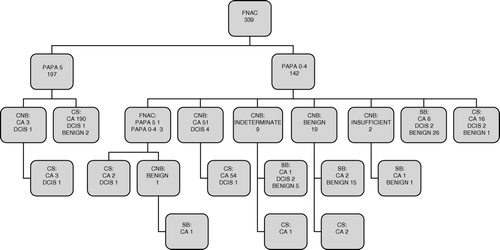
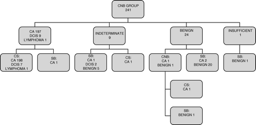
In the CNB group, a subsequent CNB was necessary for two lesions. A surgical biopsy was performed for 33 lesions, revealing four invasive cancers and 2 DCIS. In addition, invasion was detected in the surgical specimen in two lesions with DCIS in the initial CNB. Cancer surgery without further biopsies was performed in one patient with an indeterminate finding in their initial CNB and a definitely malignant finding in imaging. The surgical specimen revealed invasive cancer ().
Figure 2. Diagnostic work-up in the 241 breast lesions with core needle biopsy (CNB) as the initial biopsy. Abbreviations as in .
Delay in cancer surgery
In both groups, when the biopsy showed cancer, the median time from initial biopsy to definite surgical treatment was 31 days. The extra needle and surgical biopsies delayed the definite surgical treatment for 75 of the 283 patients with FNAC but for only 5 of the 207 patients with CNB, 27% vs. 2%. All cancer patients who had FNAC as their initial biopsy method had their operation a mean of 3 days later than did patients with CNB. In addition, one patient with bilateral cancer had FNAC for the one and CNB for the other breast. Both the FNAC and the CNB failed to detect cancer, and surgical cancer treatment was delayed accordingly (). Six patients with neoadjuvant treatment or co-morbidity postponing surgery were excluded from this analysis.
Table V. Time interval in days between first biopsy and definite surgical treatment for 490 women with in situ or invasive breast cancers with fine needle aspiration cytology (FNAC) or core needle biopsy (CNB).
Cost aspects
In the FNAC group, the extra expenses resulted from 93 supplemental needle biopsies, 11 surgical biopsies of malignant lesions, and 8 surgical biopsies of benign lesions. Three patients underwent unnecessary axillary surgery, and these costs were charged. One of these three women had 3 extra inpatient care days because of a wound infection; the costs of these extra days were also included.
In the CNB group, the extra expenses were due to two supplemental needle biopsies, four surgical biopsies of malignant lesions, and five surgical biopsies of benign lesions plus one unnecessary sentinel node biopsy performed in a patient with a postoperative diagnosis of lymphoma instead of ductal carcinoma.
The cost of the initial biopsy was 150 € per lesion for FNAC and 176 € per lesion for CNB. With the expenses caused by the additional needle biopsies included, corresponding costs were 210 € for FNAC and 177 € per lesion in the CNB group. The need for surgical biopsies and the unnecessary axillary operations due to false-positive findings raised the costs to 294 € in lesions with FNAC as the initial biopsy and to 223 € in lesions with CNB.
Discussion
In the present study, both the preoperative rate of definitely malignant diagnosis and also the rate of malignant or suspicious diagnosis CNB were superior to that of FNAC. According to Quality assurance in the diagnosis of breast disease by EUSOMA, at least 70% of patients with breast cancer should have a preoperative diagnosis of malignancy, that is FNAC or CNB reported as definitely malignant Citation[8]. In our series FNAC failed to achieve this target. Our retrospective, non-randomized study setting is the major limitation of the present study and therefore is a potential source of bias. On the other hand, our FNAC and CNB groups were rather well balanced as regards to the clinical, radiological and histological features of these lesions. Furthermore, CNB was better than FNAC regardless of these tumor characteristics although, the difference failed to reach significance in some lesion groups with a very small number of cases. Spiculated mass lesions in MG are regarded as highly predictive for invasive carcinoma Citation[9]. We combined this radiological feature with a suspicious finding on ultrasound. In the present study, almost all, (99%) of these lesions proved to be invasive carcinomas. We can assume that all this kind of lesions was referred to our clinic because of their malignant radiological finding despite of their biopsy outcomes. Therefore, for these lesions we calculated sensitivities; even in these lesions, CNB was superior to FNAC regardless of lesion size.
In studies with FNAC and CNB taken from the same lesion at the same session, an equal absolute sensitivity of 72–75% has been reported Citation[10], but in another study, the absolute sensitivity for CNB was higher, 80% vs. 65% Citation[4]. The specificity has been significantly better for CNB (90% vs. 82%) Citation[10]. Together FNAC and CNB can lead to an increase in absolute sensitivity without affecting specificity, and also decrease the rate of inadequate samples Citation[4], Citation[10].
FNAC obtained by pathologists has been suggested to be more sensitive than FNAC performed by non-pathologists in making an unequivocal diagnosis of breast cancer Citation[11]. When the immediate evaluation of FNAC is made by the cytopathologist, it is possible to decrease the rate of insufficient samples to as low as 5.8% Citation[12]. Also a distinction between invasive and in situ carcinoma has been possible in 294 of 320 (91.8%) cases when the smear was fully diagnostic Citation[12]. In another study FNAC overestimated DCIS in 9% (the cytological finding was malignant cells, but in the surgical specimen there was DCIS without invasive component) which may be a problem because of the differences in treatment of in situ and invasive carcinomas with respect to the axilla Citation[13]. For highly suspicious microcalcifications the sensitivity of FNAC has been be over 70%, but over 75% of the inadequate or benign FNA smears proved to be malignant on histology Citation[14]. In a series with 758 women, a larger needle gauge and vacuum-assisted core devices reduced underestimation of malignancies as did increasing the total volume of tissue obtained at CNB Citation[15]. For these reasons it is recommendable to use vacuum-assisted biopsy or CNB in the diagnosis of microcalcifications.
It is noteworthy that most of the units at Helsinki region refer patients to our Breast Surgery Unit. Most of the private clinics or smaller public health care units have not introduced CNB at all in their practice. Consequently, only 15% of the biopsies performed in the private clinics were CNB. On some occasions, FNAC was even obtained under a coordinate grid technique for lesions not visible on breast US, such as microcalcifications. In contrast, a standard practice in the breast-imaging division of The Department of Radiology of HUCH is to use primarily CNB for suspicious breast lesions.
In the United States, a multicenter clinical trial to evaluate FNAC for non-palpable lesions performed by multiple operators was terminated early because of the high rate of insufficient samples Citation[5]. In our study the biopsies were taken and analyzed by several radiologists and pathologists with varying levels of experience. This may have influenced our results, especially the sensitivity of FNAC. On the other hand, the present study setting reflects the situation in many breast surgery centers with several referring units.
It was impossible to include all lesions considered as benign based on findings from the clinical examination, imaging, and needle biopsy, because most of patients with those lesions are not even referred to our unit. Therefore we were not able to calculate sensitivities or specificities of FNAC and CNB for all kind of lesions.
Many studies have found percutaneous biopsies to be cost-effective, sparing patients from surgical biopsies without compromising breast cancer detection Citation[16–18]. In this study, FNAC by itself was less expensive than CNB, but the frequent need for additional biopsies and the costs due to unnecessary operations raised the total expenses of FNAC over those of CNB. The savings would have been more than 24%, had the first biopsy method been CNB instead of FNAC in all cases. To our best knowledge, this is the first study comparing FNAC and CNB in order to evaluate the need for additional biopsies and to address the consequences of these supplemental diagnostic procedures. Comparison of true costs generated by the biopsy methods is however demanding, and our results may not be generalizable, due to differences in costs of services and in the quality of technical performance and in analysis of needle biopsy specimens. We do not have annual statistics concerning the biopsies taken in our region; therefore we can not calculate the overall costs of the whole FNAC series or CNB series. Measuring all costs from repeated biopsies, unnecessary axillary surgery, the cost of additional sick leave days, or possible future problems in cases of true cancer is also demanding. In all patients referred to our unit with benign findings in FNAC, except in those with simple cysts, the diagnosis is confirmed with CNB before surgical biopsy is omitted. This practise raises further the overall costs of FNAC.
In our study only one of the private clinics referring patients to our unit offered an immediate result from FNAC. In general, it takes from 3 to 7 days for the result. This and the frequent need for repeat biopsies diminished the advantage of quick results expected of FNAC. In addition to generating extra expenses, multiple biopsies also delayed surgical cancer treatment. However, the delay in surgery was also lengthened due to the long waiting lists for additional biopsies and outpatient clinic and surgical treatment, especially during vacation times. Regrettably, some patients had to wait as long as 3 months for surgery.
In units with immediate reporting of FNAC and further biopsies readily available, the use of FNAC does not cause such a delay in cancer surgery. Nevertheless, many other clinics also have limited radiological, cytological, and surgical resources and may thus encounter similar problems as addressed here.
In a single centre study by Bulgaresi et al. Citation[19] the positive predictive value of FNAC was high when abnormal cytology categories Papa 3–5 were combined with the clinical and imaging findings. Therefore FNAC was suggested to be a useful test in breast diagnosis, especially in assisting clinical decision-making whether to take additional biopsies or to progress to surgical management Citation[19]. Although FNAC was highly sensitive in detecting abnormality, resulting in Papa 3–5 in 94% of the malignant breast lesions in present study, that is not enough for optimal treatment planning. Before the surgeon can discuss about different treatment options, like sentinel node biopsy, immediate breast reconstruction or neoadjuvant therapy, with the patient, not only the definitely malignant diagnosis is crucial, but also the knowledge about the invasiveness and the type of the cancer. Had the first biopsy method been CNB instead of FNAC in all cases, the saving would have been more than 24%. The difficulties in the demanding evaluation of cytological findings were also apparent; false-positive FNAC findings led to unnecessary axillary surgery in three patients. The need for additional biopsies also means more appointments with radiologists and surgeons, which strains the capacity of the public health service. Multiple procedures and delay at any stage in obtaining a definitive diagnosis of both benign and malignant conditions are undesirable outcomes causing patient discomfort and anxiety.
In the preoperative breast cancer detection CNB was better than FNAC. The frequent need for additional diagnostic work-up raised the total expenses of FNAC over those of CNB and seemed to extend time to treatment. It is therefore recommended to use CNB as the initial needle biopsy method.
References
- Gordon PB, Goldenberg SL, Chan NHL. Solid breast lesions: Diagnosis with US-guided Fine-needle aspiration biopsy. Radiology 1993; 198: 573–80
- Prker SH, Burbank F, Jackman RJ, Aucreman CJ, Gardenosa G, Cink TM, et al. Percutaneous large-core breast biopsy: A multi-institutional study. Radiology 1994; 193: 359–64
- Britton PD. Fine needle aspiration or core biopsy. The Breast 1999; 8: 1–4,5
- Lieske B, Ravichandran D, Wright D. Role of fine-needle aspiration cytology and core biopsy in the preoperative diagnosis of screen-detected breast carcinoma. Br J Cancer 2006; 95: 62–6
- Pisano ED, Fajardo LL, Caudry DJ, Sneige N, Frable WJ, Berg WA, et al. Fine-needle aspiration biopsy lesions in a multicenter clinical trial: Results from the radiologic diagnostic oncology group V. Radiology 2001; 219: 785–92
- Britton PD, Flower CD, Freeman AH, Sinntamby R, Warren R, Goddard MJ, et al. Changing to core biopsy in an NHS breast screening unit. Clin Radiol 1997; 52: 764–7
- Shannon J, Douglas-Jones AG, Dallimore NS. Conversion to core biopsy in preoperative diagnosis of breast lesions: Is it justified by results?. J Clin Pathol 2001; 54: 762–5
- Perry N on behalf of the EUSOMA Working Party. Quality assurance in the diagnosis of breast disease. Eur J Cancer 2001;37:1159–72.
- Thurfjell MG, Lindgren A, Thurfjell E. Nonpalpable breast cancer: Mammographic appearance as predictor of histologic type. Radiology 2002; 222: 165–70
- Westenend P, Sever A, Beekman-de Volder H, Liem S. A comparison of aspiration cytology and core needle biopsy in the evaluation of breast lesions. Cancer Cytopathol 2001; 93: 146–50
- Sun W, Li A, Abreo F, Turbat-Herrera E, Grafton W. Comparison of fine-needle aspiration cytology and core biopsy for diagnosis of breast cancer. Diagnostic Cytopathol 2000; 24: 421–5
- Sauer T, Young K, Østerbø Thoresen S. Fine needle aspiration cytology in the work-up of mammographic and ultrasonographic findings in breast cancer screening: An attempt at differentiating in situ and invasive carcinoma. Cytopathol 2002; 13: 101–10
- Pijnappel R, van den Donk M, Holland R, Mali W, Peterse J, Hendrikc J, et al. Diacnostic accuracy for different srategies of image-guided breast interventions in cases of nonpalpable breast lesions. Br J Cancer 2004; 90: 595–600
- Farshid G, Rush G. The use of fine-needle aspiration cytology and core biopsy in the assessment of highly suspicious mammographic microcalcifications. Analysis of outcome for 182 lesions detected in the setting of a population-based breast cancer screening program. Cancer Cytopathol 2003; 99: 357–64
- Houssami N, Ciatto S, Ellis I, Ambrogetti D. Underestimation of malignancy of breast core-needle biopsy. Cancer 2007; 109: 487–95
- Liberman L, Kaplan JB. Percutaneous core biopsy of nonpalpable breast lesions: Utility and impact on cost of diagnosis. Breast Disease 2001; 13: 49–57
- Altomare V, Guerriero G, Giacomelli L, Battista C, Carino R, Montesano M, et al. Management of nonpalpable breast lesions in a modern functional breast unit. Breast Cancer Res Treat 2005; 93: 85–9
- Logan-Young W, Dawson AE, Wilbur DC, Avila EE, Tomkiewicz ZM, Sheils LA, et al. The cost-effectiveness of fine-needle aspiration cytology and 14-gauge core needle biopsy compared with open surgical biopsy in the diagnosis of breast carcinoma. Cancer 1998; 82: 1867–73
- Bulgaresi P, Cariaggi P, Ciatto S, Houssami N. Positive predictive value of breast fine needle aspiration cytology (FNAC) in combination with clinical and imaging findings: A series of 2334 subjects with abnormal cytology. Breast Cancer Res Treat 2006; 97: 319–21