Abstract
Purpose. The purpose of this study is to determine the inter- and intra-fractional respiration induced tumour movements as well as setup accuracy in a stereotactic body frame for stereotactic treatments of NSCLC patients. Patients and methods. From August 2005 to March 2008, 26 patients with NSCLC where given a stereotactic treatment. The patients were scanned with normal and uncoached respiration without use of abdominal compression. Each patient had CT-scans performed at four occasions throughout the treatment: As part of the CT-simulation and before the three radiotherapy treatments. At every occasion five individual CT-scans covering the tumour volume were obtained. In this way 20 scans where obtained from each patient. In each CT-scan the maximum positions of the tumour where located in all six directions, represented by the top, bottom, anterior, posterior, left and right part of the tumour. These coordinate constitute the data of this study. Results. The standard deviations of the respiration induced intra-fractional movements were: LR: 0.9 mm, AP: 1.6 mm and CC: 2.0 mm (1 SD). The inter-fractional movements were: LR: 1.1 mm, AP: 1.3 mm and CC: 1.7 mm (1 SD). Finally the set up accuracies in the body frame were LR: 1.5 mm, AP: 1.1 mm and CC: 1.7 mm (1 SD). Discussion and conclusions. Consecutive CT scans can be used to evaluate the respiration induced tumour movement. For patients immobilized in a stereotactic body frame, large movements of the tumour are rarely seen within the lung. With consecutive scans, using a conventional CT-scanner, it is possible to select those patients in whom the tumour movement is large. Application of 4D CT and Cone beam verification is strongly encouraged to minimize the requested treatment margin.
Extracranial stereotactic radiotherapy involves large doses per fraction and relies on the use of limited field sizes to decrease normal tissue toxicity. It is important to understand the individual uncertainties related to stereotactic treatment such as: 1) Patient immobilization 2) Tumour and organ of risk outlines 3) Dose planning/calculation and 4) Positioning of the patient on the treatment couch. Each of these uncertainties needs to be minimized to facilitate the smallest treatment port possible. To complicate matters the tumour position varies during treatment due to respiration. The respiration related movements can be divided into two parts 1) respiration during irradiation – intrafractional movements and 2) change of mean tumour position between two treatment fractions – interfractional movements.
The magnitude and importance of the different uncertainties depend on the applied planning and treatment technique. Nevertheless, in a number of situations, the respiration and patient positioning will be the uncertainties limiting the minimal size of the treatment ports. Previous studies of the movement of lung tumours have focused on peak-to-peak movement. However, margin recipes are currently mostly based on information concerning the standard deviation of the movement distribution and not on the peak-to-peak amplitude Citation[1–6]. In order to calculate the standard deviation of the movement distribution, it is necessary to know the actual tumour motion as a function of time. This motion can be studied by means of time resolved computed tomography (4D CT scanning) or by consecutive CT scans.
The aim of the present study is to investigate the inter- and intra-fractional movement of the tumour in extracranial stereotactic radiotherapy of non small cell lung cancer (NSCLC) as well as the patient positioning inside the stereotactic body frame.
Patients, materials and methods
From August 2005 to March 2008, 26 patients with a small solitary NSCLC tumour were given a stereotactic treatment. Twenty two of the patients were included in the study as described below.
Patient group and eligibility
In the patient specifics are summarized. Most of the patients suffered from poor lung function (median FEV1 was below 1 l/min). A majority of the patients were current smokers. None of the patients had nodal disease, but one patient had received stereotactic radiotherapy for single synchronous brain metastasis prior to the stereotactic treatment of her lung tumour.
Table I. Patient specifics.
Simulation, prescription, treatment planning and data collection
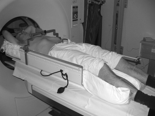
The patients were immobilized in a Lax-Blomgreen stereotactic body frame (SBF) using a VacFix vacuum bag (see ). A Siemens Somatom 4 scanner was used to acquire the CT scans. The spatial resolution was 1 mm in the craniocaudal (CC) direction and 0.98 mm in the left-right (LR) and anterior-posterior (AP) directions. The patients were scanned with normal and uncoached respiration and without the use of abdominal compression (We have not been able to reduce the respiration motion in a systematic way by using the abdominal press). The prescribed dose was 45 Gy in 3 fractions to the GTV. The Pinnacle3® system was used for treatment planning and the doses were calculated with the Collapsed-cone algorithm. The treatment duration was nine days (whenever possible). The preferable treatment technique was at least six different beam directions with no overlapping skin entries to avoid severe skin toxicity.
Figure 1. Patient immobilization using Lax-Blomgreen stereotactic body frame.
Every patient had CT-scans performed at four occasions throughout the treatment: As part of the CT-simulation and before each of the three radiotherapy treatments. At every occasion five individual CT-scans covering the tumour volume were obtained. In total 20 CT-scans where obtained for each patient. In all CT-scans the extreme positions of the tumour where identified in all six directions, represented by the top, bottom, anterior, posterior, left and right part of the tumour. These coordinates constitute the data of this study. For a few of the patients, the preferred number of CT-scans was not successfully obtained due to technical issues regarding the CT scanner. Only patients who were scanned at least three times were included in the analysis-this criterion was fulfilled by 22 of the patients. In total this study is based on analysis of 430 CT-scans.
A common reference system between the individual CT sessions is obtained by fusion of the patient anatomy of a given CT session with the patient anatomy of the first CT session. The base frame was the spinal column, which is stable during respiration and movements of the diaphragm. The fusion was performed automatically with a cross correlation or a mutual information algorithm, and afterwards manually checked to validate the fusion. The fusion of the patient anatomy makes it possible to measure the interfractional movement within the patient. To measure the patient position relative to the stereotactic body frame a common reference system located in the SBF is needed. Therefore a fusion of the SBF geometry was made similar to the fusion of the patient geometry. The interfractional movement measured in the SBF fixed system is the sum of the patient position in the frame and the interfractional movement inside the patient. Thus based on these two coordinate systems it is possible to evaluate the uncertainty of the tumour position relative to the SBF. All fusions were performed in the Syntegra module in the Pinnacle3® dose planning system.
Tumour position
To facilitate measurements of correlations between tumour movements and position in the lung, the tumour position has been measured as suggested by Onimaru Citation[7]. Onimaru measured the longitudinal tumour position in the lung as the distance from the apex to the tumour relative to the length of the lung. Likewise are the two other directions measured as the relative distance from the center line of the patient and the posterior part of the lung.
Four-dimensional CT simulation
During the study it became possible to perform 4D CT scans at our institution. To check the validity of the consecutive CT scans six patients had an additional 4D CT scan of the tumour region. The 4D CT scan was performed at the same scanner as the consecutive scans.
For the 4D CT scan 10 detector rotations were completed for each table position in a consecutive scan mode. The respiration signal was obtained by means of a thermocouple placed in a facial mask Citation[8]. In-house developed software was used to analyze the respiration signal, sort the CT slices into 10 phase bins, and to perform space-and-phase interpolations to complete each of the 10 phase bins. The tumour was contoured in one of the phases and translated to the remaining 9 respiration phases by rigid registration greyscale values matching.
Results
The standard deviation of the tumour movement measured by the consecutive CT scans is shown in . It is seen that all three components; intrafractional and interfractional movement, and positioning of the patient in the SBF are approximately of the same size. The uncertainties of the standard deviations are measured by bootstrap as described in Citation[9]. The interfractional movement given in is measured in the patient fixed coordinate system. The line ‘Sum of Inter and SBF’ in is the interfractional movement measured in the SBF coordinate system. The interfractional movement in the SBF coordinate system is the sum of the interfractional movement in the patient system plus the positioning uncertainty of the patient in the SBF. Thus the ‘Accuracy in SBF’ is calculated as the square root of the differences of the squared interfractional movement in the SBF and patient coordinate system. Likewise, the line ‘Sum of Inter, Intra and SBF’ is the corresponding sum of intra- and interfractional as well as the SBF uncertainty.
Table II. Standard deviations of the tumour movements (mm).
To facilitate comparison of the tumour motion with previous studies shows a few characteristics of the peak-to-peak intrafractional movement.
Table III. Charcteristics of the peak-to-peak intra-fractional movement (mm).
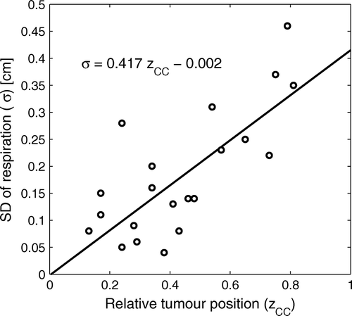
Linear regression between the intrafractional movement and tumour position has been performed. The only statistical significant result (on the 5% level) is the correlation between the intrafractional movement in the Cranio-Caudal direction and the tumour position in the Cranio-Caudal direction. A scatter plot of the two variables is shown in .
Figure 2. Standard deviation of the intrafractional movement in the Cranio-Caudal direction as function of the tumour position in the Cranio-Caudal direction. The Pearson correlation coefficient is 0.74. The fit is a linear regression. The uncertainty on the slope coefficient and on the constant is 0.085 and 0.041 (1SD), respectively.
To compare the results from the consecutive CT scans and the 4D CT scan, the intrafractional uncertainty was calculated for each individual patient. These values were compared by a pair test. The difference was not statistically significant in any of the directions.
Discussion
Consecutive CT scans of patients scheduled for stereotactic treatment of a lung tumour can be used to evaluate the respirational movement of the tumour. The results of the consecutive CT scans in this study are not significantly different from the results obtained from the 4D CT scan. A 4D CT scan has some obvious advantages compared to consecutive CT scans. One of the advantages is that it is possible directly to track the tumour movement as a function of the respiration phase. The downside of 4D CT scanning is that the quality of the scan depends on the constancy of the patient breathing pattern during the entire scan. Patients scheduled for stereotactic treatment of lung tumours normally have very poor lung function (the limited lung function is typically the reason that surgery is not possible). In some cases we have not been able to make a useful 4D CT scan (a scan where tracking of the tumour is possible) of patients scheduled for stereotactic treatment. For these patients consecutive CT scans can be useful to evaluate the respiration induced intrafractional tumour movement.
When using the stereotactic body frame for the immobilization of patients, the respirational movements for most of the patients are relatively small.
It should be stressed that the SBF uncertainty shown in is the uncertainty of the patient position relative to the SBF and not relative to the isocenter of the accelerator. The SBF needs to be aligned at the accelerator as well, which will introduce additional uncertainties. Grills Citation[10] reports a standard deviation on the positional uncertainty before image verification of approximately 2–3 mm for treatments in SBF. These values are in close agreement with what we find at our institution. To eliminate such setup errors conventional imaging of the bony anatomy is needed. However, imaging of the bony anatomy will not eliminate the interfractional uncertainties listed in since this uncertainty is related to the tumour position relative to the bony anatomy (the spine). Differences between matching on bony anatomy and soft tissue have previously been reported by Guckenberger Citation[11] and Purdie Citation[12].
The necessary margin between the CTV and PTV to fulfil dose coverage of the CTV is by van Herk Citation[4] defined as k1Σ+k2σ. The value of k1 and k2 depends on the fractions of the patient that should receive at least a specific dose to the CTV and on the shape of the penumbra of the treatment fields. Typically k1 is set to 2.5 (90% of the patient receives at least 95% of the prescribed dose in the CTV). In Citation[4] k2 is given as 0.7 for prostate treatments. The beam penumbra for treatment in lungs is broader than the penumbra for treatment in the abdominal region. Therefore k2 for treatment in lungs is not 0.7 but in our case close to 0.5 (for detail on calculation of k2 see van Herk Citation[4]).
If stereotactic dose planning and treatment are based on a single standard CT scan and image verification of the bony anatomy, both the intrafractional and interfractional movement will contribute to the systematic uncertainty (the tumour may have been scanned in an extreme position at simulation) Citation[4], and the intrafractional movement will contribute to the random uncertainty. Thus, as an example a margin of approximately 9 mm (2.5×3.1 + 0.5×2.0) will be needed in the CC direction. Introduction of 4D CT scanning and application of the mid-ventilation phase for dose planning Citation[6] will almost remove the intrafractional component from the systematic uncertainty (except for a small residual uncertainty) and thereby reducing the margin to approximately 7 mm. The relatively modest reduction of margin is due to the interfractional uncertainty component which is still present. However, a combination of a 4D CT scan and Cone beam CT image verification will be able to reduce the margin considerable. A combined 4D CT scan and Cone beam CT image verification based on soft tissue registration would be able to eliminate most of the systematic uncertainties except for a small residual component. Preliminary studies at our department show that we will have a residual uncertainty with a standard deviation slightly lower than 1 mm. Similar values have recently been published by Grills Citation[10]. Using a residual uncertainty of 1 mm the margin related to the application of 4D CT scanning and image verification by Cone beam CT would be approximately 3.5 mm.
Further reduction of the margin can be achieved by the use of respiratory gating of the accelerator. Gating of the accelerator may potentially reduce the random uncertainty (the intrafractional movement). The random uncertainty is responsible for a margin contribution of approximately 1 mm (0.5×2.0 mm). Gating will therefore at the maximum be able to reduce the margin with an additional millimeter. Due to the interfractional tumour movement, which in this study is of the same size as the intrafractional movement, a daily correlation between internal and external markers is needed to facilitate gating Citation[13]. Since gating introduces additional complexity and QA to the treatment and prolongs the needed treatment slots it is, in our opinion, not advisable to use accelerator gating as a standard modality for stereotactic treatment of lung tumours, except for a small subset of the patient population who have large respiration induced intrafractional movements. As seen from most of the potential candidates for gating has tumours located close to the diaphragm.
Several reports have been published on the respiratory movement of the lungs. Some of them have used repeated electronic portal images. Erridge Citation[14] analyzed 574 AP images and 407 lateral images from 25 patients. The mean amplitude was determined to Left-Right 7.3 mm, Anterior-Posterior 9.4 mm and Cranio-Caudal 12.5 mm, but their methodology actually measures the combined effect of inter- and intrafractional motion. Ekberg Citation[15] measured the mean amplitude of the respiratory movement using fluoroscopic measurements in 20 patients Left-Right 2.4 mm, Anterior-Posterior 2.4 mm, and Cranio-Caudal 3.9 mm. An elegant way to determine the amplitude has been the respiratory movements of an implanted gold marker using a real time tracking system. Data from this system has been published by e.g. Seppenwoolde Citation[16], Harada Citation[17], and Onimaru Citation[7]. Onimaru studied 42 markers in 39 patients and determined the median movement to be Left-Right: 1.1 mm, Anterior-Posterior: 2.3 mm, and Cranio-Caudal: 5.4 mm. Our corresponding values () are contained within the range of the above stated values and are thus in line with other reported studies on peak-to-peak amplitudes of respiration induced movements. However, the variability in the values, which is also seen in , calls for the use of 4D CT such that treatment margins can be applied based on the size of the respiration induced tumour movement of the individual patient.
As a note of precaution it should be stressed that other uncertainties in the whole planning/treatment process than those discussed in this article exist. As mentioned previously there is typically a residual error in most of the individual steps from planning CT to treatment of the patient. Theses steps include contouring of the target, precision of the dose planning system, transfer of the planning data to the treatment unit, and alignment of the image verification equipment relative to the isocenter of the accelerator. All these needs to be evaluated before deciding on margins used clinically. Thus it would not be advisable to use the margins reported in this article without an evaluation of the other uncertainties in the individual institution.
Conclusion
This study concludes that consecutive CT scans can be used to evaluate the respiration induced tumour movement.
When using the stereotactic body frame for the immobilization of patients, the respirational movements are for most patients relatively small.
Three possible ways of reducing the CTV to PTV margin could be: 1) the use of mid-ventilation phase (4D CT) for treatment planning 2) the use of Cone Beam treatment position verification (registration of tumour rather than bone) 3) the use of gating. Simple estimates of the effect of these changes are that 4D CT scanning could reduce the margin from 9 mm to 7 mm. Cone beam combined with 4D CT scanning could reduce the margin further to 3.5 mm. Addition of respiratory gating would at most reduce the margin with 1 mm and is only useful for a very limited number of patients with substantial tumour movements, while introduction of 4D CT scanning and Cone beam verification is strongly encouraged for all patients. Declaration of interest: The authors report no conflicts of interest. The authors alone are responsible for the content and writing of the paper.
References
- Bel A, van Herk M, Lebesque JV. Target margins for random geometrical treatment uncertainties in conformal radiotherapy. Med Phys 1996; 23: 1537–45
- Stroom JC, de Boer HC, Huizenga H, Visser AG. Inclusion of geometrical uncertainties in radiotherapy treatment planning by means of coverage probability. Int J Radiat Oncol Biol Phys 1999; 43: 905–19
- Antolak JA, Rosen II. Planning target volumes for radiotherapy: How much margin is needed?. Int J Radiat Oncol Biol Phys 1999; 44: 1165–70
- van Herk M, Remeijer P, Rasch C, Lebesque JV. The probability of correct target dosage: Dosepopulation histograms for deriving treatment margins in radiotherapy. Int J Radiat Oncol Biol Phys 2000; 47: 1121–35
- Parker BC, Shiu AS, Maor MH, Lang FF, Liu HH, White RA, et al. PTV margin determination in conformal SRT of intracranial lesions. J Appl Clin Med Phys 2002; 3: 176–89
- Wolthaus JWH, Sonke JJ, van Herk M, Belderbos JSA, Rossi MMG, Lebesque JV, et al. Comparison of different strategies to use four-dimensional computed tomography in treatment planning for lung cancer patients. Int J Radiat Oncol Biol Phys 2008; 70: 1229–38
- Onimaru R, Shirato H, Fujino M, Suzuki K, Yamazaki K, Nishimura M, et al. The effect of tumor location and respiratory function on tumor movement estimated by real-time tracking radiotherapy (RTRT) system. Int J Radiat Oncol Biol Phys 2005; 63: 164–9
- Wolthaus JWH, van Herk M, Muller SH, Belderbos JSA, Lebesque JV, de Bois JA, et al. Fusion of respiration-correlated PET and CT scans: Correlated lung tumour motion in anatomical and functional scans. Phys Med Biol 2005; 50: 1569–83
- Petruccelli JD, Nandram B, Chen M. Applied statistics for engineers and scientists. Prentice Hall; 1999.
- Grills IS, Hugo G, Kestin LL, Galerani AP, Chao KK, Wloch J, et al. Image-guided radiotherapy via daily online cone-beam CT substantially reduces margin requirements for stereotactic lung radiotherapy. Int J Radiat Oncol Biol Phys 2007; 7: 1045–56
- Guckenberger M, Meyer J, Wilbert J, Baier K, Mueller G, Wulf J, et al. Cone-beam CT based image-guidance for extracranial stereotactic radiotherapy of intrapulmonary tumors. Acta Oncol 2006; 45: 897–906
- Purdie TG, Bissonnette JP, Franks K, Bezjak A, Payne D, Sie F, et al. Cone-beam computed tomography for on-line image guidance of lung stereotactic radiotherapy: Localization, verification, and intrafraction tumor position. Int J Radiat Oncol Biol Phys 2007; 68: 243–52
- Korreman SS, Juhler-Nttrup T, Boyer AL. Respiratory gated beam delivery cannot facilitate margin reduction, unless combined with respiratory correlated image guidance. Radiother Oncol 2008; 86: 61–8
- Erridge SC, Seppenwoolde Y, Muller SH, van Herk M, Jaeger KD, Belderbos JSA, et al. Portal imaging to assess set-up errors, tumor motion and tumor shrinkage during conformal radiotherapy of non-small cell lung cancer. Radiother Oncol 2003; 66: 75–85
- Ekberg L, Holmberg O, Wittgren L, Bjelkengren G, Landberg T. What margins should be added to the clinical target volume in radiotherapy treatment planning for lung cancer?. Radiother Oncol 1998; 48: 71–7
- Seppenwoolde Y, Shirato H, Kitamura K, Shimizu S, van Herk M, Lebesque JV, et al. Precise and real-time measurement of 3D tumor motion in lung due to breathing and heartbeat, measured during radiotherapy. Int J Radiat Oncol Biol Phys 2002; 53: 822–34
- Harada T, Shirato H, Ogura S, Oizumi S, Yamazaki K, Shimizu S, et al. Real-time tumor-tracking radiation therapy for lung carcinoma by the aid of insertion of a gold marker using bronchofiberscopy. Cancer 2002; 95: 1720–7