To the Editor
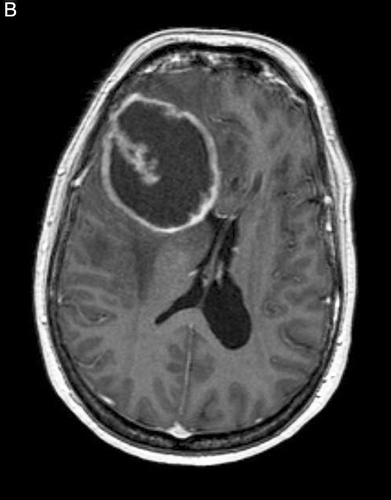
Glioblastomas are considered fast-growing tumors, but there is little information on their growth kinetics prior to diagnosis. This is because most patients develop symptoms only when their tumors have reached a size of several centimetres Citation[1]. We have followed a 58-year-old female patient with multiple sclerosis whose monitoring MRI in August 2007 is shown in . Because of worsening of her condition with apathy, the MRI was repeated in April 2008. T1-weighted MRI with gadolinium revealed a large right frontal lesion with ring enhancement which turned out to be a glioblastoma (). This 6.5 cm glioblastoma was not detectable in August 2007 even in retrospect and thus must have developed de novo within less than 8 months.
Figure A. Axial T1 MRI with contrast shows no evidence of a brain tumor.
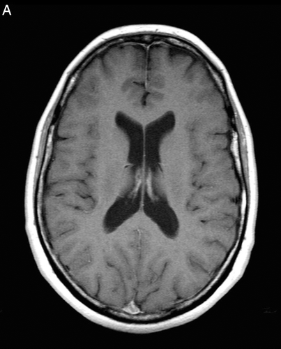
Figure B. Eight months later, a large glioblastoma has developed.
References
- Wen P, Kesari S. Malignant gliomas in adults. N Engl J Med 2008; 359: 492–507