
Introduction
Proton beam therapy is a relatively novel radiation treatment modality increasingly used in solid tumours in order to reduce radiation dose to tumour-surrounding normal tissues [Citation1,Citation2]. Among the indications for proton beam is the treatment of non-small cell lung cancer as well as of oesophageal cancer patients, albeit mostly in the context of prospective clinical trials [Citation3–5]. Intrathoracic tumours are, however, highly complex when treated with proton beams: on the one hand due to the various tissues and properties protons encounter along the beam path, on the other hand due to the tissues’ intra- or even inter-fractional movement. Consequently, for intrathoracic targets, several approaches compensating for those resulting range uncertainties have been recommended and widely implemented [Citation6,Citation7]. One approach is four-dimensional (4D) robust treatment planning and delivery taking into account the anatomical changes during several phases of the breathing cycle. Another approach is frequent anatomical imaging during the course of fractionated proton delivery in order to re-calculate the radiation dose and to adapt the treatment plan in case organ at risk (OAR) dose constraints are violated [Citation8]. The combination of the two as well as the world’s first plan-of-the-day is reported here in a patient with an atypical carcinoid of the left lung.
Patient history
In August 2020, the 32 years-old male presented at University Hospital Carl Gustav Carus, Dresden, Germany. In September 2019, he had undergone a left-sided pneumonectomy and extensive, bilateral lymphadenectomy for an atypical carcinoid, staged pT3N0. In June 2020, he had developed a lymph node recurrence at the mediastinal levels 1, 2, 3a and 4 with extranodal tumour spread. Due to the pneumonectomy on the left side he opted to have proton beam therapy to the involved lymph node sites in order to maximally reduce the radiation dose to the remaining right lung.
Plan-of-the-day proton beam therapy
For 4D robust treatment planning, a 4D-CT was obtained on 25th August and reconstructed in eight breathing cycles. On this, the clinical target volume (CTV) containing the above mentioned lymph node stations as well as novel macroscopic lymph nodes in station 6 were included. Moreover, relevant OARs, i.e. right lung, heart, brachial plexus, spinal cord, thyroid, and oesophagus were contoured. On the average phase of this 4D-CT scan, the initial proton treatment, termed R1, was calculated to a total dose of 66 Gy(RBE) in 2 Gy(RBE)-fractions using a 4D-robust multi-field optimised (MFO) approach and the RayStation treatment planning system (RaySearch, Stockholm, Sweden). To further improve robustness low-weighted SFUD-like (single-field uniform dose) objectives were added into the optimisation and reduced spot and energy layer distances were chosen.
On the fourth day of proton beam therapy, 8th September, a control 4D-CT on the in-room CT scanner of the proton facility was obtained in order to verify the patient anatomy. On this 4D-CT, the level of pleural effusion in the left-sided pneumonectomy cavity had increased from 253,4 mL at the treatment planning state (25th August 2020) to 498,7 mL (8th September 2020) and started to deviate the mediastinum to the patient’s right. The coverage of the target volumes was still fulfilled, but borderline.
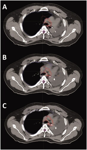
Only two days later, on 10th September 2020, another 4D-CT was obtained revealing that the pleural effusion had again increased, to now 814,0 mL. Together with the patient, it was decided to stop proton beam delivery and to assess the cause for the increasing volume. The subsequently performed analysis of the pleural fluid obtained by a pulmonologist, however, revealed no sign of malignant cells. Thus, in order to monitor the decrease of pleural fluid, repeat 4D-CTs were obtained on 16th September, 28th September and 2nd October (levels of pleural effusion 1146,2 mL, 536,7 mL and 473,3 mL, respectively). Moreover, it was decided to use three of the obtained 4D-CTs for treatment planning of the plan-library-based plan-of-the-day approach in particle therapy. For these three plans, termed R2A, R2B and R2C (), respectively, the 4D-CTs of 25th August (least pleural effusion), 2nd October (intermediate level of effusion) and 28th September (largest level of pleural effusion) were used. On the average CT scans of these 4D-CTs, a simultaneously integrated boost concept (SIB) was prescribed, in total 25 fractions of 2.0/2.3 Gy(RBE) to the elective volume and macroscopic lymph nodes. This concept was chosen to partially compensate for the prolonged overall treatment time caused by the treatment interruption.
Figure 1. A representative transverse slice of the CT scans acquired on 25th August (A; least pleural effusion), 8th September (B; most pleural effusion) and 2nd October (C; intermediate level of pleural effusion), which served as basis for treatment plans R1/R2A, R2C and R2B, respectively. The deviation of the contours of right lung (purple), CTV elective (red) and CTV boost (orange) with varying positions of the mediastinum can be appreciated.
Subsequently, from 12th October until 16th November 2020, the remaining 25 prescribed fractions were delivered as planned. During this time, 14 control 3D-CTs were obtained leading to the use of plan R2A in 19 fractions and of plan R2B in 6 fractions (fractions 13, 14, 22–25; see ). Plan R2C was never used. The treatment was tolerated well, the patient only complained about dysphagia grade 1 and erythema of the skin grade 3.
Table 1. Overview of the applied treatment plans and acquired control CT during the course of the plan-library-based plan-of-the-day approach.
Dose summation of the plan-of-the-day approach
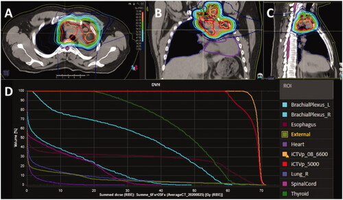
When deforming all 4D-CTs and treatment plans to the 4D-CT of the initial treatment plans R1 and R2A, the cumulative doses to the high-dose CTV [68,9 Gy(RBE)] and OARs were kept within tolerance limits (). The mean doses to right lung, the heart, the oesophagus and the thyroid were 5.0 Gy(RBE), 0.9 Gy(RBE), 19.3 Gy(RBE) and 46.5 Gy(RBE), respectively. The dose delivered to 2% of the volume of an OAR, D2%, was 58.2 Gy(RBE) to the left brachial plexus required to cover the lymph node in level 1, and 38.1 Gy(RBE) to the right brachial plexus. The maximum dose to the spinal cord was 32.9 Gy(RBE).
Figure 2. The summed dose distribution of the proton beam treatment plans R1, R2A and R2B covering the high-dose clinical target volume (CTV; iCTVp_08_6600) and the elective CTV (iCTVp_5000) is shown in transverse (A), coronal (B) and sagittal (C) views. The 62 Gy(RBE) isodose line is depicted in orange. The dose-volume histogram for the summed dose is presented in (D).
Discussion
We here report on the logistical and technical feasibility of delivering proton beam therapy using a plan-of-the-day approach. The time required on the gantry was insignificantly prolonged for the acquisition of the control CTs. This is due to the unique equipment at our facility enabling the scan acquisition and subsequent irradiation on a robotic couch. The radiation technicians were successfully trained in matching the acquired CT to one of the three 4D-CTs used for the plan-of-the-day approach and to decide on the optimal plan to use. Thus, this procedure can be used in particle facilities equipped with either an on-gantry conebeam-CT (CBCT) or an in-room CT. The latter is ideal, since it can be directly used for proton dose re-calculation.
The plan-of-the-day approach was first introduced for targets exhibiting a large inter-fraction variation, e.g., the uterine cervix and bladder [Citation9–12]. For this approach, cervical cancer patients receive two to three CTs with varying levels of bladder filling and intermediate positions of its filling are warped or simulated. Various treatment plans covering the extreme positions of the position of the uterine cervix and of one to three positions in between are calculated and selected based on the actual anatomy of the day, assessed by CBCT. Using this approach, centres have started to decrease the safety margins commonly applied and thus to reduce the irradiated volume. Taking into account the experience obtained with the patient reported here, the plan-of-the-day approach in protons may well be extended to other body sites, e.g., gynaecological or urological tumours with extensive elective volumes or tumour of the upper abdomen.
With the wider introduction of better resolution CBCT and hybrid machines combining magnetic resonance imaging (MRI) and a linear accelerator, MR-LINAC, the plan-the-day-approach is increasingly being substituted by an online-adaptive approach using real-time imaging and plan adaptation on the MR-LINAC [Citation13,Citation14]. This treatment technique has been successfully introduced for a variety of solid tumours benefitting from the superb soft-tissue contrast of MR and from the real-time imaging capabilities. The next step may well be to integrate MRI and proton beam therapy in order to online-adaptive proton beam therapy, as is currently pioneered by scientists of our institute [Citation15–17].
Conclusions
This case report on the plan-of-the-day approach for a thoracic target volume treated with proton therapy underlines its logistic and technical feasibility. This is a major step towards online adaptive proton therapy.
References
- Grau C, Durante M, Georg D, et al. Particle therapy in Europe. Mol Oncol. 2020;14(7):1492–1499.
- Chang JY, Jabbour SK, De Ruysscher D, et al. Consensus statement on proton therapy in Early-Stage and locally advanced Non-Small cell lung cancer. Int J Radiat Oncol Biol Phys. 2016;95(1):505–516.
- Zschaeck S, Simon M, Löck S, et al. PRONTOX – proton therapy to reduce acute normal tissue toxicity in locally advanced non-small-cell lung carcinomas (NSCLC): study protocol for a randomised controlled trial. Trials. 2016;17(1):4.
- Wink KCJ, Roelofs E, Simone CB, 2nd, et al. Photons, protons or carbon ions for stage I non-small cell lung cancer – results of the multicentric ROCOCO in silico study. Radiother Oncol. 2018;128(1):139–146.
- Troost EGC, Wink KCJ, Roelofs E, et al. Photons or protons for reirradiation in (non-)small cell lung cancer: results of the multicentric ROCOCO in silico study. Br J Radiol. 2020;93(1107):20190879 Mar
- Chang JY, Zhang X, Knopf A, et al. Consensus guidelines for implementing Pencil-beam scanning proton therapy for thoracic malignancies on behalf of the PTCOG thoracic and lymphoma subcommittee int. Int J Radiat Oncol Biol Phys. 2017;99(1):41–50.
- Meijers A, Jakobi A, Stützer K, et al. Log file-based dose reconstruction and accumulation for 4D adaptive pencil beam scanned proton therapy in a clinical treatment planning system: implementation and proof-of-concept. Med Phys. 2019;46(3):1140–1149.
- den Otter LA, Anakotta RM, Weessies M, et al. Investigation of inter‐fraction target motion variations in the context of pencil beam scanned proton therapy in non‐small cell lung cancer patients. Med Phys. 2020;47(9):3835–3844.
- Heijkoop ST, Langerak TR, Quint S, et al. Clinical implementation of an online adaptive plan-of-the-Day protocol for nonrigid motion management in locally advanced cervical cancer IMRT. Int J Radiat Oncol Biol Phys. 2014;90(3):673–679.
- Ahmad R, Bondar L, Voet P, et al. A margin-of-the-day online adaptive intensity-modulated radiotherapy strategy for cervical cancer provides superior treatment accuracy compared to clinically recommended margins: a dosimetric evaluation. Acta Oncol. 2013;52(7):1430–1436.
- Murthy V, Master Z, Adurkar P, et al. ‘Plan of the day’ adaptive radiotherapy for bladder cancer using helical tomotherapy. Radiother Oncol. 2011;99(1):55–60.
- Huddart R, Hafeez S, Lewis RA, et al. Clinical outcomes of a randomized trial of adaptive plan-of-the-Day treatment in patients receiving ultra-hypofractionated weekly radiation therapy for bladder cancer. Int J Radiat Oncol Biol Phys. 2021;110(2):412–424.
- Corradini S, Alongi F, Andratschke N, et al. MR-guidance in clinical reality: current treatment challenges and future perspectives. Radiat Oncol. 2019;14(1):92.
- Yock AD, Ahmed M, Ayala-Peacock DN, et al. Initial analysis of the dosimetric benefit and clinical resource cost of CBCT-based online adaptive radiotherapy for patients with cancers of the cervix or rectum. J Appl Clin Med Phys. 2021;22(10):210–221.
- Schellhammer SM, Gantz S, Luhr A, et al. Technical note: experimental verification of magnetic field-induced beam deflection and Bragg peak displacement for MR-integrated proton therapy. Med Phys. 2018;45(7):3429–3434.
- Schellhammer SM, Hoffmann AL. Prediction and compensation of magnetic beam deflection in MR-integrated proton therapy: a method optimized regarding accuracy, versatility and speed. Phys Med Biol. 2017;62(4):1548–1564.
- Schellhammer SM, Hoffmann AL, Gantz S, et al. Integrating a low-field open MR scanner with a static proton research beam line: proof of concept. Phys Med Biol. 2018;63(23):23LT01.