Abstract
Background. Flow cytometry and cell culture, the two main laboratory techniques employed for counting endothelial progenitor cells (EPCs), have serious limitations. Mononuclear cells cultured in media favouring endothelial growth allow cells to replicate and differentiate/mature. EPCs under these circumstances tend to form groups of cells called endothelial colony forming units (EC‐CFUs). EC‐CFUs are widely accepted as a surrogate as an estimate of EPC number and function in cell culture. However, some important limitations may restrict the assumption that EC‐CFUs reflect EPC numbers accurately.
Our findings. Our own experience of EPC culture in atrial fibrillation has demonstrated that: 1) the size of EC‐CFUs and proportion of single cells fluctuate significantly, even on the same culture plate; 2) the ability of EPCs to migrate towards one another to form EC‐CFUs varies; and 3) the rate of EPC differentiation and proliferation may significantly affect the number of EC‐CFUs, despite similarities in EPC counts on separate plates. In contrast, the count of differentiated cultured EPCs by flow cytometry with specific mature endothelial markers (e.g. CD146, vascular endothelial (VE) cadherin) is a potentially more objective alternative.
Summary. Endothelial CFU counts represent the cumulative characteristics of EPC quantity and their functional characteristics, and cannot be reliably used for the estimation of EPC numbers in peripheral blood or the bone marrow. Until stronger definition(s) of bone marrow or peripheral blood population(s) of EPCs are developed, flow cytometry may be the more optimal technique for EPC quantification.
Introduction
The discovery of a number of progenitor cell lineages (precursors to a variety of different cardiac and vascular cells) that demonstrate developmental plasticity has stimulated interest and excitement for the prospects of a new era of ‘regenerative’ interventions Citation1–5. The pertinent role of the endothelium in many cardiovascular disease states has focused many to investigate the role of endothelial progenitor cells (EPCs). Indeed, it is generally assumed that greater numbers of these cells at baseline reflects greater regenerative potential and thereby a more favourable prognosis Citation6.
Flow cytometry and cell culture are the two main laboratory techniques employed for counting and functional assessment of endothelial progenitor cells (EPCs). Both of these techniques, however, have serious limitations, often overlooked by the scientific community. This document explores the potential pitfalls encountered in counting EPCs following cell culture.
Definition of EPCs
EPCs are believed to predominantly originate from haematopoietic stem cells in bone marrow and, indeed, recent evidence suggests that the haemangioblast is a common progenitor for both haematopoietic and endothelial cells Citation7. However, the definition of EPCs continues to evolve and has shifted significantly following their initial characterization by Asahara et al. in 1997 Citation5. EPCs were first identified and enumerated by their expression of both haematopoietic stem cell and endothelial cell markers, such as CD34, CD133 and vascular endothelial growth factor receptor‐2 (KDR) Citation5. Later expression of other endothelial markers (e.g. vascular endothelial (VE) cadherin, von Willebrand factor (vWf), CD146) in the process of maturation of endothelial progenitors was demonstrated Citation8. However, given the heterogeneity of the EPC population, a robust definition still does not yet exist Citation9. The situation is further complicated as expression of cell surface markers changes with EPC maturation Citation10–13. There is also some evidence that cells of other lineages can cross‐differentiate in to EPCs Citation14–16. These markers are easily detected using flow cytometry, which essentially estimates the number of cells from peripheral blood, bone marrow, or other tissues that possess a specific cell type profile of surface markers.
The expression of common markers by haematopoietic and endothelial progenitor cells in embryonic development and the trans‐differentiation potential of monocytes into cells with endothelial characteristics would suggest a possible common origin from a bone marrow precursor, perhaps a putative haemangioblast Citation7. Bone marrow also contains mesenchymal cells, which can differentiate into endothelial cells Citation15 and improve vascularization in vivoCitation17, Citation18, as well as release a variety of angiogenic growth factors Citation19 and contribute to tissue repair Citation20. In addition to bone marrow‐derived cells, other cell populations, such as fat tissue Citation16, Citation21, cardiac tissue Citation1, and neural stem cells Citation22 can also give rise to endothelial cells, suggesting that tissue‐resident stem/progenitor cells can contribute to vascular growth in the adult. Thus, whilst flow cytometry is able to accurately estimate the number of different cell types with a specific marker profile, this method can neither determine all cell populations with the potential to differentiate into endothelial cells nor a total EPC count.
Cell culture
Cultivation of peripheral blood mononuclear cells (MNCs) in media favouring endothelial growth is another widely used approach for the definition and quantitative analysis of EPCs Citation6, Citation14, Citation23 (Figure ). Adherent cells have been shown to possess endothelial characteristics, such as the expression of vWf and staining for Dil‐acetylated low‐density lipoprotein (DiLDL) and fluorescein isothiocyanate‐labelled lectin from Bandeiraea simplicifolia (FITC‐BS‐lectin) Citation6, Citation24–26. Despite the relatively low numbers of CD34+ of circulating endothelial precursors in peripheral blood (100–500 per mL), relatively large numbers of adherent cells are found during culture (≈100 000 from 1 mL of blood). This raises some controversy with respect to the identification and the origin of isolated EPCs, as these cells appear to reflect a functional subpopulation within the blood MNC that have the potential to differentiate into an endothelial phenotype in vivoCitation6, Citation13, Citation23, Citation27–29. Indeed, early endothelial‐like cells are often derived from peripheral blood monocytes Citation23. Nonetheless, a clear advantage then of the cell culture approach is that it permits quantification of all cells capable differentiating into endothelial cells. Differentiated endothelial cells tend to form groups of cells, which are called endothelial colony forming units (EC‐CFUs) (Figure ). As the number of single endothelial cells in culture is too large to count under light or fluorescent microscopy, the number of EC‐CFUs is a widely accepted surrogate estimation for the quantity of EPCs in cell culture. However, do EC‐CFUs really reflect the number of EPCs following cell culture?
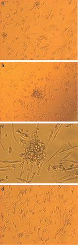
Figure 1 Endothelial progenitor cells and endothelial colony forming units under light microscopy. Note that the size of colony forming units(CFUs) varies widely. a: small CFUs consisting of a few cells (40×); b and c: bigger CFUs (40×, 100×); d: predominantly single located endothelial cells without accumulation in CFUs (40×).
Key messages
Endothelial progenitor cell colony forming units (EPC‐CFUs) are frequently used as a surrogate estimate for EPC quantity and functional characteristics.
EPC‐CFUs are often highly variable in size and appearance even across the same culture plate.
EPC‐CFUs do not appear to accurately reflect the overall EPC count following cell culture.
EPC cell count by flow cytometry following cell culture is potentially more objective and reliable.
Methods
We investigated whether EC‐CFUs really reflect the number of EPCs following cell culture in 60 patients with different forms of atrial fibrillation (AF) (paroxysmal, persistent, permanent), a significant proportion of whom had coronary artery disease (42%). This cohort of patients with AF was chosen, due to the relative ease of recruitment, given that our unit runs a large AF clinic. As the main focus of the present study was to compare and contrast methods for EPC enumeration, AF represented an ideal disease group choice due to the frequent association of this arrhythmia with various cardiovascular co‐morbidities.
Following informed consent, 20 mL peripheral blood was drawn from each patient into ethylene diamine tetraacetic acid (EDTA) tubes. MNCs were isolated by density gradient centrifugation. In brief, 4 mL peripheral blood was diluted with an equal volume of Hank's balanced salt solution (Sigma). This was then carefully layered on to 3 mL Ficoll® (GE‐Healthcare) in a 15 mL centrifuge tube, ensuring that the two solutions did not mix. The tube was centrifuged at 400 g for 40 minutes at 18°C with no brake. The resultant buffy cell layer was removed and washed twice with 6 mL Hank's balanced salt solution, followed by centrifugation at 100 g for 10 minutes at 18°C to remove excess Ficoll, serum and platelets. The resultant cell fraction was then re‐suspended in 1 mL endothelial specific culture medium and, following an initial cell count, was plated on 24‐well culture plates pre‐coated with human fibronectin (Sigma). This process was repeated twice for each patient. The culture medium used was Medium M199 (Gibco) supplemented with 5 mg/100 mL endothelial growth supplement (Upstate), 15% foetal bovine serum (Sigma) and penicillin/streptomycin (Sigma). At 48 hours media were changed. After 6 days in culture, media and non‐adherent cells were removed and adherent cells underwent cytochemical analysis.
Cells were consequently incubated with DiLDL (Invitrogen) (2.4 mg/mL) for 2 hours and with FITC‐labelled lectin Ulex europaeus (Sigma) (10 mg/mL) for 1 hour and then fixed with 2% paraformaldehyde for 20 minutes. Dual‐staining cells positive for both lectin and DiLDL were considered as EPCs. Additionally, expression of vascular endothelial (VE) cadherin was measured. Adherent cells were detached from the culture plate using 1 mmol/L EDTA diluted in phosphate buffered saline. Following a wash, cells were then incubated first with 10 µL Octagam (Octapharma) and 200 µL 10% mouse serum (Sigma), and then in the dark for 20 minutes with 10 µL phycoerythrin (PE)‐conjugated VE cadherin (Becton‐Dickinson). Following a cell wash to remove excess antibody, the sample was immediately run on a BD FACScan flow cytometer.
Our experience
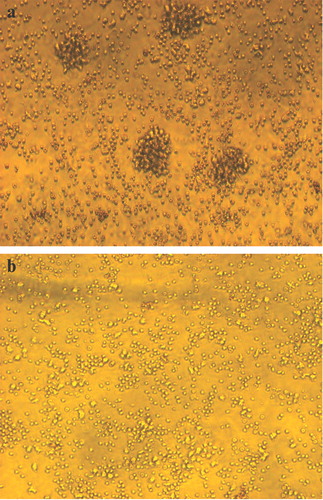
We consistently demonstrated that the size and number of EC‐CFUs greatly fluctuates, even on the same cell culture plate, varying from only a few cells (Figure ) to large colonies (Figures ). The latter are often demonstrated on research publications, and sometimes represent very large cell accumulations. However, thin, spindle shaped endothelial‐like cells are often located separately seen within the same plate (Figure ). Variations in the size of EC‐CFUs amongst different samples are common, and the migratory capacity of EPCs can be changed in different pathological states Citation26, Citation30, Citation31. Indeed, the ability of EPCs to migrate to each other to form a colony may significantly affect the number of EC‐CFUs formed in culture experiments, despite a similar number of EPCs. The importance of culture cell migration for colony forming can be proven visibly by the poor CFU forming capacity of MNCs before their differentiation into endothelial cells (Figure ) and the fact that EC‐CFUs can include different proportions of total EPCs (Figure ). The subjectivity regarding which clusters should be considered as EC‐CFUs makes this approach even less reliable. Furthermore, recent studies have demonstrated that EC‐CFUs have limited differentiation capacity into endothelial cells and may retain some myeloid progenitor activity Citation32.
Figure 2 Endothelial progenitor cells(EPCs) migration and colony forming units (CFUs). a: Migration of EPCs to each other before forming CFUs (40×). b: This process may be impaired in some patients (40×).
Discussion
Most researchers perform EPC counts (based on numbers of CFUs) relative to the day of initiation of cell culture (generally between day 4 and day 7). The rate of EPC differentiation depends on the functional condition of EPCs, culture media used, and endothelial growth supplements. Simultaneous with EPC differentiation, the process of cell proliferation (another functional characteristic of EPCs) begins. Thus, the number of CFUs depends not only on EPC counts and migratory activity, but also on the rate of EPC proliferation and differentiation. Amongst adherent MNCs, there may also be some mature circulating endothelial cells (CECs), and thus some researchers prefer to perform pre‐plating of MNCs to reduce contamination with CECs, which also express endothelial markers, including CD34 Citation9. How effective this approach is remains unclear, but the loss of some EPCs during the pre‐plating stage may significantly affect total EPC counts.
Possible alternatives to CFU counts
A count of the total number of cultured endothelial cells (single cells plus cells inside CFU) may be more accurate in terms of EPC quantification, but numbers of single EPCs in cell culture are too great for an accurate manual count under light microscope. As an alternative, the count of differentiated cultured EPCs by flow cytometry can be used, with specific mature endothelial markers (e.g. CD146, CD144 (VE cadherin) or vWf). This approach has been tested in our department (with VE cadherin as a marker) and permits count of both single EPCs and EPCs grouped in CFUs together. CD146 is expressed on non‐differentiated endothelial progenitors, although in small amounts Citation33. The rate of vWf expression is slower, and its use can thus underestimate the true EPC count. Thus, VE cadherin may be the optimal marker, from those currently available. Indeed, VE cadherin is an adhesion molecule that plays a fundamental role in microvascular permeability and in the morphogenic/proliferative events associated with angiogenesis, suggesting its importance for endothelial integrity. VE cadherin is exclusively expressed by endothelial cells in adults Citation34–36. In addition, embryos' yolk sac cells with haemoangiogenic potential have been shown to lose VE cadherin expression when differentiating to haematopoietic progenitor cells Citation37.
This analytical approach permits the exclusion of the effect of migratory activity on EPC counts, but cannot exclude the effect of EPC proliferation and CEC numbers. It is impossible to fully eliminate the contamination of peripheral blood MNCs with CECs, but we can estimate the proportion of CECs by flow cytometric count of cells with pure markers of mature endothelial cells (e.g. CD146 or VE cadherin) after the first media change (in 24–48 hours), when non‐adherent MNCs are removed and EPCs have still not differentiated into mature endothelial cells. At this stage, CD146 or VE cadherin+ cells will reflect CEC contamination. Any increase in VE cadherin+ or CD146+ cells will mainly represent differentiated EPCs, as CECs proliferation seems to be relatively low or rare. As there is no marker which can reliably distinguish mature EPCs from their generic endothelial cells as a result of EPC proliferation, it seems reasonable to standardize the day of the EPC count (e.g. days 4–7) and use media/supplement to make the impact of EPC proliferation less significant.
In conclusion, endothelial CFU counts represents the cumulative characteristics of EPC quantity and their functional characteristics, including rate of differentiation, proliferation and senescence, and migration activity. We suggest that CFU counts cannot be reliable used for the estimation of EPC numbers in peripheral blood nor the bone marrow. Until stronger definition(s) of bone marrow or peripheral blood population(s) of EPCs are developed, flow cytometry may be the more optimal technique for EPC quantification. However simultaneous measurement of EPCs by flow cytometry and CFU counts will continue to provide complementary data and allow further cross‐validation of both these methods.
Acknowledgements
Drs Shantsila and Watson contributed equally to this work. Dr Shantsila was recipient of a European Society of Cardiology research fellowship.
References
- Beltrami A. P., Barlucchi L., Torella D., Baker M., Limana F., Chimenti S., et al. Adult cardiac stem cells are multipotent and support myocardial regeneration. Cell 2003; 114: 763–76
- Urbanek K., Torella D., Sheikh F., De Angelis A., Nurzynska D., Silvestri F., et al. Myocardial regeneration by activation of multipotent cardiac stem cells in ischemic heart failure. Proc Natl Acad Sci U S A 2005; 102: 8692–7
- Nygren J. M., Jovinge S., Breitbach M., Sawen P., Roll W., Hescheler J., et al. Bone marrow‐derived hematopoietic cells generate cardiomyocytes at a low frequency through cell fusion. Nat Med 2004; 10: 494–501
- Laflamme M. A., Myerson D., Saffitz J. E., Murry C. E. Evidence for cardiomyocyte repopulation by extracardiac progenitors in transplanted human hearts. Circ Res 2002; 90: 634–40
- Asahara T., Murohara T., Sullivan A., Silver M., van der Zee R., Li T., et al. Isolation of putative progenitor endothelial cells for angiogenesis. Science 1997; 275: 964–7
- Hill J. M., Zalos G., Halcox J. P., Schenke W. H., Waclawiw M. A., Quyyumi A. A., et al. Circulating endothelial progenitor cells, vascular function, and cardiovascular risk. N Engl J Med 2003; 348: 593–600
- Vogeli K. M., Jin S. W., Martin G. R., Stainier D. Y. A common progenitor for haematopoietic and endothelial lineages in the zebrafish gastrula. Nature 2006; 443: 337–9
- Lin Y., Weisdorf D. J., Solovey A., Hebbel R. P. Origins of circulating endothelial cells and endothelial outgrowth from blood. J Clin Invest 2000; 105: 71–7
- Friedrich E. B., Walenta K., Scharlau J., Nickenig G., Werner N. CD34‐/CD133+/VEGFR‐2+ endothelial progenitor cell subpopulation with potent vasoregenerative capacities. Circ Res 2006; 98: e20–5
- Fina L., Molgaard H. V., Robertson D., Bradley N. J., Monaghan P., Delia D., et al. Expression of the CD34 gene in vascular endothelial cells. Blood 1990; 75: 2417–26
- Peichev M., Naiyer A. J., Pereira D., Zhu Z., Lane W. J., Williams M., et al. Expression of VEGFR‐2 and AC133 by circulating human CD34(+) cells identifies a population of functional endothelial precursors. Blood 2000; 95: 952–8
- Yin A. H., Miraglia S., Zanjani E. D., Almeida‐Porada G., Ogawa M., Leary A. G., et al. AC133, a novel marker for human hematopoietic stem and progenitor cells. Blood 1997; 90: 5002–12
- Miraglia S., Godfrey W., Yin A. H., Atkins K., Warnke R., Holden J. T., et al. A novel five‐transmembrane hematopoietic stem cell antigen: isolation, characterization, and molecular cloning. Blood 1997; 90: 5013–21
- Fernandez Pujol B., Lucibello F. C., Gehling U. M., Lindemann K., Weidner N., Zuzarte M. L., et al. Endothelial‐like cells derived from human CD14 positive monocytes. Differentiation 2000; 65: 287–300
- Oswald J., Boxberger S., Jorgensen B., Feldmann S., Ehninger G., Bornhauser M., et al. Mesenchymal stem cells can be differentiated into endothelial cells in vitro. Stem Cells 2004; 22: 377–84
- Zuk P. A., Zhu M., Mizuno H., Huang J., Futrell J. W., Katz A. J., et al. Multilineage cells from human adipose tissue: implications for cell‐based therapies. Tissue Eng 2001; 7: 211–28
- Reyes M., Dudek A., Jahagirdar B., Koodie L., Marker P. H., Verfaillie C. M. Origin of endothelial progenitors in human postnatal bone marrow. J Clin Invest 2002; 109: 337–46
- Al‐Khaldi A., Eliopoulos N., Martineau D., Lejeune L., Lachapelle K., Galipeau J. Postnatal bone marrow stromal cells elicit a potent VEGF‐dependent neoangiogenic response in vivo. Gene Ther 2003; 10: 621–9
- Kinnaird T., Stabile E., Burnett M. S., Shou M., Lee C. W., Barr S., et al. Local delivery of marrow‐derived stromal cells augments collateral perfusion through paracrine mechanisms. Circulation 2004; 109: 1543–9
- Kuznetsov S. A., Mankani M. H., Gronthos S., Satomura K., Bianco P., Robey P. G. Circulating skeletal stem cells. J Cell Biol 2001; 153: 1133–40
- Planat‐Benard V., Silvestre J. S., Cousin B., Andre M., Nibbelink M., Tamarat R., et al. Plasticity of human adipose lineage cells toward endothelial cells: physiological and therapeutic perspectives. Circulation 2004; 09: 656–63
- Wurmser A. E., Nakashima K., Summers R. G., Toni N., D'Amour K. A., Lie D. C., et al. Cell fusion‐independent differentiation of neural stem cells to the endothelial lineage. Nature 2004; 430: 350–6
- Rehman J., Li J., Orschell C. M., March K. L. Peripheral blood ‘endothelial progenitor cells’ are derived from monocyte/macrophages and secrete angiogenic growth factors. Circulation 2003; 107: 1164–9
- Shintani S., Murohara T., Ikeda H., Ueno T., Honma T., Katoh A., et al. Mobilization of endothelial progenitor cells in patients with acute myocardial infarction. Circulation 2001; 103: 2776–9
- Vasa M., Fichtlscherer S., Adler K., Aicher A., Martin H., Zeiher A. M., et al. Increase in circulating endothelial progenitor cells by statin therapy in patients with stable coronary artery disease. Circulation 2001; 103: 2885–90
- Vasa M., Fichtlscherer S., Aicher A., Adler K., Urbich C., Martin H., et al. Number and migratory activity of circulating endothelial progenitor cells inversely correlate with risk factors for coronary artery disease. Circ Res 2001; 89: E1–7
- Harraz M., Jiao C., Hanlon H. D., Hartley R. S., Schatteman G. C. CD34(‐) blood‐derived human endothelial cell progenitors. Stem Cells 2001; 19: 304–12
- Schmeisser A., Garlichs C. D., Zhang H., Eskafi S., Graffy C., Ludwig J., et al. Monocytes coexpress endothelial and macrophagocytic lineage markers and form cord‐like structures in Matrigel under angiogenic conditions. Cardiovasc Res 2001; 49: 671–80
- Rookmaaker M. B., Vergeer M., van Zonneveld A. J., Rabelink T. J., Verhaar M. C. Endothelial progenitor cells: mainly derived from the monocyte/macrophage containing CD34‐ but also partly derived from the hematopoietic stem cell containing CD34+ mononuclear population. Circulation 2003; 108: 150e
- Chen J. Z., Zhang F. R., Tao Q. M., Wang X. X., Zhu J. H. Number and activity of endothelial progenitor cells from peripheral blood in patients with hypercholesterolaemia. Clin Sci (Lond) 2004; 107: 273–80
- Heeschen C., Lehman R., Honold J., Assmus B., Aicher A., Walter D. H., et al. Profoundly reduced neovascularization capacity of bone marrow mononuclear cells derived from patients with chronic ischemic heart disease. Circulation 2004; 109: 1615–22
- Yoder M. C., Mead L. E., Prater D., Krier T. R., Mroueh K. N., Li F., et al. Re‐defining endothelial progenitor cells via clonal analysis and hematopoietic stem/progenitor cell principals. Blood 2007; 109: 1801–9
- Delorme B., Basire A., Gentile C., Monsonis F., Desouches C., Blot‐Chabaud M., et al. Presence of endothelial progenitor cells, distinct from mature endothelial cells, within human CD146+ blood cells. Thromb Haemost 2005; 94: 1270–9
- Dejana E., Bazzoni G., Lampugnani M. G. Vascular endothelial (VE)‐cadherin: only an intercellular glue?. Exp Cell Res 1999; 252: 13–19
- Dejana E., Spagnuolo R., Bazzoni G. Interendothelial junctions and their role in the control of angiogenesis, vascular permeability and leukocyte transmigration. Thromb Haemost 2001; 86: 308–15
- Stevens T., Garcia J. G. N., Shasby D. M., Bhattacharya J., Malik A. B. Mechanisms regulating endothelial cell barrier function. Am J Physiol Lung Cell Mol Physiol 2000; 279: L419–22
- Nishikawa S. I., Nishikawa S., Hirashima M., Matsuyoshi N., Kodama H. Progressive lineage analysis by cell sorting and culture identifies FLK1 VE‐cadherin cells at a diverging point endothelial and hemopoietic lineages. Development 1998; 125: 1747–57