Abstract
Calcium (Ca2+) and phosphate (Pi) are essential to many vital physiological processes. Consequently the maintenance of Ca2+ and Pi homeostasis is essential to a healthy existence. This occurs through the concerted action of intestinal, renal, and skeletal regulatory mechanisms. Ca2+ and Pi handling by these organs is under tight hormonal control. Disturbances in their homeostasis have been linked to pathophysiological disorders including chronic renal insufficiency, kidney stone formation, and bone abnormalities. Importantly, the kidneys fine‐tune the amount of Ca2+ and Pi retained in the body by altering their (re)absorption from the glomerular filtrate. The ion transport proteins involved in this process have been studied extensively. Recently, new key players have been identified in the regulation of the Ca2+ and Pi balance. Novel regulatory mechanisms and their implications were introduced for the antiaging hormone klotho and fibroblast growth factor member 23 (FGF23). Importantly, transgenic mouse models, exhibiting disturbances in Ca2+ and Pi balance, have been of great value in the elucidation of klotho and FGF23 functioning. This review highlights the current knowledge and ongoing research into Ca2+ and Pi homeostasis, emphasizing findings from several relevant knockout mouse models.
Introduction
The tight control of plasma calcium (Ca2+) and phosphate (Pi) levels is essential to the performance of many vital physiological functions. Muscle contraction, blood clotting and neuronal excitation all require Ca2+, whereas Pi is vital to intracellular signaling, as a component of membrane lipids and to build the backbone of DNA. Moreover, significant elements of bone are Ca2+ and PiCitation1, Citation2. Several organs contribute to the exquisite regulation of Ca2+ and Pi homeostasis by facilitating intestinal absorption, bone (de)mineralization, and renal excretion/reabsorption of both ions. Regulation of these processes occurs by a number of hormones. The biologically active form of vitamin D (1,25‐dihydroxyvitamin D3), parathyroid hormone (PTH), and calcitonin have been extensively studied in this regard. More recently, fibroblast growth factor member 23 (FGF23) and klotho have been identified as new players essential to the regulation of Ca2+ and Pi homeostasis Citation3–5. Remarkably, large alterations in dietary Pi and Ca2+ intake produce only small alterations in the circulating levels of these ions due to the combined action of these signaling molecules on bone, intestinal, and renal Ca2+ and Pi transport Citation2, Citation6, Citation7. At times these regulatory processes are overwhelmed producing disturbances in Ca2+ and Pi homeostasis. Patients with chronic renal insufficiency (CRI) highlight pathophysiology complicated by altered Ca2+ and Pi handling. These individuals have a decreased glomerular filtration rate, a decreased circulating plasma 1,25‐dihydroxyvitamin D3 (1,25(OH)2D3) level and consequently develop secondary hyperparathyroidism Citation8. This review highlights the physiological mechanisms governing Ca2+ and Pi transport, two intimately related processes, and the clinical implications of their altered regulation.
Key messages
Maintenance of normal phosphate (Pi) and calcium (Ca2+) homeostasis is crucial to many vital physiologic processes including cellular signaling, DNA structure, bone mineralization, muscle contraction, blood clotting, and neuronal excitation.
Fibroblast growth factor member 23 (FGF23), klotho, parathyroid hormone, and 1,25‐dihydroxyvitamin‐D3 are key regulators of Ca2+ and Pi homeostasis. Their concerted action alters (re)absorption from intestine, kidney, and bone via recently identified pathways.
The concerted interplay of klotho and FGF23 contributes substantially to the regulation of renal Pi handling and may provide a therapeutic target for chronic renal insufficiency‐related hyperphosphatemia and other disorders involving disturbances of Pi balance.
Physiology of Ca2+ and Pi homeostasis
Ingested Ca2+ and Pi are absorbed by different segments of the small intestine. The active absorption of sodium (Na+) throughout the entire course of the intestine results in a large net water absorption. This mainly occurs in the small intestine, where Ca2+ and Pi are concomitantly taken up in a passive, paracellular manner, down their concentration gradient. The active transcellular absorption of Pi occurs predominately from the ileum Citation9, whereas active Ca2+ transport takes place largely from the duodenum Citation10.
The vast majority of whole body Ca2+ and Pi is stored as the mineral hydroxylapatite in the skeleton. In blood 45% of Ca2+ is present in a free, ionized form, 45% is bound to proteins, and a small fraction, 10%, forms complexes with anions including citrate, sulphate, and phosphate. Plasma Pi is present in its inorganic form and as a component of several organic substances including sugars, phosphoproteins, and high‐energy phosphates. The tightly regulated renal elimination of electrolytes, including Ca2+ and Pi, maintains a near constant plasma level of ions and significantly contributes to whole body homeostasis. Free Ca2+ and Pi are filtered by the glomerulus and gain entry into the renal tubule. In contrast to the gastrointestinal system, Pi is largely absorbed in a transcellular, Na+‐dependent manner in the proximal tubule (PT). This process is dependent on the electrochemical gradient present for Na+Citation11. Renal Ca2+ absorption occurs paracellularly in the PT and the thick ascending limb of Henle (TAL). A small (10%–15%) highly regulated amount of Ca2+ is actively absorbed from the distal convoluted tubule (DCT) and the connecting tubule (CNT) Citation10, Citation11. A diverse array of ion transport proteins, localized to the apical and basolateral membranes of intestinal and renal epithelia, mediate this active transport of Ca2+ and Pi. The entry of these ions, from lumina into the cytosol of the specific epithelium, is tightly regulated and represents the rate‐limiting step in transcellular Ca2+ and Pi (re)absorption Citation10.
Ca2+ and Pi transport proteins
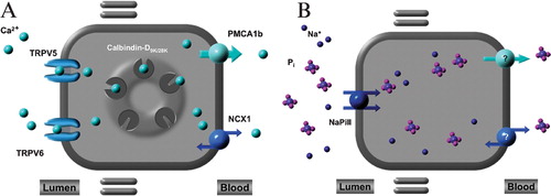
The ability of the small intestine to actively absorb Ca2+ and Pi is hormonally regulated and occurs from the duodenum and ileum, respectively. Active intestinal Ca2+ absorption begins with its luminal entry mediated by the epithelial Ca2+ channel, TRPV6 Citation12. Ca2+ is subsequently shuttled to the basolateral side of the cell via the Ca2+‐binding protein, calbindin‐D9K. This process is completed by transport of Ca2+ back into the bloodstream via the plasma membrane Ca2+‐adenosine triphosphatase (PMCA1b) Citation13.
The Na+‐dependent Pi cotransporter type IIb (NaPi‐IIb) is localized to the brush border of ileum. There it is responsible for active Pi transport from the intestinal lumen into the blood Citation9, Citation14. NaPi‐IIb is also present, although to a significantly decreased extent, in duodenum and jejunum, consistent with less overall active Pi reabsorption from these locales Citation9. The mechanism and identity of the molecules responsible for the extrusion of Pi back into the blood is not known at present.
After intestinal absorption into the blood, Ca2+ and Pi are filtered across the glomerulus into the tubular lumen of the nephron. Along the course of the nephron passive, paracellular Ca2+ absorption occurs from the PT and TAL. A three‐step process mediates the active Ca2+ reabsorption mechanism from the DCT and CNT. First, Ca2+ enters the cell via the epithelial Ca2+ channel TRPV5 (and to a lesser extent TRPV6). Then Ca2+ diffuses through the cytosol via calbindin‐D28K, to the basolateral side of the cell. There, Ca2+ is extruded into the peritubular capillary by the Na+‐Ca2+‐exchanger (NCX1) and PMCA1b Citation10. Active Pi absorption occurs in the PT of the kidney via the Na+‐dependent Pi cotransporters, NaPi‐IIa and NaPi‐IIc. Both display high expression levels in the early segments (S1 and S2) of the PT Citation2, Citation11, Citation15–17. Figure summarizes the transcellular pathways of Ca2+ (A) and Pi (B) transport in kidney and intestine.
Figure 1 Transcellular Ca2+ and Pi transport. Active Ca2+ and Pi transport across renal and intestinal epithelium. A: Ca2+ influx is mediated by TRPV5 in the renal distal convoluted and connecting tubules and TRPV6 in the duodenum. Subsequently, Ca2+ is transported across the cell to the basolateral membrane by Ca2+‐binding proteins calbindin‐D28K (kidney) and calbindin‐D9K (intestine). Extrusion into the blood takes place via the plasma membrane Ca2+‐ATPase (PMCA1b) in the intestine and sodium‐Ca2+ exchanger (NCX1) and PMCA1b in the kidney. B: The ileal brush border, sodium‐dependent Pi cotransporter, NaPi‐IIb, mediates entry of Pi into enterocytes. In the proximal tubules, NaPi‐IIa and NaPi‐IIc are responsible for Pi entry into the tubular epithelial cells. The transport mechanisms involved in basolateral extrusion of Pi into blood are unknown both in the intestine and kidney.
Hormonal regulation of transcellular Ca2+ (re)absorption
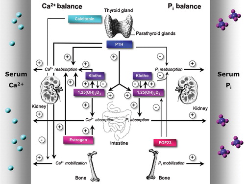
Exquisite regulation of TRPV5 and TRPV6 activity is essential to whole body Ca2+ homeostasis. Here, we review the regulatory effects of the classically described hormones: PTH, 1,25(OH)2D3, and calcitonin on active Ca2+ transport processes. Later we will discuss the newly identified regulators of Ca2+ homeostasis: tissue kallikrein (TK), the antiaging hormone klotho, and estrogen. Central to this process is the Ca2+‐sensing receptor (CaSR), which is expressed in the parathyroid gland where it senses blood Ca2+ levels Citation18, Citation19. In response to changes in blood Ca2+ concentrations, the CaSR regulates the release of PTH into the circulation (Figure ). Specifically, hypocalcemia inhibits the CaSR that in turn promotes the secretion of PTH. This hormone stimulates Ca2+ mobilization from bone and the conversion of inactive vitamin D to 1,25(OH)2D3 by the renal cytochrome P450 enzyme 25‐hydroxyvitamin D3‐1α‐hydroxylase (1αOHase) Citation20. Increased 1,25(OH)2D3 levels activate vitamin D receptor (VDR)‐mediated gene transcription, resulting in an increased transcription of Ca2+ transport proteins. Ultimately this results in the stimulation of Ca2+ (re)absorption Citation3, Citation21–24. Hypercalcemia has the opposite effect. This state activates the CaSR, thereby inhibiting PTH release and stimulating the secretion of calcitonin from the parafollicular cells of the thyroid. Calcitonin decreases osteoclast‐mediated bone resorption and promotes lowering the blood Ca2+ concentration Citation25.
Figure 2 Key players in Ca2+ and Pi homeostasis. The concerted interplay of intestinal uptake, reabsorption in kidney, and bone (de)mineralization establishes the maintenance of a normal Ca2+ and Pi balance. The calcium sensing receptor (CaSR), present in the thyroid gland, senses blood Ca2+ levels and triggers the secretion of the calcitropic hormones parathyroid hormone (PTH) and calcitonin. Ovarian‐produced estrogen stimulates Ca2+ reabsorption also. Similar hormones are involved in the regulation of Pi balance. Blood Pi levels are controlled by PTH, 1,25(OH)2D3, klotho, and fibroblast growth factor member 23 (FGF23). A negative feedback mechanism prevents the accumulation of FGF23 and klotho since 1αOHase‐mediated 1,25(OH)2D3 production is inhibited by FGF23 and klotho.
Estrogen contributes importantly to Ca2+ homeostasis by promoting bone mineralization and stimulating renal TRPV5 expression. This results in increased active Ca2+ reabsorption, independently of 1,25(OH)2D3Citation26–28. PTH, 1,25(OH)2D3, and estrogen exert their effect on renal‐mediated Ca2+ handling by altering, at the transcriptional level, the expression of Ca2+ transporters Citation7. This has been confirmed, in vivo, where a direct stimulatory effect of PTH on renal Ca2+ transporter expression levels was demonstrated in PTH‐supplemented parathyroidectomized rats Citation29, and where the dietary supplementation of Ca2+ and the administration of estrogen to ovariectomized rats was shown to alter renal TRPV5 expression, all independently of 1,25(OH)2D3Citation7, Citation23, Citation28.
The TK knockout (TK−/−) mouse displays significant renal Ca2+ wasting compared to control mice, implicating an important role for TK in the regulation of active Ca2+ reabsorption Citation30. In 2006, Gkika et al. described the molecular mechanism through which TK stimulates TRPV5‐mediated active Ca2+ reabsorption. TK, a proteolytic enzyme that is secreted into the tubular fluid by renal epithelial cells, activates the bradykinin receptor stimulating the phospholipase C/1,2‐diacylglycerol/protein kinase C (PLC/DAG/PKC) pathway. This results in increased plasma membrane expression of TRPV5, possibly due to an inhibition of TRPV5 endocytosis. Regardless of the exact mechanism, TK increases TRPV5‐mediated Ca2+ transport Citation31.
Recently the antiaging hormone klotho has also been implicated in the regulation of the Ca2+ balance. Klotho knockout (klotho−/−) mice display hypercalcemia, hyperphosphaturia, hypercalciuria, and other manifestations resembling aging Citation32. Chang et al. demonstrated that klotho stimulates TRPV5‐mediated Ca2+ transport in vitro, revealing a novel regulatory mechanism of active Ca2+ transport Citation33. Klotho is a type I transmembrane protein with β‐glucuronidase activity. This activity is contained in the extracellular domain that can be secreted into the blood, urine, and cerebrospinal fluid Citation32, Citation34. Klotho hydrolyzes extracellular N‐linked oligosaccharides present on TRPV5, entrapping the channel in the plasma membrane. This stimulatory effect of klotho on TRPV5 substantiates the important role of this antiaging hormone in Ca2+ homeostasis, specifically by altering the luminal membrane Ca2+ permeability of the DCT and the cortical collecting duct (CCD) Citation33. Alternatively, the mechanism of hypercalciuria observed in klotho−/− mice has been suggested to be the result of decreased proximal Ca2+ absorption because of a decreased Na+K+‐ATPase activity Citation35.
Hormonal regulation of transcellular Pi (re)absorption
Our understanding of the hormonal control of Pi balance is rapidly evolving. Klotho and FGF23 were recently identified as new key players involved in Pi regulation, adding to what had already been delineated about the classical PTH/1,25(OH)2D3 regulatory pathway. PTH, 1,25(OH)2D3, and dietary Pi intake are known to alter Pi (re)absorption Citation36. An increased intestinal NaPi‐IIb expression can be stimulated by either 1,25(OH)2D3 and/or low Pi intake, ultimately resulting in elevated blood PiCitation6. Conversely, PTH decreases blood Pi levels by inhibiting Pi reabsorption from the PT. This occurs via an acute redistribution of NaPi‐IIa proteins from the brush border to intracellular lysosomes, targeting them for degradation. The overall effect of PTH is therefore to lower blood Pi levels by stimulating Pi excretion Citation37.
Recently, FGF23 has been labeled a phosphatonin, due to its role in Pi homeostasis Citation5, Citation38–40. Injection of FGF23 into mice results in hypophosphatemia by stimulating renal Pi excretion. This is achieved by decreasing the expression of renal NaPi‐IIa and NaPi‐IIc and inhibiting 1αOHase production. The role of FGF23 in human Ca2+ and Pi homeostasis has been firmly established by genetic linkage analysis. Inactivating mutations in FGF23 have been identified in patients with tumoral calcinosis causing hyperphosphatemia, and activating mutations have been shown to cause autosomal dominant hypophosphatemic rickets (ADHR) Citation41–44. FGF23 is a secreted, transmembrane protein produced in bone osteoblasts Citation45, Citation46, which can bind to and activate FGF receptors (FGFRs) Citation47, Citation48. Further, the involvement of klotho in FGF23 signaling was suggested as klotho−/− mice and FGF23 knockout (FGF23−/−) mice exhibit many common phenotypic features including hyperphosphatemia Citation32, Citation38, Citation49. This was confirmed in 2006, when Kurosu et al. demonstrated that klotho forms a complex with FGFRs. This new complex binds FGF23 with a higher affinity than either the FGFR or klotho alone Citation50. Two separate groups extended this observation by showing that direct binding of klotho to the FGFR1(IIIc) subtype converts this receptor into a specific FGF23 receptor Citation48, Citation51. In addition, Ogawa et al. demonstrated that another member of the klotho family, β‐klotho, can function as a cofactor in FGF21 signaling, through a similar interaction as with klotho and FGF23 Citation52. FGF21 is expressed predominantly in the liver and is a metabolic regulator of glucose uptake in adipocytes, consequently it provides a potential therapeutic target in diabetes mellitus Citation53. Other FGF family members have been implicated in varying signal transduction pathways throughout the body Citation54. A recent report revealed that FGF7 is overexpressed in tumors associated with renal Pi wasting and osteomalacia Citation55. FGF7 inhibits Pi transport in renal epithelial cells, although the exact molecular mechanisms involved still need to be elucidated. Further investigation is necessary to determine whether other FGF superfamily members are involved in the regulation of Ca2+ and Pi homeostasis.
Hypercalcemia and hypocalcemia
Disturbances in both serum and whole body Ca2+ levels can cause severe pathological conditions, the etiology of which is both complex and variable. Hypercalcemia can result from Ca2+ hyperabsorption from the gastrointestinal (GI) tract, decreased urinary excretion, or an increased resorption from bone. Elevated serum PTH levels, secondary to hyperparathyroidism or a hypophosphatemic state, will cause increased Ca2+ absorption from the GI tract. Increased Ca2+ loss from bone is caused by elevated PTH and/or 1,25(OH)2D3 levels or skeletal metastasis, while severe dehydration will increase serum Ca2+ concentration without altering the total amount in blood. Symptoms and findings of hypercalcemia include fatigue, electrocardiogram abnormalities, nausea, vomiting, constipation, anorexia, abdominal pain, hypercalciuria, and consequently kidney stone formation. Treatment of hypercalcemia depends on the severity of the abnormality and ranges from dietary adaptation to the administration of calcimimetic compounds that activate the CaSR in the parathyroid glands, reducing blood PTH levels Citation56. Hypocalcemia can result in muscle cramping, depression, psychosis, and seizures. Causes include decreased Ca2+ absorption due to a poor intake, 1,25(OH)2D3 deficiency or resistance, lack of sunlight, decreased bone resorption, a complication of thyroid surgery (i.e. parathyroidectomy), or renal Ca2+ wasting. Oral Ca2+ and 1,25(OH)2D3 supplementation and ultraviolet light exposure are the current treatments for hypocalcemia.
Hypercalciuria
Hypercalciuria is a risk factor for renal Ca2+ stone formation and therefore contributes to this significant health and socioeconomic problem Citation57, Citation58. In the United States (US) more than 5% of the population will develop a clinically significant episode of kidney stone disease in their lifetime of which the economic impact is approximately $2 billion annually Citation59, Citation60. Hypercalciuria has been classified into at least three different forms. Absorption hypercalciuria is due to Ca2+ hyperabsorption from the GI tract, renal hypercalciuria is the result of a defect in renal Ca2+ reabsorption, whereas resorptive hypercalciuria manifests urinary Ca2+ wasting secondary to increased bone degradation. Other than these three types of hypercalciuria, the largest group of patients with this disorder lack an explanation for their increased Ca2+ excretion and have been classified as having idiopathic hypercalciuria. Individuals with a family history of nephrolithiasis are themselves prone to develop kidney stones. This strongly suggests that genetic factors are involved in the pathogenesis of idiopathic hypercalciuria Citation61. There is a myriad of potential disturbances in Ca2+ homeostasis that can cause hypercalciuria. Thus, hypercalciuria may be monogenic, polygenic, or multifactorial in its etiology. As TRPV5 as well as TRPV6 gene ablation in mice leads to severe forms of hypercalciuria, these channels were proposed as candidate genes for hypercalciuria Citation1, Citation62. To date, mutation analyses of the TRPV5‐encoding gene has not revealed a primary role for TRPV5 in autosomal dominant idiopathic hypercalciuria Citation63. However, the involvement of TRPV5 or TRPV6 in hypercalciuria has not been definitively excluded. Specific single nucleotide polymorphisms (SNPs) or combinations of SNPs in TRPV5/TRPV6 may modulate channel activity, and might therefore be responsible for altered renal Ca2+ excretion Citation64. Further investigation is necessary to identify mutations in TRPV5 and TRPV6 associated with disease.
Hyperphosphatemia and hypophosphatemia
Autosomal dominant hypophosphatemic rickets (ADHR) is a hereditary disorder characterized by bone malformation and renal Pi wasting. Activating mutations in the FGF23 gene have been identified in these patients Citation41, Citation65. Related diseases include X‐linked hypophosphatemia (XLH) and hereditary hypophosphatemic rickets with hypercalciuria (HHRH); both are characterized by disturbances in Pi homeostasis. Tumor‐induced osteomalacia (TIO) is characterized by hypophosphatemia secondary to renal Pi wasting and reduced blood 1,25(OH)2D3 levels Citation66, Citation67. Whilst the molecular identity of all the molecules responsible for these clinical disorders have yet to be identified, in a few instances genes involved in the maintenance of Pi homeostasis have been implicated other than FGF23 Citation68, Citation69. Hypophosphatemia causes decreased bone mineralization and subsequently bone fragility, pain, rickets, and growth retardation Citation69. Treatment is aimed at the replacement of Pi and/or 1,25(OH)2D3.
Deactivating mutations in FGF23 cause hyperphosphatemic tumoral calcinosis, characterized by hypervitaminosis D and increased intestinal and renal Pi absorption Citation43. Consistent with this, FGF23−/− mice exhibit severe hyperphosphatemia further substantiating the regulatory role of FGF23 in Pi homeostasis Citation49. In humans, renal insufficiency, malignancy, drug abuse, or hypoparathyroidism can lead to hyperphosphatemia. Treatment is with Pi binders or directed at the primary cause.
CRI
CRI is the progressive loss of renal function evinced by a decreasing glomerular filtration rate. Clinical symptoms and findings include hypertension, edema, hyperkalemia, hypocalcemia secondary to a decreased serum 1,25(OH)2D3 level, secondary hyperparathyroidism, and hyperphosphatemia. Disorders with disturbances in both Ca2+ and Pi homeostasis, of which CRI is just one, can lead to severe bone, cardiovascular, and other systemic diseases. CRI patients ultimately develop end stage renal disease and require renal replacement therapy, i.e. dialysis or kidney transplantation. In order to minimize the burden of this disease it is imperative to attempt normalization of the Ca2+ and Pi balance in these patients Citation70.
Elevated blood FGF23 levels are detectable in CRI patients with secondary hyperparathyroidism. This is thought to represent a compensatory mechanism in an attempt to excrete excess Pi and thus lower blood Pi levels. The klotho−/− mice exhibit increased blood FGF23 levels as well, possibly due to their apparent hyperphosphatemia Citation71, Citation72. As klotho has been implicated in the stimulation of TRPV5‐mediated Ca2+ reabsorption and in the regulation of FGF23‐mediated Pi reabsorption, both klotho and FGF23 may play significant roles in the pathogenesis, treatment options and prediction of the prognosis of CRI Citation73–75.
Lessons from TRPV5 and TRPV6 knockout mice
The generation and characterization of TRPV5 knockout (TRPV5−/−) mice confirmed this epithelial Ca2+ channel to be the gatekeeper of active Ca2+ reabsorption Citation1. TRPV5−/− mice display severe hypercalciuria compared to control (TRPV5+/+) mice that are normocalcemic. The knockout mice display hypervitaminosis D and upregulation of intestinal TRPV6 and calbindin‐D9K, as a compensatory mechanism for their severe renal Ca2+ wasting. Cross‐breeding of TRPV5−/− and 1αOHase knockout (1αOHase−/−) mice was performed in order to address the role of the increased blood 1,25(OH)2D3 levels in these animals. TRPV5/1αOHase double knockout mice have decreased intestinal TRPV6 and calbindin‐D9K expression, hypocalcemia, and severe bone abnormalities compared to TRPV5−/− mice. These findings support the notion that the hypervitaminosis D observed in TRPV5−/− mice is responsible for the upregulation of intestinal Ca2+ transport proteins, and the consequent intestinal Ca2+ hyperabsorption, likely as compensation for the renal Ca2+ leak Citation76. Notably, TRPV5−/− mice have acidic urine and are polyuric relative to their control littermates Citation1. Both of these symptoms would act to promote the excretion of large amounts of Ca2+ without it being precipitated in the collecting ducts. TRPV5−/− mice display significant hyperphosphaturia, predisposing them to an increased risk of Ca2+ phosphate precipitation. The molecular mechanism responsible for the renal Pi leak in TRPV5−/− mice remains unknown.
The TRPV5−/− mice show a greatly diminished expression of calbindin‐D28K. To assess whether the absence of TRPV5 or the downregulation of calbindin‐D28K was responsible for the phenotype observed in the TRPV5−/− animals, double knockout mice, for both TRPV5 and calbindin‐D28K, were generated. These mice do not display a further increase in their Ca2+ loss relative to TRPV5−/− mice, confirming that TRPV5 and not calbindin‐D28K is the rate‐limiting transporter in active Ca2+ reabsorption Citation77.
In vivo studies of specific knockout mice models significantly contribute to our knowledge of renal and intestinal Ca2+ handling. Recently, Bianco et al. generated the TRPV6 knockout (TRPV6−/−) mouse in order to assess the role of this channel in intestinal Ca2+ absorption in vivo and its involvement in other organ systems Citation62. Surprisingly, these mice display skin abnormalities due to a decreased Ca2+ content in their epidermis and have impaired fertility. Consistent with known TRPV6 localization the knockout animals exhibit defective intestinal Ca2+ absorption as well as significant hypercalciuria and decreased bone mineralization compared to control littermates Citation62. Further, TRPV6−/− mice have secondary hyperparathyroidism and hypervitaminosis D such that they are normocalcemic. These findings indicate that TRPV6 is central to 1,25(OH)2D3‐regulated Ca2+ absorption and overall Ca2+ homeostasis. Finally, the exhibition of abnormalities in multiple organs in TRPV6−/− mice underlines the importance of this channel in other body tissues. While TRPV5 clearly functions as a gatekeeper of active Ca2+ reabsorption from the lumen of DCT and CNT, these recent findings suggest that TRPV6 may be an important regulator of Ca2+ transport during embryonic and placental development. Maintenance of normal Ca2+ levels is crucial for physiological functioning of the uterus and placenta, including both smooth muscle contraction and embryo implantation. Recently, Lee et al. demonstrated that estrogen regulates TRPV6 expression levels during pregnancy, which is important for normal uterine function Citation78. Studies of TRPV6−/− mice during embryogenesis and development will be important to elucidate the exact role of TRPV6 and of its contribution to Ca2+ homeostasis during different stages of organogenesis, especially in the intestine and kidney.
Insights into Pi and Ca2+ homeostasis from FGF23 and klotho knockout mice
The recent generation of FGF23−/− mice, by two independent research groups, confirmed a central role for this hormone in Pi homeostasis Citation38, Citation49. The FGF23−/− mice display hyperphosphatemia, hypervitaminosis D, growth retardation and limb deformation. Crossing the FGF23−/− mice with 1αOHase‐deficient animals rescued much of this phenotype Citation79. Specifically the atherosclerosis and ectopic skeletal and soft tissue calcifications observed in the FGF23−/− animals are absent from the FGF23/1αOHase double knockout mice. The double knockout animals also have increased survival. Of note the phenotype is not rescued completely, the FGF23/1αOHase double knockout mice have a further reduction in bone mineral density in comparison to the FGF23−/− mice. Together these findings strongly suggest that the aging‐like features, disturbed electrolyte levels, and impaired skeletogenesis in FGF23−/− mice are the result of increased blood 1,25(OH)2D3 levels Citation79, Citation80. To further elucidate the role of 1,25(OH)2D3 action in FGF23−/− mice, Hesse and co‐workers cross‐bred FGF23−/− mice with VDR knockout (VDR−/−) mice Citation81. FGF23/VDR double knockout mice display a similar phenotype to the VDR−/− mice, suggesting that the aging‐like symptoms in FGF23−/− mice depend upon intact signaling through the VDR Citation82. It is known that FGF23 expression is itself regulated by dietary Pi and 1,25(OH)2D3, suggesting a regulatory feedback mechanism in which 1,25(OH)2D3 stimulates FGF23 production, that in turn inhibits 1αOHase, thereby decreasing circulating 1,25(OH)2D3 levels Citation83, Citation84.
Klotho−/− mice display a phenotype similar to FGF23−/− mice, which includes hyperphosphatemia, hypervitaminosis D and bone abnormalities Citation32. The fact that the interplay of klotho and FGFRs is essential for FGF23 functioning and the finding that both klotho−/− and FGF23−/− mouse strains display comparable symptoms indicates that this newly identified klotho/FGF23 regulatory pathway is essential to Pi homeostasis Citation48, Citation50. Klotho−/− mice also demonstrate other phenotypic features not described in FGF23−/− mice, including pulmonary, neuronal, and skin disorders, implying that klotho may interact with other FGFRs throughout the body Citation32, Citation50. How klotho causes these additional phenotypic features and whether other FGF family members mediate them requires further investigation.
Outlook
The work described in this review has greatly increased our knowledge of Ca2+ and Pi homeostasis; however, several questions remain. Identification of the antiaging hormone klotho as a modulator of TRPV5 activity and its interaction with FGFRs permitting increased FGF23 signaling have been major breakthroughs in the understanding of both Ca2+ and Pi handling. An important tool in the elucidation of these research questions has been the use of knockout animal models. In this regard, it remains a challenge to determine the exact role of 1,25(OH)2D3 in both klotho‐ and FGF23‐mediated signaling. It will be through further investigation of these newly identified regulatory pathways, including klotho, FGF23, and 1,25(OH)2D3, that a clearer understanding of how the body controls Ca2+ and Pi balance will be achieved. This knowledge will be crucial for the manipulation of either klotho or FGF23 to a therapeutic end point.
Acknowledgements
This work was financially supported in part by grants from the Dutch Kidney Foundation (C03.6017 and C06.2170), the Netherlands Organization for Scientific Research (Zon‐Mw 016.006.001, NWO‐ALW 814.02.001, NWO‐CW 700.55.302), and the Dutch Stomach‐Intestine‐Liver foundation (MWO 03‐19). J. Hoenderop and R. T. Alexander are supported by an EURYI and a CIHR Clinician‐Scientist award, respectively. We thank the members of our laboratory for valuable discussion and advice.
References
- Hoenderop J. G., van Leeuwen J. P., van der Eerden B. C., Kersten F. F., van der Kemp A. W., Merillat A. M., et al. Renal Ca2+ wasting, hyperabsorption, and reduced bone thickness in mice lacking TRPV5. J Clin Invest 2003; 112: 1906–14
- Takeda E., Yamamoto H., Nashiki K., Sato T., Arai H., Taketani Y. Inorganic phosphate homeostasis and the role of dietary phosphorus. J Cell Mol Med 2004; 8: 191–200
- Hurwitz S. Homeostatic control of plasma calcium concentration. Crit Rev Biochem Mol Biol 1996; 31: 41–100
- Torres P. U., Prie D., Molina‐Bletry V., Beck L., Silve C., Friedlander G. Klotho: An antiaging protein involved in mineral and vitamin D metabolism. Kidney Int 2007; 71: 730–7
- Shimada T., Hasegawa H., Yamazaki Y., Muto T., Hino R., Takeuchi Y., et al. FGF‐23 is a potent regulator of vitamin D metabolism and phosphate homeostasis. J Bone Miner Res 2004; 19: 429–35
- Capuano P., Radanovic T., Wagner C. A., Bacic D., Kato S., Uchiyama Y., et al. Intestinal and renal adaptation to a low‐Pi diet of type II NaPi cotransporters in vitamin D receptor‐ and 1alphaOHase‐deficient mice. Am J Physiol Cell Physiol 2005; 288: C429–34
- van Abel M., Hoenderop J. G., van der Kemp A. W., van Leeuwen J. P., Bindels R. J. Regulation of the epithelial Ca2+ channels in small intestine as studied by quantitative mRNA detection. Am J Physiol Gastrointest Liver Physiol 2003; 285: G78–85
- Slatopolsky E., Delmez J. A. Pathogenesis of secondary hyperparathyroidism. Am J Kidney Dis 1994; 23: 229–36
- Radanovic T., Wagner C. A., Murer H., Biber J. Regulation of intestinal phosphate transport. I. Segmental expression and adaptation to low‐Pi diet of the type IIb Na+‐Pi cotransporter in mouse small intestine. Am J Physiol Gastrointest Liver Physiol 2005; 288: G496–500
- Hoenderop J. G., Nilius B., Bindels R. J. Calcium absorption across epithelia. Physiol Rev 2005; 85: 373–422
- Murer H., Hernando N., Forster I., Biber J. Proximal tubular phosphate reabsorption: molecular mechanisms. Physiol Rev 2000; 80: 1373–409
- Peng J. B., Chen X. Z., Berger U. V., Vassilev P. M., Tsukaguchi H., Brown E. M., et al. Molecular cloning and characterization of a channel‐like transporter mediating intestinal calcium absorption. J Biol Chem 1999; 274: 22739–46
- Hoenderop J. G., Bindels R. J. Epithelial Ca2+ and Mg2+ channels in health and disease. J Am Soc Nephrol 2005; 16: 15–26
- Hilfiker H., Hattenhauer O., Traebert M., Forster I., Murer H., Biber J. Characterization of a murine type II sodium‐phosphate cotransporter expressed in mammalian small intestine. Proc Natl Acad Sci U S A 1998; 95: 14564–9
- Collins J. F., Ghishan F. K. Molecular cloning, functional expression, tissue distribution, and in situ hybridization of the renal sodium phosphate (Na+/Pi) transporter in the control and hypophosphatemic mouse. FASEB J 1994; 8: 862–8
- Hernando N., Gisler S. M., Pribanic S., Deliot N., Capuano P., Wagner C. A., et al. NaPi‐IIa and interacting partners. J Physiol 2005; 567: 21–6
- Forster I. C., Hernando N., Biber J., Murer H. Proximal tubular handling of phosphate: A molecular perspective. Kidney Int 2006; 70: 1548–59
- Chang W., Pratt S., Chen T. H., Nemeth E., Huang Z., Shoback D. Coupling of calcium receptors to inositol phosphate and cyclic AMP generation in mammalian cells and Xenopus laevis oocytes and immunodetection of receptor protein by region‐specific antipeptide antisera. J Bone Miner Res 1998; 13: 570–80
- Riccardi D., Park J., Lee W. S., Gamba G., Brown E. M., Hebert S. C. Cloning and functional expression of a rat kidney extracellular calcium/polyvalent cation‐sensing receptor. Proc Natl Acad Sci U S A 1995; 92: 131–5
- Brown E. M., Gamba G., Riccardi D., Lombardi M., Butters R., Kifor O., et al. Cloning and characterization of an extracellular Ca2+‐sensing receptor from bovine parathyroid. Nature 1993; 366: 575–80
- Muller D., Hoenderop J. G., Merkx G. F., van Os C. H., Bindels R. J. Gene structure and chromosomal mapping of human epithelial calcium channel. Biochem Biophys Res Commun 2000; 275: 47–52
- Weber K., Erben R. G., Rump A., Adamski J. Gene structure and regulation of the murine epithelial calcium channels ECaC1 and 2. Biochem Biophys Res Commun 2001; 289: 1287–94
- Hoenderop J. G., Dardenne O., Van Abel M., Van Der Kemp A. W., Van Os C. H., St Arnaud R., et al. Modulation of renal Ca2+ transport protein genes by dietary Ca2+ and 1,25‐dihydroxyvitamin D3 in 25‐hydroxyvitamin D3‐1alpha‐hydroxylase knockout mice. FASEB J 2002; 16: 1398–406
- Hoenderop J. G., Muller D., Van Der Kemp A. W., Hartog A., Suzuki M., Ishibashi K., et al. Calcitriol controls the epithelial calcium channel in kidney. J Am Soc Nephrol 2001; 12: 1342–9
- Fudge N. J., Kovacs C. S. Physiological studies in heterozygous calcium sensing receptor (CaSR) gene‐ablated mice confirm that the CaSR regulates calcitonin release in vivo. BMC Physiol 2004; 4: 5
- Prince R. L. Counterpoint: estrogen effects on calcitropic hormones and calcium homeostasis. Endocr Rev 1994; 15: 301–9
- Young M. M., Nordin B. E. The effect of the natural and artificial menopause on bone density and fracture. Proc R Soc Med 1969; 62: 242
- Van Abel M., Hoenderop J. G., Dardenne O., St Arnaud R., Van Os C. H., Van Leeuwen H. J., et al. 1,25‐dihydroxyvitamin D3‐independent stimulatory effect of estrogen on the expression of ECaC1 in the kidney. J Am Soc Nephrol 2002; 13: 2102–9
- van Abel M., Hoenderop J. G., van der Kemp A. W., Friedlaender M. M., van Leeuwen J. P., Bindels R. J. Coordinated control of renal Ca2+ transport proteins by parathyroid hormone. Kidney Int 2005; 68: 1708–21
- Picard N., Van Abel M., Campone C., Seiler M., Bloch‐Faure M., Hoenderop J. G., et al. Tissue kallikrein‐deficient mice display a defect in renal tubular calcium absorption. J Am Soc Nephrol 2005; 16: 3602–10
- Gkika D., Topala C. N., Chang Q., Picard N., Thebault S., Houillier P., et al. Tissue kallikrein stimulates Ca2+ reabsorption via PKC‐dependent plasma membrane accumulation of TRPV5. EMBO J 2006; 25: 4707–16
- Kuro‐o M., Matsumura Y., Aizawa H., Kawaguchi H., Suga T., Utsugi T., et al. Mutation of the mouse klotho gene leads to a syndrome resembling ageing. Nature 1997; 390: 45–51
- Chang Q., Hoefs S., van der Kemp A. W., Topala C. N., Bindels R. J., Hoenderop J. G. The beta‐glucuronidase klotho hydrolyzes and activates the TRPV5 channel. Science 2005; 310: 490–3
- Imura A., Iwano A., Tohyama O., Tsuji Y., Nozaki K., Hashimoto N., et al. Secreted Klotho protein in sera and CSF: implication for post‐translational cleavage in release of Klotho protein from cell membrane. FEBS Lett 2004; 565: 143–7
- Imura A., Tsuji Y., Murata M., Maeda R., Kubota K., Iwano A., et al. alpha‐Klotho as a regulator of calcium homeostasis. Science 2007; 316: 1615–8
- Katai K., Miyamoto K., Kishida S., Segawa H., Nii T., Tanaka H., et al. Regulation of intestinal Na+‐dependent phosphate co‐transporters by a low‐phosphate diet and 1,25‐dihydroxyvitamin D3. Biochem J 1999; 343(Pt 3)705–12
- Bacic D., Lehir M., Biber J., Kaissling B., Murer H., Wagner C. A. The renal Na+/phosphate cotransporter NaPi‐IIa is internalized via the receptor‐mediated endocytic route in response to parathyroid hormone. Kidney Int 2006; 69: 495–503
- Shimada T., Kakitani M., Yamazaki Y., Hasegawa H., Takeuchi Y., Fujita T., et al. Targeted ablation of Fgf23 demonstrates an essential physiological role of FGF23 in phosphate and vitamin D metabolism. J Clin Invest 2004; 113: 561–8
- Riminucci M., Collins M. T., Fedarko N. S., Cherman N., Corsi A., White K. E., et al. FGF‐23 in fibrous dysplasia of bone and its relationship to renal phosphate wasting. J Clin Invest 2003; 112: 683–92
- Berndt T. J., Schiavi S., Kumar R. "Phosphatonins" and the regulation of phosphorus homeostasis. Am J Physiol Renal Physiol 2005; 289: F1170–82
- ADHR Consortium. Autosomal dominant hypophosphataemic rickets is associated with mutations in FGF23. Nat Genet 2000; 26: 345–8
- White K. E., Carn G., Lorenz‐Depiereux B., Benet‐Pages A., Strom T. M., Econs M. J. Autosomal‐dominant hypophosphatemic rickets (ADHR) mutations stabilize FGF‐23. Kidney Int 2001; 60: 2079–86
- Benet‐Pages A., Orlik P., Strom T. M., Lorenz‐Depiereux B. An FGF23 missense mutation causes familial tumoral calcinosis with hyperphosphatemia. Hum Mol Genet 2005; 14: 385–90
- Larsson T., Yu X., Davis S. I., Draman M. S., Mooney S. D., Cullen M. J., et al. A novel recessive mutation in fibroblast growth factor‐23 causes familial tumoral calcinosis. J Clin Endocrinol Metab 2005; 90: 2424–7
- Masuyama R., Stockmans I., Torrekens S., Van Looveren R., Maes C., Carmeliet P., et al. Vitamin D receptor in chondrocytes promotes osteoclastogenesis and regulates FGF23 production in osteoblasts. J Clin Invest 2006; 116: 3150–9
- Mirams M., Robinson B. G., Mason R. S., Nelson A. E. Bone as a source of FGF23: regulation by phosphate?. Bone 2004; 35: 1192–9
- Yu X., Ibrahimi O. A., Goetz R., Zhang F., Davis S. I., Garringer H. J., et al. Analysis of the biochemical mechanisms for the endocrine actions of fibroblast growth factor‐23. Endocrinology 2005; 146: 4647–56
- Urakawa I., Yamazaki Y., Shimada T., Iijima K., Hasegawa H., Okawa K., et al. Klotho converts canonical FGF receptor into a specific receptor for FGF23. Nature 2006; 444: 770–4
- Sitara D., Razzaque M. S., Hesse M., Yoganathan S., Taguchi T., Erben R. G., et al. Homozygous ablation of fibroblast growth factor‐23 results in hyperphosphatemia and impaired skeletogenesis, and reverses hypophosphatemia in Phex‐deficient mice. Matrix Biol 2004; 23: 421–32
- Kurosu H., Ogawa Y., Miyoshi M., Yamamoto M., Nandi A., Rosenblatt K. P., et al. Regulation of fibroblast growth factor‐23 signaling by klotho. J Biol Chem 2006; 281: 6120–3
- Drueke T. B., Prie D. Klotho spins the thread of life—what does Klotho do to the receptors of fibroblast growth factor‐23 (FGF23)?. Nephrol Dial Transplant 2007; 22: 1524–6
- Ogawa Y., Kurosu H., Yamamoto M., Nandi A., Rosenblatt K. P., Goetz R., et al. betaKlotho is required for metabolic activity of fibroblast growth factor 21. Proc Natl Acad Sci U S A 2007; 104: 7432–7
- Kharitonenkov A., Shiyanova T. L., Koester A., Ford A. M., Micanovic R., Galbreath E. J., et al. FGF‐21 as a novel metabolic regulator. J Clin Invest 2005; 115: 1627–35
- Powers C. J., McLeskey S. W., Wellstein A. Fibroblast growth factors, their receptors and signaling. Endocr Relat Cancer 2000; 7: 165–97
- Carpenter T. O., Ellis B. K., Insogna K. L., Philbrick W. M., Sterpka J., Shimkets R. Fibroblast growth factor 7: an inhibitor of phosphate transport derived from oncogenic osteomalacia‐causing tumors. J Clin Endocrinol Metab 2005; 90: 1012–20
- Nemeth E. F., Steffey M. E., Hammerland L. G., Hung B. C., Van Wagenen B. C., DelMar E. G., et al. Calcimimetics with potent and selective activity on the parathyroid calcium receptor. Proc Natl Acad Sci U S A 1998; 95: 4040–5
- Lloyd S. E., Pearce S. H., Fisher S. E., Steinmeyer K., Schwappach B., Scheinman S. J., et al. A common molecular basis for three inherited kidney stone diseases. Nature 1996; 379: 445–9
- Clark J. Y., Thompson I. M., Optenberg S. A. Economic impact of urolithiasis in the United States. J Urol 1995; 154: 2020–4
- Stamatelou K. K., Francis M. E., Jones C. A., Nyberg L. M., Curhan G. C. Time trends in reported prevalence of kidney stones in the United States: 1976–1994. Kidney Int 2003; 63: 1817–23
- Pearle M. S., Calhoun E. A., Curhan G. C. Urologic diseases in America project: urolithiasis. J Urol 2005; 173: 848–57
- Curhan G. C., Willett W. C., Rimm E. B., Stampfer M. J. Family history and risk of kidney stones. J Am Soc Nephrol 1997; 8: 1568–73
- Bianco S. D., Peng J. B., Takanaga H., Suzuki Y., Crescenzi A., Kos C. H., et al. Marked disturbance of calcium homeostasis in mice with targeted disruption of the Trpv6 calcium channel gene. J Bone Miner Res 2007; 22: 274–85
- Muller D., Hoenderop J. G., Vennekens R., Eggert P., Harangi F., Mehes K., et al. Epithelial Ca2+ channel (ECAC1) in autosomal dominant idiopathic hypercalciuria. Nephrol Dial Transplant 2002; 17: 1614–20
- Akey J. M., Swanson W. J., Madeoy J., Eberle M., Shriver M. D. TRPV6 exhibits unusual patterns of polymorphism and divergence in worldwide populations. Hum Mol Genet 2006; 15: 2106–13
- Econs M. J., McEnery P. T. Autosomal dominant hypophosphatemic rickets/osteomalacia: clinical characterization of a novel renal phosphate‐wasting disorder. J Clin Endocrinol Metab 1997; 82: 674–81
- Econs M. J., Drezner M. K. Tumor‐induced osteomalacia—unveiling a new hormone. N Engl J Med 1994; 330: 1679–81
- Cai Q., Hodgson S. F., Kao P. C., Lennon V. A., Klee G. G., Zinsmiester A. R., et al. Brief report: inhibition of renal phosphate transport by a tumor product in a patient with oncogenic osteomalacia. N Engl J Med 1994; 330: 1645–9
- Sabbagh Y., Jones A. O., Tenenhouse H. S. PHEXdb, a locus‐specific database for mutations causing X‐linked hypophosphatemia. Hum Mutat 2000; 16: 1–6
- Tieder M., Modai D., Samuel R., Arie R., Halabe A., Bab I., et al. Hereditary hypophosphatemic rickets with hypercalciuria. N Engl J Med 1985; 312: 611–7
- Hsu Y. J., Hoenderop J. G., Bindels R. J. TRP channels in kidney disease. Biochim Biophys Acta 2007; 1772: 928–36
- Shigematsu T., Kazama J. J., Yamashita T., Fukumoto S., Hosoya T., Gejyo F., et al. Possible involvement of circulating fibroblast growth factor 23 in the development of secondary hyperparathyroidism associated with renal insufficiency. Am J Kidney Dis 2004; 44: 250–6
- Pande S., Ritter C. S., Rothstein M., Wiesen K., Vassiliadis J., Kumar R., et al. FGF‐23 and sFRP‐4 in chronic kidney disease and post‐renal transplantation. Nephron Physiol 2006; 104: 23–32
- Miyamoto K., Ito M., Segawa H., Kuwahata M. Molecular targets of hyperphosphataemia in chronic renal failure. Nephrol Dial Transplant 2003; 18(Suppl 3)iii79–80
- Locatelli F., Cannata‐Andia J. B., Drueke T. B., Horl W. H., Fouque D., Heimburger O., et al. Management of disturbances of calcium and phosphate metabolism in chronic renal insufficiency, with emphasis on the control of hyperphosphataemia. Nephrol Dial Transplant 2002; 17: 723–31
- Fliser D., Kollerits B., Neyer U., Ankerst D. P., Lhotta K., Lingenhel A., et al. Fibroblast Growth Factor 23 (FGF23) Predicts Progression of Chronic Kidney Disease: The Mild to Moderate Kidney Disease (MMKD) Study. J Am Soc Nephrol 2007; 18: 2600–8
- Renkema K. Y., Nijenhuis T., van der Eerden B. C., van der Kemp A. W., Weinans H., van Leeuwen J. P., et al. Hypervitaminosis D mediates compensatory Ca2+ hyperabsorption in TRPV5 knockout mice. J Am Soc Nephrol 2005; 16: 3188–95
- Gkika D., Hsu Y. J., van der Kemp A. W., Christakos S., Bindels R. J., Hoenderop J. G. Critical role of the epithelial Ca2+ channel TRPV5 in active Ca2+ reabsorption as revealed by TRPV5/calbindin‐D28K knockout mice. J Am Soc Nephrol 2006; 17: 3020–7
- Lee G. S., Jeung E. B. Uterine TRPV6 expression during estrous cycle and pregnancy in a mouse model. Am J Physiol Endocrinol Metab 2007; 293: E132–8
- Razzaque M. S., Sitara D., Taguchi T., St‐Arnaud R., Lanske B. Premature aging‐like phenotype in fibroblast growth factor 23 null mice is a vitamin D‐mediated process. FASEB J 2006; 20: 720–2
- Sitara D., Razzaque M. S., St Arnaud R., Huang W., Taguchi T., Erben R. G., et al. Genetic ablation of vitamin D activation pathway reverses biochemical and skeletal anomalies in Fgf‐23‐null animals. Am J Pathol 2006; 169: 2161–70
- Erben R. G., Soegiarto D. W., Weber K., Zeitz U., Lieberherr M., Gniadecki R., et al. Deletion of deoxyribonucleic acid binding domain of the vitamin D receptor abrogates genomic and nongenomic functions of vitamin D. Mol Endocrinol 2002; 16: 1524–37
- Hesse M., Frohlich L. F., Zeitz U., Lanske B., Erben R. G. Ablation of vitamin D signaling rescues bone, mineral, and glucose homeostasis in Fgf‐23 deficient mice. Matrix Biol 2007; 26: 75–84
- Saito H., Maeda A., Ohtomo S., Hirata M., Kusano K., Kato S., et al. Circulating FGF‐23 is regulated by 1alpha,25‐dihydroxyvitamin D3 and phosphorus in vivo. J Biol Chem 2005; 280: 2543–9
- Barthel T. K., Mathern D. R., Whitfield G. K., Haussler C. A., Hopper H. A 4th., Hsieh J. C., et al. 1,25‐Dihydroxyvitamin D3/VDR‐mediated induction of FGF23 as well as transcriptional control of other bone anabolic and catabolic genes that orchestrate the regulation of phosphate and calcium mineral metabolism. J Steroid Biochem Mol Biol 2007; 103: 381–8