Abstract
Background. Prolongation of P‐wave durations and increased P‐wave dispersion are independent predictors of atrial fibrillation (AF). AF is the most common arrhythmia of the general population. Prehypertension, including those with systolic blood pressure ranging from 120–139 mmHg or diastolic blood pressure ranging from 80–89 mmHg was described by JNC7. Prehypertension is the predictor of development of hypertension in the future. Prehypertension is associated with excess cardiovascular morbidity and mortality. In this study, we evaluated relationship between prehypertension and P‐wave dispersion. Methods. Seventy‐eight prehypertensive patients (group 1: mean age 44.6±11.2 years; 45 male) and 78 normotensive patients (group 2: mean age 43.3±7.0 years; 43 male) were enrolled in this study. Standard 12‐lead ECGs were recorded in all patients using a paper speed of 50 mm/s. In all patients, transthoracic echocardiographic examination was performed. Results. Pmax and P‐wave dispersion were significantly higher in group 1 compared with group 2 (103.59±19.8 ms vs 93.59±13.4 ms, p<0.001; 50.51±18.6 ms vs 39.85±10.6 ms, p<0.001, respectively). Conclusion. Pmax and P‐wave dispersion increase in prehypertensive patients compared with normotensive patients. This data might show increased risk of AF in prehypertension.
Introduction
P‐wave dispersion (PWD) is defined as the differences between the maximum (Pmax) and the minimum (Pmin) P‐wave duration on standard electrocardiogram (ECG). PWD shows heterogeneous conduction in atria Citation[1]. Previous trials demonstrated that prolongation of PWD is an independent risk factor for atrial fibrillation (AF) Citation[2–4]. AF is the most common sustained arrhythmia in the general population, and it causes cardiovascular morbidity and mortality Citation[5]. Previous studies showed that the risk of AF increased in hypertension (HT) with prolongation of PWD Citation[4],Citation[6]. Prehypertension is defined as systolic blood pressure (SBP) of 120–139 mmHg or diastolic blood pressure (DBP) of 80–89 mmHg Citation[7]. Prehypertension is the predictor of development of hypertension in the future Citation[8]. Also, the relation between prehypertension and increased cardiovascular morbidity and mortality has been demonstrated Citation[9]. The aim of this study was to investigate the relationship between prehypertension and P‐wave duration.
Methods
Study population
A total of 200 volunteer subjects were recruited for the study. After evaluation for exclusion criteria (described below), remaining 156 patients were included in the study. Of these, 78 were prehypertensive patients (group 1: mean age 44.6±11.2 years) and 78 were normotensive patients (group 2: mean age 43.3±7.0 years). Prehypertension was diagnosed if the average of the three blood pressure measurements at the two clinic visits was consistently found between 120–139 mmHg systolic and/or 80–89 mmHg diastolic. Blood pressure was measured twice in each patient with a standard mercury sphygmomanometer by the same person blinded to the study in the same room after a 5‐min resting period. Systolic and diastolic blood pressures were recorded at Korotkoff phases I and V, respectively. An average of two measurements was recorded. The diagnosis of prehypertension was based on the criteria of the Seventh Report of the National Joint Committee of Prevention, Detection, Evaluation and Treatment of High Blood Pressure (JNC7) Citation[7].
Patients who had stage 1 or 2 hypertension, diabetes mellitus, coronary artery disease, lung diseases and/or pulmonary HT, valvular heart diseases, renal diseases, collagen vascular diseases, rhythms other than sinus, any cardiovascular drug use, abnormal thyroid function or serum electrolyte values were excluded from the study.
Transthoracic echocardiography was performed using by Vingmed Vivid 3 echocardiograph and a 2.5‐MHz transducer for measurement of left ventricular ejection fraction (LVEF), left atrial (LA) diameter and diastolic function analysis.
A written consent was obtained from all patients and our local ethical committee approved the study.
Twelve‐lead electrocardiogram and P‐wave duration analysis
Twelve‐lead ECG of all patients was recorded in the supine position (Trismed, Cardipia 400, Korea). ECG recordings were obtained at a paper speed of 50 mm/s and 10 mm/mV amplitude. In a previous study, it is demonstrated that on‐screen manual measurements were more reliable than the measurements of paper printouts Citation[10]. Because of this, P‐wave analysis was done on the computer. The beginning of the P‐wave was defined as the point where the first atrial deflection crossed the isoelectric line and the end of the P‐wave was defined as point where the atrial deflection returned the isoelectric line. The P‐wave durations (Pmax, Pmin) were calculated in all 12 leads of ECG. The difference between Pmax and Pmin was defined as PWD. Two ECG readers blinded to the study (i.e. blood pressures of subjects) evaluated the ECGs.
Statistical analysis
Results are reported as mean±standard deviation (SD) and percentages. Continuous variables were analyzed with Student's t‐test. Categorical variables were compared using χ2 test. Linear regression analysis was used for associations between study parameters. Multiple logistic regression analysis was used to evaluate the independent associates of risk of prehypertension. The odds ratios (OR) and 95% confidence intervals (CI) were calculated. A value of p<0.05 was considered statistically significant.
SPSS 15.0 for Windows statistical software package program was used for statistical analysis.
Results
Demographic characteristics of the groups 1 and 2 are shown in . There were no statistically significant differences in age, gender, cigarette smoking, dyslipidemia, height, weight, body mass index (BMI) and heart rate between groups.
Table I. Demographic characteristics of the study population.
Compared with group 2, mean systolic and diastolic blood pressures were significantly higher in group 1 (130.8±1.8 mmHg vs 112.9±7.0 mmHg, p< 0.001; 81.9±3.2 mmHg vs 75.5±6.1 mmHg, p<0.001, respectively).
Echocardiographic parameters including left ventricular ejection fraction (LVEF), left atrial (LA) dimension and diastolic Doppler indexes were not statistically different between groups ().
Table II. Echocardiographic parameters of the study population.
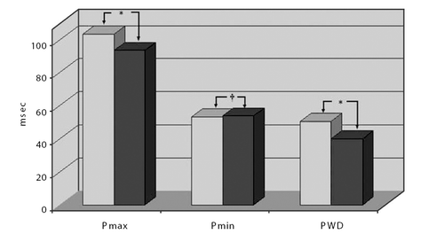
Pmax and PWD were significantly higher in group 1 compared with group 2 (103.59±19.8 ms vs 93.59±13.4 ms, p<0.001; 50.51±18.6 ms vs 39.85±10.6 msec, p<0.001, respectively). However, no statistically significant difference in Pmin duration was found between group 1 and group 2 (53.08±12.20 ms vs 53.74±12.10 ms, p = 0.73, respectively) (; ).
Table III. Comparison of P-wave values of the groups.
Figure 1. Associations of P‐wave characteristics between two groups. Darker bars indicate normotensive group, lighter bars indicate prehypertensive group. Pmax, maximum P‐wave; Pmin, minimum P‐wave; PWD, P‐wave dispersion. *p<0.001; †p>0.05.
In univariate logistic regression analysis, each 1‐ms increase in PWD increased the risk of prehypertension 1.021 times (95% CI 1.004–1.038, p = 0.014). After adjustment for age, sex, BMI, diabetes mellitus, smoking, LVEF, and heart rate, the multiple‐adjusted OR of the risk of prehypertension was 1.023 (95% CI 1.005–1.041, p = 0.011) for each 1‐ms increase in PWD.
In addition, there was a significant positive correlation between systolic blood pressure and PWD (r = 0.165, p = 0.015). However, no significant correlation was found between diastolic blood pressure and PWD (p>0.05).
There were no significant correlations between LA diameter and Pmax/PWD (p>0.05). There were also no significant relations between Pmax and thickness of IVS/PW, PWD and IVS/PW (p>0.05).
Discussion
AF is one of the most common arrhythmias that increases cardiovascular morbidity and mortality and decreases quality of life Citation[5]. PWD is non‐invasive marker that shows intra‐atrial conduction heterogenicity Citation[11]. This heterogeneous electrical activity causes atrial re‐entry. Atrial re‐entry is one of the pathophysiological mechanisms of AF Citation[12]. Maximum P‐wave duration and PWD are commonly used to determine the risk of AF in different patient populations Citation[13–17]. In hypertensive patients, Dilaveris et al. demonstrated that PWD was significantly higher in patients with a history of AF than control group. They concluded that hypertensive patients at risk for paroxysmal AF (pAF) could be detected by P‐wave analysis while in sinus rhythm Citation[18].
Studies have shown that prehypertension is associated with excess cardiovascular morbidity and mortality Citation[9]. The relation between BP and cardiovascular risk is graded and continuous Citation[19]. In the follow‐up of the Multiple Risk Factor Intervention Trial (MRFIT), age‐adjusted relative risk of 1.61 for fatal coronary events and 2.14 for strokes was observed in men who had SBP of 130–139 mmHg and DBP of 85–89 mmHg when compared with men with optimal blood pressure Citation[20]. In the Strong Heart Study, prehypertensive population had 1.8‐fold increased risk of cardiovascular events compared with normotensive population Citation[21].
To our knowledge, this is the first study to evaluate whether there is an association between PWD and prehypertension. We found that PWD and Pmax values were significantly higher in prehypertension compared with normotensive patients. Because PWD shows heterogeneous electrical activity in the left atrium, this data can be important for the prediction of AF in prehypertension.
Elevations of plasma renin and norepinephrine concentrations have been described in prehypertension Citation[22],Citation[23]. Increased angiotensin (Ang) II in plasma relates to arrhythmogenic electrical dispersion Citation[24]. Ang II and catecholamine were also demonstrated to increase atrial fibrosis Citation[25],Citation[26]. Excess atrial fibrosis causes heterogeneous and different atrial conduction Citation[27]. Increased Ang II and atrial fibrosis can explain the increase in PWD and Pmax in prehypertension in this study.
Tükek et al. Citation[28] showed that PWD was significantly higher in patients with pAF compared in patients without pAF even though left atrium sizes of both groups were equal. In this study, there was no significant difference in left atrial size between groups.
Study limitations
The small number of patients was the most important limitation of this study. The other limitation was absence of rhythm follow‐up in both groups. In addition, we measured left atrial maximal diameter in our study, this may not accurately reflect atrial size because atrial dilatation can be eccentric.
Conclusion
As a result, we concluded that prehypertension might cause prolongation of PWD and Pmax.
This data might show increased risk of AF in prehypertension.
References
- Gur M, Yilmaz R, Demirbag R, Akyol S, Altiparmak H. Relation between P‐wave dispersion and left ventricular geometric patterns in newly diagnosed essential hypertension. J Electrocardiology 2008; 41: 54.e1–54.e6
- Li Z, Hertervig E, Carlson J, Johansson C, Olsson SB, Yuan S. Dispersion of refractoriness in patients with paroxysmal atrial fibrillation: evaluation with simultaneous endocardial recordings from both atria. J Electrocardiol 2002; 35: 227–234
- Li Z, Hertervig E, Yuan S, Yang Y, Lin Z, Olsson SB. Dispersion of atrial repolarization in patients with paroxysmal atrial fibrillation. Europace 2001; 3: 285–291
- Ozer N, Aytemir K, Atalar E, Sade E, Aksöyek S, Ovünç K, et al. P wave dispersion in hypertensive patients with paroxysmal AF. Pacing Clin Electrophysiol 2000; 23: 1859–1862
- Kannel WB, Wolf PA, Benjamin EJ, Levy D. Prevalence, incidence, prognosis and predisposing condition for atrial fibrillation: population‐based estimates. Am J Cardiol 1998; 82: 2N–9N
- Ciaroni S, Cuenoud L, Bloch A. Clinical study to investigate the predictive parameters for the onset of atrial fibrillation in patients with essential hypertension. Am Heart J 2000; 139: 814–819
- Chobanian AV, Bakris GL, Black HR, Cushman WC, Green LA, Izzo JL, Jr, et al. The Seventh Report of the Joint National Committee on prevention, detection, evaluation, and treatment of high blood pressure: the JNC7 report. JAMA 2003; 289: 2560–2572
- Leitschuh M, Cupples LA, Kannel W, Gagnon D, Chobanian A. High‐normal blood pressure progression to hypertension in the Framingham Heart Study. Hypertension 1991; 17: 22–27
- Vasan RS, Larson MG, Leip EP, Evans JC, O'Donnell CJ, Kannel WB, et al. Impact of high‐normal blood pressure on the risk of cardiovascular disease. N Engl J Med 2001; 345: 1291–1297
- Dilaveris P, Batchvarov V, Gialafos J, Malik M. Comparison of different methods for manual P‐wave duration measurement in 12‐lead electrocardiograms. Pacing Clin Electrophysiol 1999; 22: 1532–1538
- Perzanowski C, Ho AT, Jacobson AK. Increased P‐wave dispersion predicts recurrent atrial fibrillation after cardioversion. J Electrocardiol 2005; 38: 43–46
- Dagli N, Karaca I, Yavuzkir M, Balin M, Arslan N. Are maximum P wave duration and P wave dispersion a marker of target organ damage in the hypertensive population?. Clin Res Cardiol 2008; 97: 98–104
- Yilmaz R, Demirbag R. P‐wave dispersion in patients with stable coronary artery disease and its relationship with severity of the disease. J Electrocardiology 2005; 38: 279–284
- Tükek T, Akkaya V, Demirel S, Sözen AB, Kudat H, Atilgan D, et al. Effect of Valsalva maneuver on surface electrocardiographic P‐wave dispersion in paroxysmal atrial fibrillation. Am J Cardiol 2000; 85: 896–899
- Senen K, Turhan H, Riza Erbay A, Basar N, Saatci Yasar A, Sahin O, et al. P‐wave duration and P‐wave dispersion in patients with dilated cardiomyopathy. Eur J Heart Fail 2004; 6: 567–569
- Erbay AR, Turhan H, Yasar AS, Bicer A, Senen K, Sasmaz H, et al. Effects of long‐term beta‐blocker therapy on P‐wave duration and dispersion in patients with rheumatic mitral stenosis. Int J Cardiol 2005; 102: 33–37
- Akdemir R, Ozhan H, Gunduz H, Tamer A, Yazici M, Erbilen E, et al. Effect of reperfusion on P‐wave duration and P‐wave dispersion in acute myocardial infarction: primary angioplasty versus thrombolytic therapy. Ann Noninvasive Electrocardiol 2005; 10: 35–40
- Dilaveris PE, Gialafos EJ, Chrissos D, Andrikopoulos GK, Richter DJ, Lazaki E, et al. Detection of hypertensive patients at risk for paroxysmal atrial fibrillation during sinus rhythm by computer assisted P wave analysis. J Hypertension 1999; 17: 1463–1470
- Stamler J, Stamler R, Neaton JD. Blood pressure, systolic and diastolic, and cardiovascular risks: US population data. Arch Intern Med 1993; 153: 598–615
- Neaton JD, Kuller L, Stamler J, Wentworth DN. Impact of systolic and diastolic blood pressure on cardiovascular mortality. Hypertension: pathophysiology, diagnosis and management, JH Laragh, BM Brenner 2nd edn. Vol. 1. Raven Press, New York 1995; 127–144
- Zhang Y, Lee ET, Devereux RB, Yeh J, Best LG, Fabsitz RR, et al. Prehypertension, diabetes, and cardiovascular disease risk in a population‐based: the Strong Heart Study. Hypertension 2006; 47: 410–414
- Esler M, Julius S, Zweifler A, Randall O, Harburg E, Gardiner H, et al. Mild high‐renin essential hypertension: Neurogenic human hypertension?. N Engl J Med 1977; 296: 405–411
- Julius S, Krause L, Schork NJ, Mejia AD, Jones KA, van de Ven C, et al. Hyperkinetic borderline hypertension in Tecumseh, Michigan. J Hypertens 1991; 9: 77–84
- Allessie MA, Boyden PA, Camm J, et al. Pathophysiology and prevention of atrial fibrillation, et al. Pathophysiology and prevention of atrial fibrillation. Circulation 2001; 103: 769
- Brilla CG, Funck RC, Rupp H. Lisinopril‐mediated regression of myocardial fibrosis in patients with hypertensive heart disease. Circulation 2000; 102: 1388–1393
- Kelm M, Schafer S, Mingers S, Heydthausen M, Vogt M, Motz W, et al. Left ventricular mass is linked to cardiac noradrenaline in normotensive and hypertensive patients. J Hypertens 1996; 14: 1357–1364
- Cha YM, Dzeja PP, Shen WK, Jahangir A, Hart CY, Terzic A, et al. Failing atrial myocardium: Energetic deficits accompany structural remodeling and electrical instability. Am J Physiol Heart Circ Physiol 2003; 284: H1313–h1320
- Tükek T, Akkaya V, Atilgan D, Demirel S, Sözen AB, Kudat H, et al. Changes in P wave dispersion, left atrial size and function in hypertensive patients with paroxysmal atrial fibrillation. Arch Turkish Soc Cardiol 2000; 28: 538–542