Abstract
The case of a 65-year-old woman presenting with Guillain-Barré syndrome is herein reported. Tomographic investigation revealed abdominal and retroperitoneal fibromatosis. During her hospitalization, renal involvement ensued, and subsequent renal biopsy demonstrated findings of crescentic pauci-immune glomerulonephritis negative for ANCA antibodies and with characteristics indicative of necrotic angiitis. The simultaneous existence of the three diseases in the same patient as well as the relation between necrotic vasculitis and G-B syndrome is speculated, and the relevant literature is reviewed.
INTRODUCTION
Fibromatosis, also known as a desmoid tumor, is a rare neoplasm of the connective tissue that arises from skeletal muscle, aponeuroses, and tendinous tissue. It is usually found in the trunk, mainly within the abdominal cavity but also in the extremities,Citation[1] and differs from fibrosarcoma and adiposarcoma by a lack of metastatic potential. However, it does possess two properties typical of malignant tumors: the ability to infiltrate neighboring structures, and frequent recurrence after surgical removal.Citation[2] Histologically, it is characterized by the presence of abundant fibroblasts devoid of malignant characteristics, such as nuclear divisions. Fibromatosis represents 3% of mesenchymal tissue neoplasms and 0.03% of all tumors.Citation[3] Rarely, this tumor is located in the retroperitoneal space, but it is not related in any way to retroperitoneal fibrosis, a disorder characterized by an inflammatory response of connective tissue.Citation[4] Clinically, fibromatosis manifests itself as an obstructive disease of the alimentary tract; its diagnosis relies on histological findings and its treatment on surgical removal, radiation therapy, and certain drugs. The frequency of local recurrence after resection ranges from 40 to 80%.Citation[1] In many patients, fibromatosis is associated with colon polyposis.Citation[5] This study examines the case of a patient suffering from histologically proven abdominal fibromatosis and, at the same time, from Guillain-Barré polyneuropathy and necrotic vasculitis, a clustering of disorders unique in the available literature.
CASE REPORT
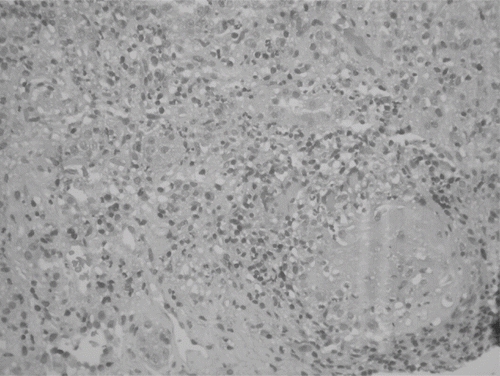
A 73-year-old female patient with a history of arterial hypertension treated with atenolol and nifedipine developed an acute right lower extremity pain syndrome that also affected the left arm within the next 24 hours. Progressively, generalized weakness ensued, together with motion abnormalities of the lower extremities. Three days later, and after being admitted in the county hospital, the patient was referred to the Neurology Department of the authors' hospital with a likely diagnosis of Guillain-Barré syndrome. Neurological examination revealed muscle weakness of the left arm and both legs, the absence of tendon reflexes in both upper and lower extremities, and dysesthesia with a “stocking-like” distribution in the lower extremities. Electromyography demonstrated findings typical of Guillain-Barré syndrome, and although lumbar puncture was inconclusive (absence of the pathognomonic finding of albuminocytologic dissociation), the diagnosis was considered certain based on the clinical presentation and electromyography. Laboratory investigation revealed an elevated white blood cell count (18 K/mm3) with leftward shift (85% neutrophils), anemia (31% hematocrit), impaired renal function (urea nitrogen 75 mg/dL and serum creatinine 1.4 mg/dL), and elevated markers of inflammation (erythrocyte sedimentation rate 125mm/h, C-reactive protein 155 mg/dL). Thorax roentgenography showed bronchopulmonary infiltrates to a small extent next to the left pulmonary hilus. Intravenous gamma-globulin therapy was initiated (30 gr/day for five days) along with a combined antibiotic treatment, including a third-generation cephalosporin and a macrolide. The respiratory infection subsided, and the patient was transferred to the Internal Medicine Department for further investigation. Upper abdominal sonography was unremarkable, as were virologic examinations and serologic tests (Wright, Widal, Toxoplasma gondii). Anti-Neutrophil-Cytoplasmic Antibodies (ANCA) were negative, whereas tumor marker CA-125 was elevated (81.6 with an upper normal limit of 35). This finding led to computed-tomographic (CT) investigation of the abdomen, which revealed large masses in the pelvis in the proximity of the cecum (see ) and not accompanied by enlarged lymph nodes, and with an overall appearance suggestive of a tumor of mesenchymal origin. Lower abdominal and transvaginal sonography ruled out the possibility of these masses being attached to the uterine wall, which would make them compatible with uterine fibroleiomyomata. Consequently, the patient underwent a CT-guided biopsy of the masses; histologic examination was indicative of a tissue consisting of neoplastic cells surrounded by bundles of collagen fibers. No nuclear divisions were recognized (see ). This histological appearance coupled with an immunohistochemistry investigation yielded the probable diagnosis of retroperitoneal fibromatosis. In a second attempt, carried out after a short discharge of the patient from the hospital, biopsies were acquired from three different neoplastic sites, this time confirming the diagnosis. However, the patient's renal function had deteriorated in the meantime, now exhibiting microscopic hematuria, a serum creatinine of 4.6 mg/dL, and marked proteinuria (750 mg/24 h urine-collection), all without any obstructive cause being demonstrable at urinary tract sonography. Renal biopsy was performed, showing a recent crescent formation and necrotic granulomas in 50% of glomeruli—findings consistent with necrotic vasculitis (see ). Therapy with intravenous corticosteroids at escalating doses was commenced, reaching 3 g methylprednisolone a day, which was followed by per os corticosteroid administration combined with 50 mg cyclophosphamide qd. A small improvement in renal function was observed (serum creatinine 3.7 mg/dL). Surgical consultation was sought, and the possibility of resection or irradiation of the tumor was discussed; however, the patient and her relatives denied it emphatically, and according to their wish, the patient was discharged. She was lost to follow-up thereafter.
Figure 1 Computed tomography revealing extended desmoid tumors in the abdominal cavity.
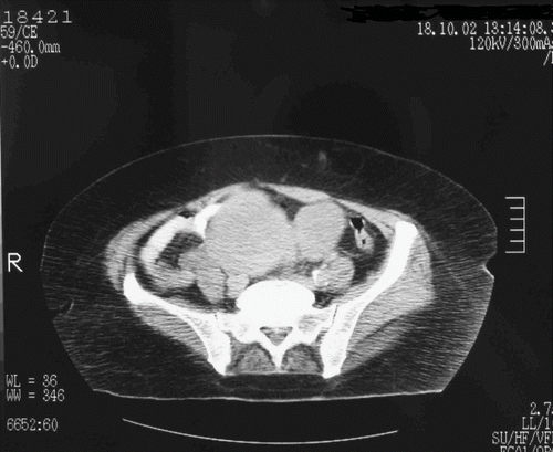
Figure 2 Biopsy of the abdominal mass, revealing desmoid connective tissue with neoplastic cells (fibroblasts) and collagen, consistent with fibromatosis. (Hematoxylin and eosin stain; original magnification, *200).
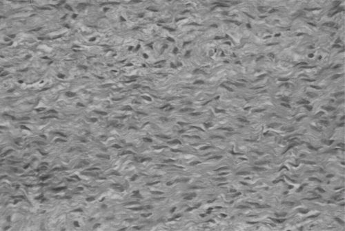
Figure 3 Renal biopsy, demonstrating an obsolescent glomerulus surrounded by epithelioid and mononuclear cells, as well as multinucleated Langhans-type giant cells. (Hematoxylin and eosin stain; original magnification, *250).
DISCUSSION
The symptomatology that led the patient to the hospital was typical of the Guillain-Barré syndrome.Citation[6] Neurologic examination and electromyography findings set the diagnosis, although no albuminocytologic dissociation was found in the cerebrospinal fluid. However, this finding is not considered necessary for the diagnosis of the syndrome, as it depends on the specific time point on the course of the disease that lumbar puncture is performed.Citation[7] Guillain-Barré syndrome is considered an autoimmune disorder, the development of which is usually triggered by a viral infection of the respiratory or gastrointestinal system preceding the neurologic manifestations by one to three weeks.Citation[8] Other factors or conditions believed to play a triggering role are certain pathogens (Campylobacter duodeni, the Epstein-Barr virus, hepatitis viruses, Mycoplasma pneumoniae, HIV-1), childhood immunizations, surgical procedures, lymphomas, and systemic lupus erythematosus.Citation[8] In the case presented here, the cause is likely to have been a febrile respiratory infection with a ten-day course and diffuse pulmonary infiltrates on chest X-ray.
Tomographic investigation revealed abdominal and retroperitoneal masses of considerable size, proven with biopsy to represent fibromatous tumors. As mentioned before, fibromatosis has a subclinical course, until the masses reach such size that they compress adjacent organs. In the present case, however, it was merely an elevation of a tumor marker and not symptoms that led to an abdominal CT-scan and eventually the diagnosis, despite the extensive spread of the tumor—which also involved the retroperitoneal space, an extremely rare site for fibromatosis to develop.Citation[1]
It is not possible to determine the exact time when the tumor first appeared; however, the extent of it reveals a course of long duration. Therefore, it should be considered unlikely that Guillain-Barré syndrome in this patient was linked pathogenetically with fibromatosis due to the absence of temporal association between the two disorders. Furthermore, fibromatosis is characterized by the neoplastic growth of fibroblasts, without signs of inflammation, clinical or subclinical (expressed as raised markers of systemic inflammation), while the histological lesions in Guillain-Barré syndrome include myelin inflammation and a consequent insult of the neural axon.Citation[8] Therefore, the development of Guillain-Barré syndrome on the ground of a non-inflammatory process, like fibromatosis, should be assumed improbable.
What is most interesting, though, is the renal insult that ensued during the patient's hospitalization. The renal involvement initially developed as a rapidly progressive glomerulopathy and occurred almost simultaneously with the neurological syndrome, as the patient's serum creatinine was close to normal upon admission. Renal biopsy, performed at the onset of the nephritic syndrome, revealed findings consistent with necrotic vasculitis, which could probably have been a manifestation of Wegener's disease, even though no cytoplasmic antibodies (ANCA) were detected (see ). The histological and immunohistochemical findings in support of this hypothesis are the following:
In 50% of glomeruli, cellular casts were found, confirming that this was a crescentic glomerulonephritis.
Immunofluorescence failed to demonstrate the deposition of immune complexes, which confirms the pauci-immune nature of the glomerulonephritis.
These two features, together with the absence of ANCA, characterize idiopathic, type V, crescentic glomerulopathy. However, the demonstration of necrotic vasculitis findings in light microscopy, accompanied by granulomas containing Langhans-type giant cells, makes the diagnosis of crescentic glomerulonephritis secondary to necrotic angiitis (probably in the context of Wegener granulomatosis) highly likely. Even the pulmonary infiltrates on chest radiography could have been attributed to Wegener's disease, should they have not responded so readily to antibiotic treatment. The association between the renal involvement and fibromatosis is not considered plausible for the same reasons mentioned above—the relation of the Guillain-Barré syndrome to the desmoid tumor and the lack of a time association and common pathogenetic ground. These two assumptions, however, are fulfilled in the case of the association between necrotic angiitis and Guillain-Barré syndrome in this patient. From a pathophysiologic point of view, both disorders are considered autoimmune. Guillain-Barré syndrome usually follows infections, and there are indications that certain gangliosides of the nervous tissue resemble viral or bacterial antigens; therefore, the produced antibodies are directed against myelin and the nerve axon, leading to inflammation and degeneration.Citation[9],Citation[10] Whether necrotic angiitis caused the Guillain-Barré syndrome or vice versa is elusive. Although the full effect of treatment could not be observed due to the early, self-chosen discharge of the patient, the improvement of renal function after commencing corticosteroids and cyclophosphamide suggests a primary renal disease. It could be the case, however, that a common, unknown factor triggered both disorders. There have been reports in world literature of Guillain-Barré syndrome following,Citation[11] preceding,Citation[12] or appearing simultaneouslyCitation[13],Citation[14] with some form of glomerulopathy, the most frequent of which appears to be membranous glomerulonephritis,Citation[14] clinically manifested as nephrotic syndrome. In some cases, interstitial nephritis is also observed in renal biopsy material.Citation[12],Citation[13] Guillain-Barré syndrome has also been described accompanying lupus nephritis.Citation[15] A rapidly progressive glomerulonephritis, however, rarely complicates Guillain-Barré syndrome,Citation[11] especially in the context of necrotic vasculitis, as in this case report. Finally, the co-existence of fibromatosis, relevant or not to the two autoimmune disorders (Guillain-Barré syndrome and glomerulonephritis), has never been reported before.
In conclusion, Guillain-Barré syndrome can be accompanied by some form of glomerulopathy, sometimes with a rapidly deteriorating course. The context in which this glomerulopathy evolves should be investigated; in this case, necrotic vasculitis was revealed, probably on the grounds of Wegener granulomatosis. Corticosteroids and cyclophosphamide remain the mainstay of treatment. The presence of fibromatosis along with the two other diseases seems to have been coincidental in this case, but is still a remarkable observation in its rarity.
This study was conducted independently; no company or institution supported it financially.
REFERENCES
- Boon-Swee O, Chien-Nien L, Thiow-Kong T, et al. Retroperitoneal fibromatosis presenting as acute duodenal obstruction. Australian and New Zealand Journal of Surgery 2001; 71: 74–76, [CSA]
- Janinis J, Patriki M, Vini L, et al. The pharmacological treatment of aggressive fibromatosis: a systematic review. Annals of Ongology 2003; 14: 181–190, [CROSSREF], [CSA]
- Overhaus M, Decker P, Fischer H, et al. Desmoid tumors of the abdominal wall: a case report. World Journal of Surgical Ongology 2003; 1: 11–16, [CROSSREF], [CSA]
- Kikkawa A, Kido A, Kumai T, et al. Extraabdominal fibromatosis in retroperitoneal space. World Journal of Surgical Ongology 2004; 2: 33–36, [CROSSREF], [CSA]
- Gronchi A, Casali P, Mariani L, et al. Quality of surgery and outcome in extra-abdominal aggressive fibromatosis: a series of patients surgically treated at a single institution. Journal of Clinical Oncology 2003; 21: 1390–1397, [INFOTRIEVE], [CROSSREF], [CSA]
- Fulgham JR, Wijdicks EF. Guillain-Barré Syndrome. Crit Care Clin 1997; 13: 1–15, [INFOTRIEVE], [CROSSREF], [CSA]
- Cortson RN, MeGale EH, Stonier C, et al. Abnormalities of cerebrospinal fluid amino acids in patients with the Guillain-Barré Syndrome. Journal of Neurology, Neurosurgery, and Psychiatry 1991; 44: 86–89, [CSA]
- Hartung HP, Willison HJ, Kieseier BC. Acute immuno-inflammatory neuropathy: update on Guillain-Barré Syndrome. Curr Opin Neurol 2002; 15: 571–577, [INFOTRIEVE], [CROSSREF], [CSA]
- Salloway S, Mermel L, Seamans M, et al. Miller-Fisher syndrome associated with Campylobacter jejuni bearing lipopolysaccharide molecules that mimic human ganglioside GD3. Infection and Immunity 1996; 64: 2945–2949, [INFOTRIEVE], [CSA]
- Creange A, Belec L, Clair B, et al. Circulating transforming growth factor beta 1 (TGF-beta 1) in Guillain-Barré Syndrome. Decreased concentrations in the early course and increase with motor function. Journal of Neurology, Neurosurgery, and Psychiatry 1998; 64: 162–165, [INFOTRIEVE], [CSA]
- Bettinelli A, Giani M, Rossi L, et al. Ex novo episodes of acute glomerulonephritis and Guillain-Barre syndrome: a case report. Clin Nephrol 1989; 31(5)269–273, [INFOTRIEVE], [CSA]
- Kanemoto K, Nakahara C, Saitoh H, et al. Renal glucosuria and membranous glomerulonephritis in chronic inflammatory demyelinating polyradiculoneuropathy: CIDP. Nippon Jinzo Gakkai Shi 1999; 41(5)511–516, [INFOTRIEVE], [CSA]
- Talamo TS, Borochovitz D. Membranous glomerulonephritis associated with the Guillain-Barre syndrome. Am J Clin Pathol 1982; 78(4)563–566, [INFOTRIEVE], [CSA]
- Careless D, Rigby R, Axelsen R, Boyle R. A case of Guillain-Barre syndrome with focal segmental glomerulosclerosis. Am J Nephrol 1993; 13(2)160–163, [INFOTRIEVE], [CSA]
- Van Laarhoven H, Rooyer F, van Engelen B, van Dalen R, Berden J. Guillain–Barré syndrome as presenting feature in a patient with lupus nephritis, with complete resolution after cyclophosphamide treatment. Nephrol Dial Transplant 2001; 16: 840–842, [INFOTRIEVE], [CROSSREF], [CSA]