Abstract
Objective. The objective of this study was to evaluate the effect of gold nanoparticles on renal cell. Intervention. This study was performed as an experimental study. A mixture of gold nanoparticle solution and renal cell sediment was prepared and further analyzed. Results. This work revealed that after mixing the renal cell sediment with gold nanoparticle solution, gold nanoparticles can be seen within the renal cells. Conclusions. Conclusively, the gold nanoparticle can penetrate into renal cell. This is the first report on this observation, and further implies the possibility of other nanoparticle nephrotoxicity in the present nanomaterial era.
Keywords:
INTRODUCTION
Present-day advances in biotechnology have brought us to new era of nanotechnology. Nanoparticles differ from the same material at larger scale in chemical and physical properties.Citation[1] The rapidly developing field of nanotechnology is likely to become yet another source of toxicity through inhalation, ingestion, skin uptake, and injection of engineered nanomaterials.Citation[2] In medicine, there is limited knowledge on the toxicity of nanoparticles, just as in the genitourinary system there has been limited knowledge on the effect of nanoparticles. In this work, the authors studied the effect of gold nanoparticles on renal cell.
MATERIALS AND METHODS
Urine Sample
Urine sample with renal cell sediment approved by routine clinical microscopic technique was used for further laboratory analysis. The sample was collected and transferred according to standard procedure of laboratory medicine.
Gold Nanoparticle Solution Preparation
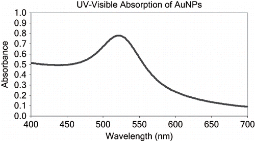
Following the classical Turkevich citrate reduction method,Citation[3] nine nanometer-sized gold nanoparticle solutions were established, and a gold nanoparticle solution concentration was prepared at 44 ppm. The gold nanoparticle is shown in . The synthesis procedure is according to that described by Hone et al.Citation[4] Briefly, HAuCl4 (12.5 mg) was dissolved in 100 mL of water, and 50 mg of sodium citrate was dissolved in 50 mL of water; both solutions were then heated to 60oC. The rapid addition of the sodium citrate solution to the gold solution was followed by increasing the temperature to 85oC and continuous stirring for 2.5 hours. The gold nanoparticle solution can be stored in the dark under 4oC for more than a month. A diagram of UV-visible absorption of gold nanoparticle solution is presented in . The exact synthetic laboratory is at Department of Chemistry, Faculty of Science, Chulalongkorn University, Bangkok, Thailand. The laboratory is under safety and quality control from both department and university organization.
Figure 1. The figure of gold nanoparticle.
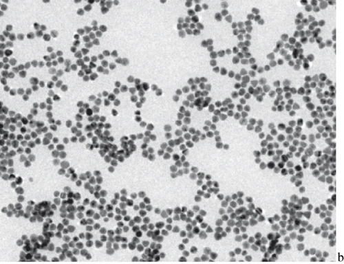
Figure 2. UV-visible absorption of gold nanoparticle solution.
Study of the Direct Effect of Gold Nanoparticle on the Renal Cell
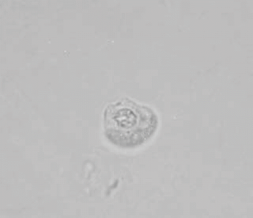
First, the mixture between 10 microliter of gold nanoparticle solution and renal cell sediment was prepared. After 15 minutes, the motility and morphological changes of spermatozoa were studied by clinical microscopy technique under a high power field. A control experiment without adding the gold nanoparticle solution was also performed. The entire laboratory analysis was performed in a reference ISO 15189-accredited laboratory of the Department of Laboratory Medicine, Faculty of Medicine, Chulalongkorn University.
RESULTS
This work revealed that after mixing the renal cell sediment with gold nanoparticle solution, the gold nanoparticle can be seen within the renal cells (see ).
Figure 3. Renal cell with intracellular gold nanoparticle.
DISCUSSION
Gold nanoparticle is presently a widely used nanoparticle. When gold is in very small particles (i.e., with diameters below 10 nm) and deposited on metal oxides or activated carbon, it becomes surprisingly active, especially at low temperatures, for many reactions such as CO oxidation and propylene epoxidation.Citation[5] However, the effect of nanoparticle has been poorly clarified. The nephrotoxicity of nanoparticle has just been proposed for a few years.Citation[6] Manil et al. reported their study on the nephrotoxicity of doxorubicin-loaded cyanoacrylate nanoparticles.Citation[7] They reported that the nephrotoxicity was related to an enhanced capture of naoparticles by cells of the mononuclear phagocyte system, including mesangial cells.Citation[7]
Here, we demonstrated the penetration of gold nanoparticles into renal cell sediment. This study confirms that nanoparticles can directly enter into the renal cell. Indeed, the nephrotoxicity of heavy metal has been proposed for a long time. Gold nephropathy is well documented in the literature.Citation[8] The nephrotoxic clinical manifestations are renal insufficiency, proteinuria and hematuria, and the nephrotic syndrome.Citation[8] The pathologic changes are tubular degeneration, acute tubular necrosis, and immune complex glomerulonephritis.Citation[8] Conclusively, the gold nanoparticle can penetrate into the renal cell. It is necessary to set up some hypothesis to explain the phenomena. The gold nanoparticle penetration might be due to the size effect. The size of gold nanoparticle is less than the pore size of cell membrane, which can permit the gold nanoparticle to enter into the cell. It can further be suggested for the possibility of other nanoparticles nephrotoxicity in the present nanomaterial era.
REFERENCES
- Seaton A. Nanotechnology and the occupational physician. Occup Med (Lond). Aug, 2006; 56(5)312–316
- Oberdörster G, Oberdörster E, Oberdörster J. Nanotoxicology: An emerging discipline evolving from studies of ultrafine particles. Environ Health Perspect. Jul, 2005; 113(7)823–839
- Enüstün BV, Turkevich J. Coagulation of colloidal gold. J Am Chem Soc. 1963; 85: 3317–3328
- Hone DC, Haines AH, David A. Russell, rapid, quantitative colorimetric detection of a lectin using mannose-stabilized gold nanoparticles. Langmuir. 2003; 19: 7141–7144
- Haruta M. When gold is not noble: Catalysis by nanoparticles. Chem Rec. 2003; 3(2)75–87
- Oberdörster G, Maynard A, Donaldson K, Castranova V, Fitzpatrick J, Ausman K, Carter J, Karn B, Kreyling W, Lai D, Olin S, Monteiro-Riviere N, Warheit D, Yang H, the ILSI Research Foundation/Risk Science Institute Nanomaterial Toxicity Screening Working Group. Principles for characterizing the potential human health effects from exposure to nanomaterials: Elements of a screening strategy. Part Fibre Toxicol. Oct 6, 2005; 2: 8
- Manil L, Couvreur P, Mahieu P. Acute renal toxicity of doxorubicin (adriamycin)-loaded cyanoacrylate nanoparticles. Pharm Res. Jan, 1995; 12(1)85–87
- Antonovych TT. Gold nephropathy. Ann Clin Lab Sci. Sep–Oct, 1981; 11(5)386–391