Abstract
Patients with chronic renal failure have a higher risk of developing tuberculosis compared to the general population, and especially extra-pulmonary tuberculosis. This pathology is often difficult to diagnose and requires a combination of multidisciplinary examinations. The confirmation of the diagnosis is important in order to quickly match the treatment with the pathology. We report a patient with primary esophageal tuberculosis for whom the interferon gamma release assay facilitated a timely diagnosis.
Patients with chronic renal failure have a relatively higher risk (6.9–25.3%) of developing tuberculosis compared to the general population because of reduced cellular immunity, and the disease is associated with increased mortality.Citation[1],Citation[2] Moreover, extra-pulmonary tuberculosis is more common than pulmonary tuberculosis in this population, but is often difficult to diagnose.Citation[1],Citation[2] We report a patient with primary esophageal tuberculosis for whom the interferon gamma release assay facilitated a timely diagnosis.
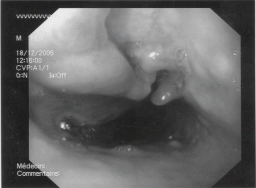
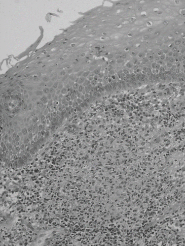
A 79-year-old Algerian male, in France for more than 40 years, with chronic renal failure due to autosomal dominant polycystic kidney disease was hospitalized in November 2006 for a low-grade fever, anorexia, significant weight loss (10 kg), and dysphagia of three months in duration. The patient had a history of hypertension, ischemic heart disease, chronic obstructive pulmonary disease, and chronic C hepatitis. A physical examination did not expose any particular signs, including no evidence of lymphadenopathy. Laboratory assessment revealed a mild normochromic and normocytic anemia (hemoglobin: 10.1 g/dL) and an elevated C-reactive protein (48 mg/L). The white blood cell count was 9.7 × 10⁹/L with a normal differential count. Tuberculin skin test (TST) was negative. An upper gastro-intestinal endoscopy revealed a deep ulcer measuring 3 cm in diameter at the upper third of the esophagus (see ). A computed tomographic scan of the thorax revealed a mass lesion in the lower esophagus and thickening the esophageal wall, associated with para-tracheal lymphadenopathy. There was no radiological evidence of active pulmonary tuberculosis, nor was pulmonary cancer (i.e., esophageal carcinoma) suspected. The histological examination of biopsy specimens indicated a sketch of granuloma without giant cells or necrosis but was not specific enough to form a conclusion, so the pathologist suggested to repeat the biopsy. A Ziehl-Neelsen stain for acid-fast bacilli and a PCR for Mycobacterium tuberculosis were negative. Looking forward to the results of the second histological examination, the clinical description and the word “granuloma” prompted us to go further in the search of tuberculosis by detecting the release of interferon-gamma in the patient's blood with the test QuantiFERON®-TB Gold (QFT-G, Cellestis, Carnegie, Victoria, Australia). QFT-G assay was positive and allowed us to start a six-month therapy with isoniazid, rifampin, pyrazinamide, and ethambutol. Finally, the second endoscopy suggested esophageal tuberculosis (see ), and cultures grew at day 20 from gastric liquid and at day 45 from the esophageal biopsy. The strain was identified as M. tuberculosis by PCR. No complaints or clinical signs remained after a follow-up period of six months.
DISCUSSION
Esophageal tuberculosis is rarely reported, and the differential diagnosis includes carcinoma of esophagus, benign stricture, viral or fungal esophagitis, and (rarely) Crohn's disease.Citation[3],Citation[4] Extra pulmonary tuberculosis is often difficult to diagnose and requires a combination of examinations, including a medical evaluation, radiological examination, histological examination of biopsy sample, TST, direct examination, and culture of clinical samples or molecular diagnosis. The confirmation of the diagnosis is important in order to quickly match the treatment with the pathology. In a recently reported similar case, all of these investigations were initially negative and the diagnosis was ascertained four weeks after the beginning of explorations with the culture of M. tuberculosis from an esophageal brushing.Citation[3] In our case, QFT-G allowed us to gain time and start the antituberculous therapy when other investigations were non-conclusive. In dialysis patients, tuberculosis is associated with increased mortality and particularly after kidney transplantation.Citation[1] In that way, a timely identification of tuberculosis is important among patients before transplantation, and new tests that facilitate the detection of patients with latent tuberculosis infection or active tuberculosis are significant. QFT-G is an enzyme-linked immunosorbent assay test that detects the release of interferon-gamma in fresh heparinized whole blood from sensitized persons when it is incubated with mixture of synthetic peptides simulating three proteins present in M. tuberculosis and pathogenic M. bovis strains. The QFT-G is more specific and is as sensitive as TST, even in immunocompromised patients such as dialysis patients.Citation[5–7] Moreover, dialysis patients studied for response to TST are often anergic.Citation[8] Our report suggests that QFT-G can be helpful in the diagnosis of tuberculosis in patients suffering from chronic renal failure when other investigations are inconclusive. This test can be performed routinely, and the response is available sooner than the culture results.
DECLARATION OF INTEREST
The authors report no conflicts of interest. The authors alone are responsible for the content and writing of the paper.
REFERENCES
- Klote MM, Agodoa LY, Abbott KC. Risk factors for Mycobacterium tuberculosis in U.S. chronic dialysis patients. Nephrol Dial Transplant. 2006; 21: 3287–3292
- Ahmed AT, Karter AJ. Tuberculosis in California dialysis patients. Int J Tuberc Lung Dis. 2004; 8: 341–345
- Ahmed W, Rylance PB, Jackson MA, Nicholas JC, Odum J. A diabetic haemodialysis patient with dysphagia and weight loss. Nephrol Dial Transplant. 2003; 18: 1018–1020
- Fujiwara Y, Osugi H, Takada N, Takemura M, Lee S, Ueno M, Fukuhara K, Tanaka Y, Nishizawa S, Kinoshita H. Esophageal tuberculosis presenting with an appearance similar to that of carcinoma of the esophagus. J Gastroenterol. 2003; 38: 477–481
- Mazurek GH, Jereb J, LoBue P, Iademarco MF, Metchock B, Vernon A. Division of Tuberculosis Elimination, National Center for HIV, STD, and TB Prevention, Centers for Disease Control and Prevention (CDC). Guidelines for using the QuantiFERON-TB Gold test for detecting Mycobacterium tuberculosis infection, United States. MMWR Morb Mortal Wkly Rep. 2005; 54: 49–55
- Kobashi Y, Mouri K, Obase Y, Fukuda M, Miyashita N, Oka M. Clinical evaluation of QuantiFERON TB-2G test for immunocompromised patients. Eur Respir J. 2007; 30: 945–950
- Pai M, Zwerling A, Menzies D. Systematic review: T-cell-based assays for the diagnosis of latent tuberculosis infection: An update. Ann Intern Med. 2008; 149: 177–184
- Poduval RD, Hammes MD. Tuberculosis screening in dialysis patients—is the tuberculin test effective?. Clin Nephrol. 2003; 59: 436–440


