
Abstract
Purpose: Symptoms and disorders related to menopause and its associated estrogen deficiency have become a considerable health concern worldwide. Ovarian hormone depletion/estrogen deficiency can be usefully studied using animal models after removal of the ovaries [ovariectomy (Ovx)]. This study assessed renal changes after Ovx-induced estrogen deficiency in a rat model.
Methods: Rats were randomly allotted into one control group (group I, healthy) and three study groups (group II, Ovx group; group III, Ovx +17β-estradiol group; and group IV, Ovx + bortezomib group).
Results: In the Ovx group (group II), thickening of glomerular capillary walls, narrowing of Bowman’s capsular space, glomerular hypertrophy, atrophic tubules, and loss of the basal membranes of the tubules were observed. Mesangial cell proliferation was observed, particularly in the glomerulus. Immunohistochemical (IHC) staining studies in this group showed dense staining in the mesangial cells, tubular cell Nf-KB/p65, and caspase-3. Groups III and IV (Ovx +17β-estradiol and Ovx + bortezomib) showed decreased NF-kB/p65 and caspase-3 expression compared with the Ovx group (p < 0.05).
Conclusion: In renal failure related to estrogen deficiency caused by Ovx, 17β-estradiol and bortezomib have a protective effect on renal tissue.
Introduction
Estrogen is an important regulator of physiological processes in women. Estrogen deficiency has been acknowledge as causing disorders in postmenopausal women and is associated with metabolic effects and conditions that include osteoporosis, cardiovascular disease, chronic kidney disease (CKD), colon cancer, and dementia.Citation1,Citation2 17β-estradiol (E2) and estrone (E1) levels have the greatest association with the onset of symptoms related to menopause.Citation3
Ovarian hormone depletion in an experimental animal model that has undergone ovariectomy (Ovx) is a useful model for studying the effects of hormone depletion in women.Citation4–6 Ovx, or removal of the ovaries (called “oophorectomy” when performed in women as a procedure), reduces estrogen levels and increases the incidence of different types of cardiovascular and renal disorders.Citation7,Citation8 Decreased synthesis of 17ß-estradiol (E2) in experimental animals after Ovx is accompanied by an increased incidence of cardiovascular disorders and accelerated progression of renal disease.
Reduced estrogen levels can reduce the amount of information one can obtain about pathologic changes in kidney ultrastructure, immunohistochemistry (IHC), and stereologic analysis. Some studies have suggested that decreased synthesis of E2 causes renal changes and inflammatory cell infiltration.Citation4,Citation9 Additionally, several studies have demonstrated that decreased estrogen levels at menopause are associated with elevated oxidative stress throughout menopause.Citation1,Citation3,Citation10,Citation11
Inflammatory cytokine production induced by reactive oxygen species (ROS) occurs with hormone depletion in postmenopausal women.Citation12,Citation13 Nuclear factor kappa-B (NF-kB) promotes the expression of a number of genes involved in inflammation, such as cytokines and adhesion molecules. NF-kB/p65 is important in the control of cell proliferation, more specifically in protecting cells from programed cell death. Renal tubular epithelial cells may express a number of NF-kB/p65-dependent genes.
Recent studies have focused on using proteasome inhibitors to prevent the development of renal inflammation and fibrosis.Citation14 Bortezomib, a ubiquitin proteasome pathway inhibitor (via ROS-induced deubiquitinase inhibition) that is used in kidney transplants, and other compounds are under investigation as inhibitors of renal fibrosis, since transforming growth factor beta (TGF-β), a key factor in renal fibrosis, is regulated by the ubiquitin proteasome inhibitors pathway through degradation of TGF-β-signaling molecules.Citation12,Citation13,Citation15,Citation16
Estrogen has been noted as an effective treatment for the prevention of osteoporosis in postmenopausal women with normal renal function.Citation17,Citation18 In this study, our aim was to determine whether inhibiting NF-kB activation with bortezomib protects the kidneys from damage caused by E2 deficiency. We assessed the protective effects of 17β-estradiol in E2 deficiency by inducing deficiency with Ovx, with a focus on its protective effects against renal damage.
Materials and methods
Animals study
A total of 32 adult Wistar albino female rats (age, about 3 months; weight, 200 ± 50 g) were procured from the Ataturk University Animal Care and Research Unit, Erzurum, Turkey. The rats were housed in clean polypropylene cages with eight rats per cage. During the experimental period, all subjects were fed with pellets containing 21% crude protein (Purina; Nestlé Purina PetCare Company, St Louis, MO) and clean daily drinking water. All animals received humane care according to the criteria outlined in the guide for the care and use of laboratory animals prepared by the National Academy of Sciences and published by the National Institutes of Health. The study was approved by the Ataturk University Institutional Animal Ethical Committee. Our subjects were divided into four groups of eight rats each (total number of rats, N = 32) and had the same biological and physiological characteristics. The rats were randomly allotted into one of four groups: a control group (group I) or one of three experimental groups (group II, Ovx; group III, Ovx +17ß estradiol; group IV, Ovx + bortezomib).
Drug preparation and experimental protocols
For the three experimental groups, ovariectomies were performed in the operation room of Ataturk University’s experimental animals research branch. Rats were anesthetized with an intraperitoneal (IP) injection of 20 mg/kg sodium thiopental. A longitudinal slit (0.5–1 cm) was made in the midline area of the lower abdomen and the ovaries were removed. After Ovx, 25 mg/kg metamizole sodium was administered as an analgesic for 2 days.Citation1,Citation19 For the purpose of administration in group III, 17β-estradiol (Estrafem; Novo Nordisk, Bagsvaerd, Denmark) was dissolved in 0.9% NaCl. Approximately 8 weeks after Ovx, 17β-estradiol with 0.9% NaCl (group III), or 0.9% NaCl alone (group II) was administered 0.2 mg/kg by oral gavage once a day until the end of the experiment. At the same time interval after Ovx, group IV (Ovx + bortezomib) was given 0.4 mg/kg bortezomib administered IP until the end of the experiment.
Histological preparation
Kidney tissue samples were fixed in 10% formalin. After the fixation, specimens were dehydrated in an ascending series of ethanol, cleared in xylene and embedded in paraffin. The sections having thickness 4–5 μg were prepared for hematoxylin-eosin (H&E) staining (Applichem GmbH, Darmstadt, Germany).
IHC preparation
Kidney tissue samples were cut into 1–2 μm semi-thick sections and prepared for NF-kB/p65 and caspase-3 staining with Ventana benchmark GX (Ventana Medical Systems; Oro Valley, AZ). Following steps were performed for IHC staining: the sections were deparaffinized and treated with proteinase K solution (20 mg/mL in PBS), washed in distilled water, and immersed in 3% hydrogen peroxide. After several washes with PBS, the sections were immersed in an equilibration buffer. The secondary kit used was the ultraview universal DAB detection kit (Ventana Medical Systems, Oro Valley, AZ).
Stereologic analysis
For stereologic examination, stereo investigator 8 (MBF Science; Williston, VT) with a camera attachment was used. Kidney tissue samples for each rat were examined at a low magnification, with a pilot study used to estimate suitable grid size and an unbiased counting frame. The lined area was sampled systematically and randomly via fractionator probe (MBF Science; Williston, VT), and cells positive for NF-kB/p65 and caspase-3 were counted at high magnification. Sections were obtained without any randomness in their orientation, and determination of immuno-positive cells were applied as described by Selli and Kalkan.Citation1,Citation10
Finally, the mean numerical density of cells positive for NF-kB/p65 and caspase-3 was estimated by the following formula:
where Nv is the numerical density, Q represents the total markers counted, S is the number of sampling sides, and A represents the counting frame area.
Statistical analysis
Data from the statistical analysis of the numerical density of cells positive for NF-kB/p65 and caspase-3 are expressed as means ± SEM (standard error of the mean). Statistical analysis was performed using one-way analysis of variance followed by Duncan’s test for each paired experiment value; p < 0.05 was considered significant. All statistics were calculated and analyzed using IBM SPSS 20 software (IBM/SPSS; Chicago, IL).
Results
Light microscopy results
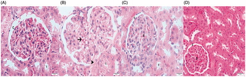
The histological samples obtained for the control group were of the normal glomerulus, the glomerular capillary wall and typical podocyte, and mesangial cells. A typical Bowman’s capsular space is also shown ().
Figures 1. H&E staining. (A) Control group. Light microscopy of a glomerulus demonstrating typically glomerular (g) structures. Bowman’s capsular space (c) × 40. (B) OVX group. Glomerular hypertrophy with capillary dilation (arrow). Atrophic tubules (t). Bowman’s capsule narrowing (arrow head). × 40. (C) Ovx + bortezomib group. Typical glomerulus (g). Bowman’s parietal line cell (arrow head). × 40 (D) OVX +17β-estradiol group. Typical glomerulus (g). Bowman’s parietal line cell (arrow head). × 40.
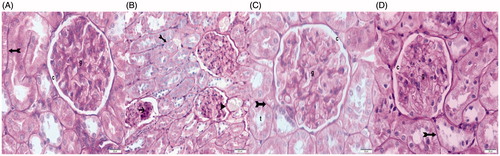
Figures 2. PAS staining. (A) Control group. Bowman’s capsule (g). Bowman’s capsular space (c). × 40. (B) OVX group. Thickening of Bowman’s parietal cell basement membrane with atypical parietal cell (arrow head). Focal glomerulosclerosis (arrow). İrregular tubular basement membrane (tailed arrow). × 20. (C) Ovx + bortezomib group. Regular Bowman’s capsule (g). Bowman’s capsular space (c). × 40. (D) OVX +17β-estradiol group. Typical Bowman’s capsule (g). Bowman’s capsular space (c). Regular tubular basement membrane (tailed arrow). × 40.
The Ovx group histological samples demonstrated hypertrophy and focal glomerulosclerosis, narrowing of Bowman’s capsular space, and glomerular capillary basement membrane thickening, with hemorrhage shown in the kidney medulla. Additionally, atrophy is shown in the proximal and distal tubules. Light microscopy samples show dense mesangial cells. The basement membrane is slightly thickened, with mild mesangial proliferation ().
For the Ovx + bortezomib group, some thickening is shown in the glomerular capillary wall, with typical podocytes and mesangial cells. Bowman’s capsule is typical; some capillary expansion also manifests. With respect to the proximal and distal tubule basal membrane structure, the histological samples were typical ().
The Ovx +17β-estradiol group histological samples demonstrated normal thickening of the glomerular capillary wall with typical podocytes and mesangial cells, along with typical proximal and distal tubule basal membrane structure ().
IHC results
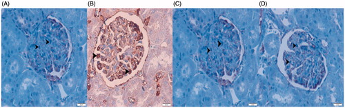
NF-kB/p65 is more or less extensively expressed in immunostaining in nuclear and/or cytoplasmic staining. The IHC studies showed Ovx group samples with densely stained mesangial cells, tubular cell NF-kB/p65, and caspase-3 ().
Figures 3. NF-kB/p65 IHC staining. Imunolocalization of NF-kB/p65 (B) and caspas-3 (F) positive mesangial cells, examined under light microscopy, from renal cortex. (A) Control group. (B) OVX group. (C) Ovx + bortezomib group. (D) OVX +17β-estradiol group. Original magnification × 40.
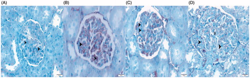
Figures 4. Caspas-3 IHC staining. Imunolocalization of caspas-3 (F) positive mesangial cells, examined under light microscopy, from renal cortex. (A) Control group. (B) OVX group. (C) Ovx + bortezomib group. (D) OVX +17β-estradiol group. Original magnification × 40.
Electron microscopy results
The micrographs from electron microscopy showed typical Bowman’s capsules in the control group, along with normal glomerular basement membrane ultrastructure. Podocyte foot processes also appear clearly ().
Figures 5. Tranmission electron microscopic micrographs. Uranyl acetate and Reynolds lead citrate stain. (A) Control group. Electron micrographs showing typical glomerular capillary with normal basal lamina (arrow). Normal podocytes with foot process. ×3000. Erythrocytes (e) × 3000. (B) OVX group. Electron micrographs showing glomerular basement membrane thickening. Capillary basement membrane lamina densa, lamina rara interna and externa disappear (asterisk). Reduction in podocyte foot process and foot process infolding (arrow head). ×6000. (C) Ovx + bortezomib group. Podocytes (P). Normal podocytes foot process (arrow head). Typical glomerular capillar basement membrane (arrow). × 5000. (D) OVX +17β-estradiol group. Normal podocytes foot process (arrow head). Typical glomerular capillar basement membrane (arrow). ×10,000. Erythrocytes (e). Capillar lumen (c). Bowman’s capsular space (b).
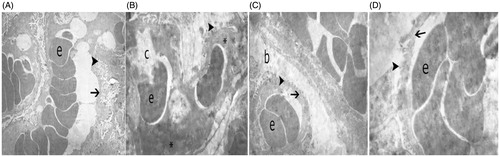
For the Ovx group, micrographs showed basement membrane thickening; not visible are the basement membrane lamina densa, lamina rara interna, and externa. Podocyte foot processes and foot process infolding are reduced ().
In the histological samples for the bortezomib group, the micrographs show some thickening of the glomerular capillary wall, with typical podocytes and mesangial cells. Some capillary expansion is shown along with typical podocyte foot processes (). The histological samples for the 17β-estradiol group demonstrate normal thickening of the glomerular capillary wall and typical podocyte ultrastructure ().
Statistical analysis
In the Ovx group, the results revealed greater immunostaining for NF-kB/p65 and caspase-3 in the renal cortex compared to the control group (p < 0.05). In the Ovx +17β-estradiol group, NF-kB/p65 and caspase-3 expression was significantly decreased compared to the Ovx group (p < 0.05). The location and amount of immunostained NF-kB/p65 and caspase-3 positive cells were similar between the 17ß-estradiol and bortezomib groups (p > 0.05) ().
Table 1. NK-κB ve caspase-3 positive cell numerical density (mm2).
Discussion
The recent research shows that Ovx (leading to E2 deficiency) results in hypertension and associated cardiovascular diseases due to the loss of the cardioprotective effect of 17ß-estradiol.Citation20 Hypertension occurs due to the increase in reabsorption of sodium through the proximal and distal tubules, as the reduction in 17ß-estradiol leads to activation between the nuclei in the hypothalamus, central nervous system, and in the renin–angiotensin system (RAAS). Hypertension causes expression of endothelin-1, which has a significant effect on kidney pathophysiology. Endothelin-1 has a vasoconstrictive effect and also causes inflammation and oxidative stress. Overexpression of the endothelin-1 gene leads to acute renal failure, followed by mesangial cell proliferation and inflammation.Citation3,Citation4,Citation21–25
It has also been reported in the literature that oxidative stress caused by E2 deficiency can reduce the activity of superoxide dismutase (SOD), catalase (CAT), and glutathione (GSH) enzymes, which cleanse free oxygen radicals in cells.Citation3,Citation21,Citation23–26 Reduction of these radical cleaning enzymes causes the accumulation of free radicals in cells (ROS). Recent studies have demonstrated that ovariectomized rats may strengthen the antioxidant defense system by reducing lipid peroxidation, and therefore they may play a role in preventing renal disorders.Citation3,Citation23
NF-kB/p65 is activated by free oxygen radicals, which can be released upon the release of interleukins as a result of inflammation. NF-kB/p65 has an important role in inflammation, cell proliferation, and apoptosis and also plays a key role in renal damage. Some studies have shown that tubular cells (especially mesangial cells) can cause NF-kB/p65 activation by infiltration of leukocytes in kidneys. Angiotensin I and II also activate NF-kB/p65.Citation10,Citation11,Citation14
This study is in accord with other studies in demonstrating that expression of NF-kB/p65 increases with changes in mesangial and tubular cells (). In the Ovx +17β-estradiol group, the expression of NF-kB/p65 decreased compared to Ovx group. Our thought is that 17β-estradiol inhibits the formation of free oxygen radicals by regulating the RAAS system. The decrease of NF-kB/p65 expression supports our idea that estrogen reduces visceral fat accumulation caused by the RAAS system or the lack of 17β-estradiol. In the Ovx + bortezomib group, expression of NF-kB/p65 decreased compared to the Ovx group. Our thought is that bortezomib ubiquitin (IkB), which is a proteasome inhibitor, prevents NF-kB/p65 expression by suppression, a finding supported by the study of Roberti et al. ().Citation13
Menopause has been linked with elevated oxidative stress.Citation1 Caspase-3 inhibits ROS and is required for efficient apoptosis. To focus on caspase-3, our kidney samples were stained with caspase-3 immunostaining. Recent studies show that caspase-9 can prevent cytochrome c release and (indirectly) the formation of ROS.Citation27 Currently available research has made clear that the triggering of caspase-3 and caspase-7 by caspase-9 is an irreversible step that leads to caspase-3 and caspase-7 apoptosis. Cell death is more efficient in the presence of caspase-3, which is the primary executioner of apoptotic death. In accordance with other studies, our research shows that (especially in mesangial and tubular cells) expression of caspase-3 increased meaningfully in the Ovx group. In the Ovx +17β-estradiol group and Ovx + bortezomib group, the expression of caspase-3 decreased compared to Ovx group () ().
Estrogens exert ROS-scavenging chain-breaking antioxidant activity as hydrogen donors from their phenol-hydroxyl ring. Estrogens can induce antioxidant enzyme expression by stimulating the antioxidant defense system. Estrogens inhibiting the formation of lipid peroxides in plasma and liver tissues in vitro.Citation3,Citation12,Citation17,Citation24,Citation28–32
Bortezomib is a proteasome inhibitor and, as such, is part of a therapeutic class that is being studied for potential use in kidney diseases and especially for treatment in cases of kidney transplantation. However, in research, it has been shown that especially high doses can cause neurotoxicity and nonselective toxicity. Due to its success in treatment and its nonselective toxicity at lower doses, a great number of phase II studies are being developed. Protocols are also being developed for a comparison between the side effects and benefits of proteasome inhibitors during use for early treatment of renal disease or of CKD (such as permanent proteinuria and glomerular diseases) without causing damage. Additionally, proteasomes may have utility in addressing the antibodies that cause rejection of transplanted kidneys. Research has shown that bortezomib can help prevent the occurrence of renal fibrosis mediated by ubiquitin proteasomes. In our research, symptoms of renal fibrosis were not found in the Ovx + bortezomib group.Citation10
Research has revealed that the effect of E2 deficiency on kidney tissue is widespread and includes infiltration of tubulointerstitial inflammatory cells, tubular atrophy and dilation, glomerulosclerosis, adhesion of Bowman’s capsule, and glomerular hypertrophy with tubulointerstitial sclerosis.Citation25,Citation33
In our research, thickening of glomerular capillary walls, narrowing of the Bowman’s capsular space, glomerular hypertrophy, atrophic tubules, and loss of the basement membrane of tubules were observed. Mesangial cell proliferation is particularly observed in glomerular cells. Glomerular hypertrophy and narrowing of Bowman’s capsular space are consistent given the assumption that obesity causes hypertension ( and ). Along these lines, we believe that obesity causes mesangial cell proliferation in the glomerular area and tubulointerstitial inflammation by causing inflammation through activation of NF-kB/p65. A dense presence of NF-kB/p65 receptors was associated with losses in tubular cells ().
Transmission electron microscopy provided less information related to Ovx-related E2 deficiency. Verlander et al. observed losses in the apical parts of tubular cells.Citation34 We believe the thickening of capillary wall is caused by the accumulation of cells from activation of the immune system in the subendothelial area, and that the increase of inflammation from NF-kB/p65 causes this activation. A significant decrease in podocyte foot processes was also observed. The mechanism of renal damage from this remains to be defined ().
Conclusions
Our results support the concept that 17β-estradiol and bortezomib have a protective effect against renal failure related to Ovx. The 17β-estradiol protects the kidney tissue by NF-kB/p65 expression, whereas bortezomib (mediated with ubiquitin [IkB]) protects kidney tissue by repressing NF-kB/p65 expression
Disclosure statement
The authors report no conflicts of interest. The authors alone are responsible for the content and writing of the paper.
References
- Selli J, Unal D, Mercantepe F, et al. Protective effects of beta glucan in brain tissues of post-menopausal rats: A histochemical and ultra-structural study. Gynecol Endocrinol. 2015;32:1–6.
- Xu S, Lu A, Wang A. [Effect of kidney deficiency caused by ovariectomy on serum osteocalcin level and tumor necrosis factor in mice with collagen induced arthritis]. Zhongguo Zhong Xi Yi Jie He Za Zhi. 1999;19:34–36.
- Ulas M, Cay M. 17β-Estradiol and vitamin E modulates oxidative stress-induced kidney toxicity in diabetic ovariectomized rat. Biol Trace Elem Res. 2011;144:821–831.
- Nissen I, Estrada FS, Nava-Kopp AT, et al. Prolame ameliorates anxiety and spatial learning and memory impairment induced by ovariectomy in rats. Physiol Behav. 2012;106:278–284.
- Shelton KA, Cline JM, Cann JA. 17-β Estradiol reduces atherosclerosis without exacerbating lupus in ovariectomized systemic lupus erythematosus-susceptible LDLr(−/−) mice. Atherosclerosis. 2013;227:228–235.
- Bosse R, Di paolo T. Dopamine and GABAA receptor imbalance after ovariectomy in rats: Model of menopause. J Psychiatr Neurosci. 1995;20:364–371.
- Sayakhot P, Vincent A, Deeks A, Teede H. Potential adverse impact of ovariectomy on physical and psychological function of younger women with breast cancer. Menopause. 2011;18:786–793.
- Albert DJ, Petrovic DM, Walsh ML. Female rats in a competitive situation: Medial hypothalamic lesions increase and ovariectomy decreases success and aggression. Physiol Behav. 1989;46:379–386.
- Shi H, Clegg DJ. Sex differences in the regulation of body weight. Physiol Behav. 2009;97:199–204.
- Kalkan Y, Kapakin KA, Kara A, et al. Protective effect of Panax ginseng against serum biochemical changes and apoptosis in kidney of rats treated with gentamicin sulphate. J Mol Histol. 2012;43:603–613.
- Unal D, Halici Z, Altunkaynak Z, Keles ON, Oral E, Unal B. A new hypothesis about neuronal degeneration appeared after a rat model of menopause. Neurodegener Dis. 2012;9:25–30.
- McGaughy J, Sarter M. Effects of ovariectomy, 192 IgG-saporin-induced cortical cholinergic deafferentation, and administration of estradiol on sustained attention performance in rats. Behav Neurosci. 1999;113:1216–1232.
- Roberti I, Vyas S. Successful treatment of severe acute antibody-mediated rejection of renal allografts with bortezomib – A report of two pediatric cases. Pediatr Transplant. 2015;19:E189–192.
- Sato T, Teramoto T, Tanaka K, Ohnishi Y, Irifune M, Nishikawa T. Effects of ovariectomy and calcium deficiency on learning and memory of eight-arm radial maze in middle-aged female rats. Behav Brain Res. 2003;142:207–216.
- Coppo R. Proteasome inhibitors in progressive renal diseases. Nephrol Dial Transplant. 2014;29:i25–i30.
- Cao MN, Zhou YB, Gao AH, et al. Curcusone D, a novel ubiquitin-proteasome pathway inhibitor via ROS-induced DUB inhibition, is synergistic with bortezomib against multiple myeloma cell growth. Biochim Biophys Acta. 2014;1840:2004–2013.
- Naves Diaz M, Rodriguez Rodriguez A, Fernandez Martin JL, Serrano Arias M, Menendez Rodriguez P, Cannata Andia JB. Effects of estradiol, calcitriol and both treatments combined on bone histomorphometry in rats with chronic kidney disease and ovariectomy. Bone. 2007;41:614–619.
- Yap FC, Taylor MS, Lin MT. Ovariectomy-induced reductions in endothelial SK3 channel activity and endothelium-dependent vasorelaxation in murine mesenteric arteries. PLoS One. 2014;9:e104686.
- Kharode YP, Sharp MC, Bodine PV. Utility of the ovariectomized rat as a model for human osteoporosis in drug discovery. Methods Mol Biol. 2008;455:111–124.
- Shively CA, Kaplan JR, Adams MR. Effects of ovariectomy, social instability and social status on female Macaca fascicularis social behavior. Physiol Behav. 1986;36:1147–1153.
- Zahedi A, Nematbakhsh M, Moeini M, Talebi A. Role of endothelin receptor antagonist; bosentan in cisplatin-induced nephrotoxicity in ovariectomized estradiol treated rats. J Nephropathol. 2015;4:134–140.
- Adams MR, Clarkson TB, Kaplan JR, Koritnik DR. Ovariectomy, social stress and coronary-artery atherosclerosis in cynomolgus monkeys. Circulation. 1983;68:189–189.
- Abrass CK. Overview: Obesity: What does it have to do with kidney disease? J Am Soc Nephrol. 2004;15:2768–2772.
- Enli Y, Oztekin O, Pinarbasili RD. The nitroxide tempol has similar antioxidant effects as physiological levels of 17beta-oestradiol in reversing ovariectomy-induced oxidative stress in mice liver and kidney. Scand J Clin Lab Invest. 2009;69:526–534.
- Tang J, Yan H, Zhuang S. Inflammation and oxidative stress in obesity-related glomerulopathy. Int J Nephrol. 2012;2012:608397.
- de Chaves G, Moretti M, Castro AA, et al. Effects of long-term ovariectomy on anxiety and behavioral despair in rats. Physiol Behav. 2009;97:420–425.
- Brentnall M, Rodriguez-Menocal L, De Guevara RL, Cepero E, Boise LH. Caspase-9, caspase-3 and caspase-7 have distinct roles during intrinsic apoptosis. BMC Cell Biol. 2013;14:32.
- Hrupka BJ, Smith GP, Geary N. Ovariectomy and estradiol affect postingestive controls of sucrose licking. Physiol Behav. 1997;61:243–247.
- Debold JF, Miczek KA. Aggression persists after ovariectomy in female rats. Horm Behav. 1984;18:177–190.
- Takahashi LK, Lisk RD. Organization and expression of agonistic and socio sexual behavior in golden hamsters over the estrous cycle and after ovariectomy. Physiol Behav. 1983;31:477–482.
- Pezeshki Z, Nematbakhsh M, Mazaheri S, et al. Estrogen abolishes protective effect of erythropoietin against cisplatin-induced nephrotoxicity in ovariectomized rats. ISRN Oncol. 2012;2012:890310.
- Xu Y, Ma XP, Ding J, et al. Treatment with qibaomeiran, a kidney-invigorating Chinese herbal formula, antagonizes estrogen decline in ovariectomized rats. Rejuvenation Res. 2014;17:372–381.
- Shively C, Kaplan JR, Adams MR. Effects of ovariectomy, social instability and social-status on female Macaca fascicularis social-behavior. Am J Primatol. 1985;8:366–366.
- Felicio LS, Nelson JF, Gosden RG, Finch CE. Restoration of ovulatory cycles by young ovarian grafts in aging mice – Potentiation by long-term ovariectomy decreases with age. Gerontologist. 1983;23:177–177.