
Abstract
Objective
The purpose of this study was to determine the effect of tripterygium glycosides (TGs) on regulating abnormal lipid deposition in nephrotic syndrome (NS) rats.
Methods
Sprague-Dawley (SD) rats were injected with 6 mg/kg doxorubicin to construct nephrotic syndrome models (n = 6 per group), and then administered with TGs (10 mg/kg·d−1), prednisone (6.3 mg/kg·d−1), or pure water for 5 weeks. Biomedical indexes, such as urine protein/creatinine ratio (PCR), blood urea nitrogen (BUN), serum creatinine (Scr), serum albumin (SA), triglycerides (TG), total cholesterol (TC)were investigated to evaluate the renal injury of rats. H&E staining experiment was used to assess the pathological alterations. Oil Red O staining was used to assess the level of renal lipid deposition. Malondialdehyde (MDA) and glutathione (GSH) were measured to assess the extent of oxidative damage to the kidney. TUNEL staining was used to assess the status of apoptosis in the kidney. Western blot analysis was performed to examine the levels of relevant intracellular signaling molecules.
Results
After treatment with TGs, those tested biomedical indexes were significantly improved, and the extent of kidney tissue pathological changes and lipid deposition in the kidney was diminished. Treatment with TGs decreased renal oxidative damage and apoptosis. Regarding the molecular mechanism, TGs significantly increased the protein expression levels of Bcl-2 but decreased the levels of CD36, ADFP, Bax, and Cleaved caspase-3.
Conclusion
TGs alleviates renal injury and lipid deposition induced by doxorubicin, suggesting that it may be a new strategy for reducing renal lipotoxicity in NS.
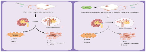
Graphical Abstract
1. Introduction
Lipotoxicity is marked by an unusual excess accumulation of lipids in non-adipose tissues, which causes deleterious effects [Citation1]. The kidney is one of the main target organs for lipotoxicity damage [Citation2]. Renal parenchymal cell lipid deposition causes cellular dysfunction and apoptosis [Citation3]. Doxorubicin-induced nephrosis in rats is a representative animal model of nephrotic syndrome [Citation4]. Doxorubicin induces nephrotoxicity via renal oxidative stress, inflammation, and apoptosis [Citation5]. In addition, doxorubicin disrupts lipid metabolism, causing the development of lipotoxicity [Citation6], which aggravates kidney damage.
TGs is multicomponent extracts from the Chinese herb, Tripterygium wilfordii (thunder god vine), which exhibit excellent immunosuppressive and anti-inflammatory activities, and it shows satisfactory efficacy in nephrotic syndrome [Citation7]. Studies have suggested that TGs plays an important role in regulating lipid metabolism. TGs effectively improves the function of impaired kidneys by promoting triglycerides (TG) catabolism via modulation of adipose triglyceride lipase [Citation8]. The two main active components of TGs, triptolide, and celastrol, could reduce excessive lipid accumulation [Citation9,Citation10]. However, it remains unknown whether TGs have a favorable therapeutic effect on lipotoxicity caused by NS. Thus, the present study performed a series of experiments to determine the effects of TGs on NS-induced lipotoxicity.
2. Methods
2.1. Animals
Eight-week-old SPF adult male Sprague–Dawley rats (200 ± 20 g) were purchased from Guangdong Medical Laboratory Animal Center (Guangzhou, China) and maintained under specific pathogen-free conditions, 20 ± 2 °C temperature, 50 ± 10% humidity, regular 12-h dark/light cycles, and the rats were allowed free access to food and water. After a week of acclimatization, the rats were randomly divided into the following four groups (n = 5–6 per group): control group, model group, TGs group, and prednisone group. All in vivo experiments were performed according to protocols approved by the Animal Care and Use Committee of Guangdong Provincial Hospital of Traditional Chinese Medicine.
2.2. Experimental design
All rats (except the control group) were given 6 mg/kg DOX via a single tail vein injection to construct a nephrotic syndrome model. After 3 weeks, the rats were regrouped according to the PCR and treated as follows: the TGs group was treated with 10 mg/kg·d−1 TGs (i.g.); the prednisone group was treated with 6.3 mg/kg·d−1 prednisone (i.g.); and the model and control groups were given equal amounts of distilled water. The body weight was recorded once a week. Rat urine was collected to detect urinary protein and creatinine on the 5th week of dosing. After 5 weeks of continuous dosing, all rats were euthanized with sodium pentobarbital. Blood was collected, and serum was separated by centrifugation at 3500 × g for 15 min. Kidneys were removed and stored according to the experimental needs.
2.3. Kidney index
All animals were weighed before being euthanized. Kidney tissues were collected and weighed immediately after the rats were sacrificed, and the kidney index was calculated using the following formula: kidney index = kidney weight (g)/body weight (g).
2.4. Serological parameters
Assay kits were used to measure the contents of TG (Cat No. A110-1-1), TC (Cat No. A111-1-1), BUN (Cat No. C013-2-1), SA (Cat No. A028-2-1), and Scr (Cat No. C011-2-1), which were all purchased from Nanjing Jincheng Institute of Biotechnology (Jiangsu, China).
2.5. Histopathological examination
Kidney tissues were immersed in 4% paraformaldehyde for 24 h, embedded in paraffin, and cut into 5-μm thick sections. Hematoxylin and eosin (H&E) staining was performed following the manufacturer’s instructions (Beijing Leagene Biotech Co., Ltd., DH0006). Three different rats’ sections from each group.
2.6. Oil Red O staining
Oil Red O solution was prepared by dissolving 0.5 g of Oil Red O powder (Sigma, O0625) in 100 mL of isopropanol, and three parts of dissolved Oil Red O were mixed with two parts of water and then filtered. Renal tissues were frozen in OTC compound, cut into 7-μm thick sections, and fixed with 4% paraformaldehyde. The sections were rinsed with ddH2O for 5 min, rinsed with 60% isopropanol for 1 min, stained with Oil Red O solution for 30 min, and finally rinsed with 60% isopropanol. Nucleus were counterstained with hematoxylin. The stained sections were observed using an Olympus microscope. Three rats in each group were randomly selected for examination.
2.7. Western blot analysis
The appropriate amount of renal cortex was placed into homogenate tubes with an equal proportion of RIPA lysis buffer (Thermo Fisher) mixed with a protease inhibitor cocktail (Roche) and homogenized using a tissue grinder. Protein lysates were then prepared for western blot analysis. Proteins were isolated by electrophoresis on 12.5% SDS–PAGE gels, transferred onto PVDF membranes, and blocked with 5% non-fat milk for 2 h. The membranes were incubated overnight at 4 °C with the following primary antibodies: Cleaved caspase-3 (CST#9661, Cell Signaling Technologies), Bcl-2 (ab196495, Abcam), Bax (CST#2772, Cell Signaling Technologies), ADFP (ab108323, Abcam), CD36 (ab133625, Abcam), and GAPDH (CST#5174, Cell Signaling Technologies). The membranes were subsequently washed and then incubated for 60 min at room temperature with HRP-conjugated anti-mouse or anti-rabbit secondary antibodies. An ECL reagent kit (Millipore, USA) and gel imaging equipment (Bio-Rad, ChemiDocTM Touch, USA) were used to detect the presence of protein bands on the membrane. The quantification of the band intensities was performed using Image Lab 5.2.1 software (BIO-RAD), and the band intensities were normalized to GAPDH.
2.8. Quantification of GSH and MDA
The homogenates were centrifuged at 4500 rpm for 15 min at 4 °C, and the supernatants were taken to determine the levels of oxidative stress biomarkers, such as GSH (Cat No. A006-2-1) and MDA (Cat No. A003-1-2), using corresponding kits according to the manufacturer’s protocols (Nanjing Jiancheng Bioengineering Institute, Nanjing, China).
2.9. TUNEL and DAPI staining
Frozen kidney tissues were cut into 7-μm thick sections and fixed in 4% paraformaldehyde for 30 min. The sections were then incubated in PBS containing 0.4% Triton X-100 for 5 min followed by incubation in TUNEL solution (Beyotime Biotechnology, Shanghai, China, Cat No. C1090) for 1 h in a humidified chamber, and the sections were counterstained with DAPI (Solarbio, Cat No.C0065). The slides were sealed with an anti-fluorescence quencher and observed under a fluorescence microscope (Olympus, Japan), and images were acquired. Three experimental animals in each group were randomly selected for this experiment.
2.10. Statistical analysis
Graphing was performed with GraphPad Prism 9 and statistical analyses were performed using IBM SPSS 25 for Windows. Multiple group comparisons were performed using one-way ANOVA, and Tukey or Dunnett T3 methods were used for post-hoc analysis. Data are expressed as the mean ± SD, and p-values ≤ 0.05 indicated statistically significant differences.
3. Results
3.1. TGs protect kidneys against doxorubicin-mediated injury
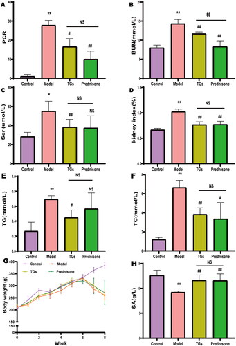
Rats were injected with DOX after 7–8 weeks, and PCR, BUN, Scr, kidney index, TG, and TC were significantly increased (), but significantly decreased body weight and SA (). These results demonstrated that the models were successful. In addition, TGs and prednisone ameliorated Dox-mediated renal injury, and the treatment effect of TGs was comparable to that of prednisone. However, TGs and prednisone did not reverse the body weight loss in rats. Compared to TGs, prednisone showed better effectiveness in reducing PCR and BUN.
Figure 1. TGs protect kidneys against doxorubicin-mediated injury. (A) 24 h Urine protein-to-creatinine ratio (PCR). (B) Blood urea nitrogen. (C) Serum creatinine concentration. (D) Kidney index. (E) Serum TG levels in rats (n = 5–6). (F) Serum TC levels in rats (n = 5–6). (G) Body weight (n= 5–6 per group). (H) Serum albumin (n= 5–6 per group). Significance between groups was determined by ANOVA followed by Dunnett’s T3. Model group vs. Control group (*p ≤ 0.05 and **p ≤ 0.01); TGs group and Prednisone group vs. Model group (#p ≤ 0.05, ##p ≤ 0.01, and NS p > 0.05); TGs group vs. Prednisone group ($$p ≤ 0.01 and NS p > 0.05).
3.2. TGs protect against alterations in the kidney architecture of NS
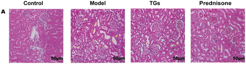
The histopathological changes in the kidney tissues of rats in each group were observed by HE staining. The results showed that severe renal tubular damage occurred after DOX injection, including renal tubular dilation with granular degeneration and tubule brush border shedding as well as vacuolar degeneration of renal tubular epithelial cells, protein casts, and inflammatory cell infiltration (). The therapeutic outcome in the TG group was equivalent to that in the prednisone group.
Figure 2. TGs protect against alterations in the kidney architecture of NS. HE staining (n = 3; scale bar: 50 µm). Yellow arrows indicate brush border detachment and absence. Blue arrows indicate protein casts. Yellow triangles indicate focal inflammatory cell infiltration.
3.3. TGs alleviate abnormal lipids in NS
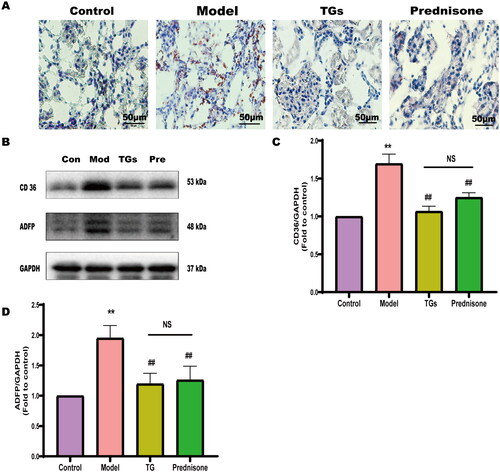
To analyze the effect of TGs on lipid deposition in kidney tissue, Oil Red O staining was performed. Lipid droplets were not observed in normal rats. However, large orange-red lipid droplets were observed in the model group, and the orange-red droplets were mainly concentrated in renal tubules (). In addition, TGs reduced lipid deposition in the kidney tissue doxorubicin-induced NS rats. WB analysis also confirmed that TGs significantly decreased the expression of the CD36 and ADFP (two lipid-related proteins) (). Thus, these findings indicated that the therapeutic outcome of TG was better than that of prednisone.
Figure 3. TGs alleviate abnormal lipids in NS. (A) Oil Red O and hematoxylin staining of kidney sections (n = 3; scale bar: 50 µm). (B–D) Expression of CD36 and ADFP in renal tissues (n = 3). Significance between groups was determined by ANOVA followed by Tukey. Model group vs. Control group (**p ≤ 0.01); TGs group or Prednisone group vs. Model group (#p ≤ 0.05 and ##p ≤ 0.01); TGs group vs. Prednisone group (NS p > 0.05).
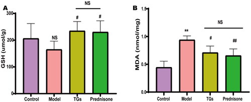
3.4. TGs inhibit renal oxidative stress in NS
Abnormal lipid metabolism induces lipid peroxidation, leading to oxidative stress. To evaluate whether TGs improve lipid-related oxidative stress, we quantified the amounts of GSH and MDA in rat kidney tissues. The results showed that DOX reduced GSH content (but not significantly) and increased MDA content in the kidneys. Treatment with TGs or prednisone restored the content of GSH and reduced the DOX-mediated increase in MDA (). These findings indicated that TGs or prednisone significantly inhibit lipid peroxidation and restore antioxidant capacity to a certain extent in the kidneys of NS model rats. The TG therapeutic outcome was equivalent to that in the prednisone group.
Figure 4. TGs inhibit renal oxidative stress in NS. (A) Levels of GSH in renal tissue homogenate. (B) Levels of MDA in renal tissue homogenate. (n = 5–6). Significance between groups was determined by ANOVA followed by Dunnett’s T3. Model group vs. Control group (**p ≤ 0.01, and NS p > 0.05); TGs group or Prednisone group vs. Model group (#p ≤ 0.05 and ##p ≤ 0.01); TGs group vs. prednisone group (NS p > 0.05).
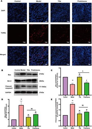
3.5. TGs inhibit renal apoptosis of NS
To determine the apoptosis levels of the kidneys, TUNEL assays and WB analyses were performed. TUNEL staining showed a significantly higher level of cell death in the model group than in the control group, TGs group and prednisone group significantly decreased the number of TUNEL-positive cells (). WB analysis indicated that the TGs and prednisone groups exhibited reduced levels of Bax and active Caspase-3 but increased levels of the Bcl-2 antiapoptotic protein compared to the model group (). The TGs therapeutic outcome was equivalent to that in the prednisone group.
Figure 5. TGs inhibit renal apoptosis of NS. (A) TUNEL and DAPI staining of kidney sections (n = 3; scale bar: 100 µm). (B–E) Bax, Bcl-2, and Cleaved caspase-3 expression in the kidney (n = 3). Significance between groups was determined by ANOVA followed by Tukey. Model group vs. Control group (**p ≤ 0.01); TGs group or Prednisone group vs. Model group (#p ≤ 0.05 and ##p ≤ 0.01); TGs group vs. prednisone group ($p ≤ 0.05, and NS p > 0.05).
4. Discussion
As early as 1982, the concept of lipid nephrotoxicity attracted much attention [Citation11]. Hyperlipidemia is prevalent in NS and is considered to be a feature of severe NS [Citation12]. Severe impairment of lipid clearance is a leading cause of abnormal lipid metabolism in NS, resulting in a surplus of fatty acids and glyceride converted to TG, which accumulate in the form of intracellular lipid droplets [Citation13]. Non-adipose tissue has a limited capacity to store TGs [Citation14], and excessive lipid deposition causes nephrotoxicity due to cellular dysfunction as well as accelerates energy metabolism, induces oxidative damage, and leads to cell death [Citation15,Citation16]. Therefore, reducing abnormal lipid deposition in the kidney mitigates kidney damage and slows the progression of NS. In the present study, DOX injection significantly increased the levels of PCR, BUN, Scr, TG, and TC but significantly decreased the levels of SA in rats compared to the model group, which indicated successful model construction. Treatment with TGs significantly reduced kidney damage in the NS model rats.
Oil Red O is a fat-soluble dye with can specifically bind to TG in tissues or cells to dye fat cells red [Citation17]. As a scavenger receptor, CD36 is a key element in fatty acid uptake [Citation18]. Overexpression of CD36 increases fatty acid uptake and directs abnormal lipid deposition and excessive oxidative stress [Citation19,Citation20]. CD36 is significantly upregulated in kidney disease and can reflect the severity of tissue injury in kidney disease to some extent [Citation21]. Adipose differentiation-related protein (ADFP) is an important lipid droplet surface protein that is mainly involved in cellular fatty acid intake, lipid droplet formation, and lipid stores [Citation22]. ADFP plays a role in preventing lipase entry into lipid droplets and slowing lipid digestion, allowing lipids to accumulate in lipid droplets [Citation23]. Without ADFP, lipid droplets are degraded by the proteasome; thus, ADFP is an indicator of lipid accumulation [Citation24,Citation25].
In the present study, Oil Red O staining and western blot analysis indicated an accumulation of lipid droplets in the kidneys of model rats, but the amount of lipid droplets was significantly reduced after treatment with TGs. Moreover, the expression of CD36 and ADFP was also downregulated in the TGs group. These results demonstrated that TGs reduce abnormal lipid deposition in the kidneys of NS rats, thus improving renal injury.
Aberrant accumulation of lipids results in induced cellular oxidative stress [Citation26]. As a representative product of lipid peroxidation, MDA directly reflects the extent of lipid peroxidation damage [Citation27]. Because GSH is an important endogenous antioxidant in the body that scavenges oxidative free radicals and prevents a variety of diseases, it reflects the ability of the tissue to resist oxidative damage [Citation28]. In the present study, the MDA content was significantly higher and the GSH level was significantly lower in the model group compared to the control group, which indicated that the kidneys of the model rats were in a state of lipid peroxidation. Moreover, treatment with TGs reduced the MDA content and increased the GSH level, thereby recovering the oxidative-antioxidative balance and reducing the cellular damage caused by lipid peroxidation.
Excessive lipid deposition may exceed the repair capacity of cells and lead to apoptosis [Citation29]. Doxorubicin has a strong cytotoxic effect and induces cell apoptosis. Combining lipid deposition and doxorubicin may exacerbate injury at the same time. Bax is an important proapoptotic protein in the Bcl-2 family [Citation30], and it is predominantly present in an inactive conformation, maintaining organismal stability in part through interaction with antiapoptotic Bcl-2 proteins; thus, Bax and Bcl-2 play a key role in cell death and survival [Citation31]. Caspase 3 is a key member of the caspase family, and activation of Caspase 3 is an indispensable step in mitochondria-dependent apoptosis [Citation32]. Its spliceosome, Cleaved caspase-3 is a key downstream factor in the apoptotic cascade and functions as an important executor of apoptosis [Citation33]. In the present study, TGs prevented apoptosis in NS rats by decreasing the expression of Bax and Cleaved caspase-3 as well as restoring the expression of Bcl-2.
In summary, the present findings suggested that TGs treatment improves renal injury induced by doxorubicin by reducing renal lipid deposition, inhibiting renal lipid peroxidation, and mitigating cell apoptosis. However, the precise mechanism and signaling pathway through which TGs improve renal lipid deposition require additional detailed studies.
Ethical approval
The animal protocol was reviewed and approved by the Experimental Animal Ethics Committee of Guangdong Provincial Hospital of Traditional Chinese Medicine, approval number (2020004).
Author contributions
Bo Liu, Aihua Wu, and Peng Xu conceived and designed the experiments. Bidan Zheng, Dongfang Lu, Xiuping Chen, Yinghua Yin, and Weiying Chen performed the experiments. Bidan Zheng, Huanmei Lin, and Dongfang Lu analyzed and interpreted the data. Bidan Zheng and Dongfang Lu wrote the manuscript. Bo Liu and Aihua Wu critically revised the manuscript. Bo Liu, Aihua Wu, Peng Xu, and Xiaowan Wang supervised the findings of the work and approved the manuscript for submission. All authors agreed with the final version of this manuscript.
Supplemental Material
Download PDF (390.8 KB)Disclosure statement
The graphical abstract was created by Figdraw. The authors declare no conflicts of interest.
Data availability statement
All data generated during the study can be obtained upon reasonable request from the corresponding author.
Additional information
Funding
References
- Engin AB. What is lipotoxicity? Adv Exp Med Biol. 2017;960:197–220.
- Nishi H, Higashihara T, Inagi R. Lipotoxicity in kidney, heart, and skeletal muscle dysfunction. Nutrients. 2019;11(7):1664.
- Li S, Nagothu K, Ranganathan G, et al. Reduced kidney lipoprotein lipase and renal tubule triglyceride accumulation in cisplatin-mediated acute kidney injury. Am J Physiol Renal Physiol. 2012;303(3):F437–F448.
- Zhang X, Cheng Y, Zhou Q, et al. The effect of Chinese traditional medicine Huaiqihuang (HQH) on the protection of nephropathy. Oxid Med Cell Longev. 2020;2020:2153912.
- Ibrahim KM, Mantawy EM, Elanany MM, et al. Protection from doxorubicin-induced nephrotoxicity by clindamycin: novel antioxidant, anti-inflammatory and anti-apoptotic roles. Naunyn-Schmiedebergs Arch Pharmacol. 2020;393(4):739–748.
- Schley G, Grampp S, Goppelt-Struebe M. Inhibition of oxygen-sensing prolyl hydroxylases increases lipid accumulation in human primary tubular epithelial cells without inducing ER stress. Cell Tissue Res. 2020;381(1):125–140.
- Liu S, Li X, Li H, et al. Comparison of Tripterygium wilfordii multiglycosides and tacrolimus in the treatment of idiopathic membranous nephropathy: a prospective cohort study. BMC Nephrol. 2015;16(1):200.
- Zhang J, Li SL, Lin W, et al. Tripterygium glycoside tablet attenuates renal function impairment in diabetic nephropathy mice by regulating triglyceride metabolism. J Pharm Biomed Anal. 2022;221:115028.
- Zhang CJ, Zhu N, Long J, et al. Celastrol induces lipophagy via the LXRα/ABCA1 pathway in clear cell renal cell carcinoma. Acta Pharmacol Sin. 2021;42(9):1472–1485.
- Huang RS, Guo F, Li YP, et al. Activation of AMPK by triptolide alleviates nonalcoholic fatty liver disease by improving hepatic lipid metabolism, inflammation and fibrosis. Phytomedicine. 2021;92:153739.
- Moorhead JF, Chan MK, El-Nahas M, et al. Lipid nephrotoxicity in chronic progressive glomerular and tubulo-interstitial disease. Lancet. 1982;2(8311):1309–1311.
- Agrawal S, Zaritsky JJ, Fornoni A, et al. Dyslipidaemia in nephrotic syndrome: mechanisms and treatment. Nat Rev Nephrol. 2018;14(1):57–70.
- Lee RG, Fu W, Graham MJ, et al. Comparison of the pharmacological profiles of murine antisense oligonucleotides targeting apolipoprotein B and microsomal triglyceride transfer protein. J Lipid Res. 2013;54(3):602–614.
- Thombare K, Ntika S, Wang X, et al. Long chain saturated and unsaturated fatty acids exert opposing effects on viability and function of GLP-1-producing cells: mechanisms of lipotoxicity. PLOS One. 2017;12(5):e0177605.
- Brennan E, Kantharidis P, Cooper ME, et al. Pro-resolving lipid mediators: regulators of inflammation, metabolism and kidney function. Nat Rev Nephrol. 2021;17(11):725–739.
- Zhang X, Li ZL, Woollard JR, et al. Obesity-metabolic derangement preserves hemodynamics but promotes intrarenal adiposity and macrophage infiltration in swine renovascular disease. Am J Physiol Renal Physiol. 2013;305(3):F265–F276.
- Laudette M, Sainte-Marie Y, Cousin G, et al. Cyclic AMP-binding protein Epac1 acts as a metabolic sensor to promote cardiomyocyte lipotoxicity. Cell Death Dis. 2021;12(9):824.
- Yang X, Wu Y, Li Q, et al. CD36 promotes podocyte apoptosis by activating the pyrin domain-containing-3 (NLRP3) inflammasome in primary nephrotic syndrome. Med Sci Monit. 2018;24:6832–6839.
- Chorner Z, Barbeau PA, Castellani L, et al. Dietary α-linolenic acid supplementation alters skeletal muscle plasma membrane lipid composition, sarcolemmal FAT/CD36 abundance, and palmitate transport rates. Am J Physiol Regul Integr Comp Physiol. 2016;311(6):R1234–R1242.
- Chen Y, Yan Q, Lv M, et al. Involvement of FATP2-mediated tubular lipid metabolic reprogramming in renal fibrogenesis. Cell Death Dis. 2020;11(11):994.
- Yang X, Okamura DM, Lu X, et al. CD36 in chronic kidney disease: novel insights and therapeutic opportunities. Nat Rev Nephrol. 2017;13(12):769–781.
- Hara Y, Goda K, Hirooka S, et al. Association between endoscopic milk-white mucosa, epithelial intracellular lipid droplets, and histological grade of superficial non-ampullary duodenal epithelial tumors. Diagnostics. 2021;11(5):769.
- Meng X, Sun R, Wang W, et al. ADFP promotes cell proliferation in lung adenocarcinoma via Akt phosphorylation. J Cell Mol Med. 2021;25(2):827–839.
- Mak KM, Ren C, Ponomarenko A, et al. Adipose differentiation-related protein is a reliable lipid droplet marker in alcoholic fatty liver of rats. Alcoholism Clin Exp Res. 2008;32(4):683–689.
- Tang W, Ma W, Ding H, et al. Adenylyl cyclase 1 as a major isoform to generate cAMP signaling for apoA-1-mediated cholesterol efflux pathway. J Lipid Res. 2018;59(4):635–645.
- Jin M, Feng H, Wang Y, et al. Gentiopicroside ameliorates oxidative stress and lipid accumulation through nuclear factor erythroid 2-Related factor 2 activation. Oxid Med Cell Longev. 2020;2020:2940746.
- Gong T, Jiang W, Gao Z, et al. Dibromoacetic acid induced hepatotoxicity in mice through oxidative stress and Toll-Like receptor 4 signaling pathway activation. Oxid Med Cell Longev. 2019;2019:5637235.
- Pompella A, Corti A. Editorial: the changing faces of glutathione, a cellular protagonist. Front Pharmacol. 2015;6:98.
- Ahowesso C, Black PN, Saini N, et al. Chemical inhibition of fatty acid absorption and cellular uptake limits lipotoxic cell death. Biochem Pharmacol. 2015;98(1):167–181.
- Spitz AZ, Gavathiotis E. Physiological and pharmacological modulation of BAX. Trends Pharmacol Sci. 2022;43(3):206–220.
- Zhou JF, Ci XX, Ma X, et al. Pterostilbene activates the Nrf2-dependent antioxidant response to ameliorate arsenic-induced intracellular damage and apoptosis in human keratinocytes. Front Pharmacol. 2019;10:497.
- Han XD, Zhou ZW, Yang W, et al. A computational and functional study elicits the ameliorating effect of the Chinese herbal formula Huo Luo Xiao Ling Dan on experimental ischemia-induced myocardial injury in rats via inhibition of apoptosis. Drug Des Devel Ther. 2015;9:1063–1102.
- Xu Q, Xi H, Chen X, et al. Milk‑derived hexapeptide PGPIPN prevents and attenuates acute alcoholic liver injury in mice by reducing endoplasmic reticulum stress. Int J Mol Med. 2020;46(3):1107–1117.