
Abstract
Background
Circular RNAs (CircRNAs) have been shown to be involved in the development of chronic kidney disease (CKD). This study aimed to investigate the role of Circ1647 in renal fibrosis, which is a hallmark of CKD.
Methods
In this study, we established a unilateral ureteral obstruction (UUO) model and delivered Circ1647 RfxCas13d knockdown plasmid into renal parenchymal cells via retrograde injection through the ureter followed by electroporation. After that, the pathological changes were determined by Hematoxylin and Eosin. Meanwhile, Immunohistochemistry, qRT-PCR and Western blot were conducted to assess the degree of fibrosis. In addition, overexpressing of Circ1647 in renal tubular epithelial cells (TCMK1) was performed to investigate the underlying mechanisms of Circ1647.
Results
Our results displayed that electroporation-mediated knockdown of Circ1647 by RfxCas13d knockdown plasmid significantly inhibited renal fibrosis in UUO mice as evidenced by reduced expression of fibronectin and α-SMA (alpha-smooth muscle actin). Conversely, overexpression of Circ1647 in TCMK1 cells promoted the fibrosis. In terms of mechanism, Circ1647 may mediate the PI3K/AKT Signaling Pathway as demonstrated by the balance of the phosphorylation of PI3K and AKT in vivo and the aggravated phosphorylation of PI3K and AKT in vitro. These observations were corroborated by the effects of the PI3K inhibitor LY294002, which mitigated fibrosis post Circ1647 overexpression.
Conclusion
Our study suggests that Circ1647 plays a significant role in renal fibrosis by mediating the PI3K/AKT signaling pathway. RfxCas13d-mediated inhibition of Circ1647 may serve as a therapeutic target for renal fibrosis in CKD.
1. Introduction
The development of chronic kidney disease (CKD) has become a major global public health problem, which has implications on the global economy. The global prevalence of CKD is estimated to be 13.4% (11.7-15.1%), while in China, it affects approximately 12% of the population [Citation1,Citation2], with limited clinical treatments available. Renal fibrosis (RF) is the key pathological basis of CKD progression to end-stage renal disease (ESRD), characterized by the excessive deposition of extracellular matrix [Citation3]. Therefore, there is an urgent need to explore the potential mechanisms of RF and develop new treatment methods.
With the development of sequencing technology, more and more researchers have begun to pay attention to the regulation of non-coding RNAs on diseases. Circular RNAs (CircRNAs) are a newly discovered class of RNA that distinguish themselves from traditional linear RNAs. Research has found that altered expression of CircRNAs is closely related to the onset of human diseases. Recent evidence suggests that CircRNAs can function as vital diagnostic or prognostic biomarkers and play an important role in the pathogenesis of acute kidney diseases [Citation4–6], kidney cancer [Citation7], kidney allograft [Citation8] and RF [Citation9]. Recent studies have demonstrated that CircRNAs exhibit diverse cellular functions, including acting as molecular sponges to interfere with microRNAs, binding to proteins to mediate protein-protein interactions, acting as scaffolds or regulators of proteins, and serving as templates for translation [Citation9]. For example, CircRNA_30032 promotes the progression of renal fibrosis in UUO models by acting as a miRNA-96-5p sponge and downregulating HBEGF expression [Citation10]. Conversely, CircRNA_37492 has the potential to attenuate renal fibrosis [Citation11].
The back-splicing (BS) of pre-mRNA exons results in the genome-wide expression of CircRNAs that share identical sequence to linear mRNA, except for the presence of unique junction sites resulting from BS [Citation12]. Despite their high abundance, the functions of CircRNAs remain largely unknown due to the lack of reliable tools to specifically target and interfere with their expression, without affecting linear RNA levels in cells. ShRNAs or specific small interfering RNAs (siRNAs) can be utilized to target BSJ sites of CircRNAs and effectively knock them down. However, it should be noted that even partial complimentarity of a half RNAi (∼10 nt) to linear mRNAs may result in altered parental gene expression. In addition, the sequence design freedom for targeting BSJ sites of CircRNAs is relatively limited, which may hinder the design of highly efficient knockdown sequences. Therefore, it is necessary to explore novel approaches for selectively knocking down CircRNAs.
Current research has demonstrated that type VI CRISPR effectors, including Cas13a, Cas13b, and Cas13d RNases, can be guided by CRISPR RNA to selectively cleave single-stranded (ss) RNA targets containing complementary protospacers in mammalian cells [Citation13,Citation14]. Among these, RfxCas13d has been shown to achieve the highest knockdown efficiency (>80%) of both CircRNAs, while linear mRNAs remain largely unaffected [Citation12]. What’s more, Yang et al. [Citation15] has recently developed High-fidelity Cas13 variants for targeted RNA degradation with minimal collateral effects which hold potential for advancing basic research and therapeutic applications.
In this study, we found that high expression of Circ1647 is specific to the UUO-CKD model. To further explore its function, we found that knocking down Circ1647 could significantly alleviate the renal fibrosis in mice and ameliorate kidney damage. It is worth noting that we used the Rfxcas13d method to knock down Circ1647 in our study, without affecting its linear RNA. Mechanistically, knocking down Circ1647 can significantly affect the activation of the PI3K/AKT signaling pathway, which may be its potential regulatory pathway.
2. Materials and methods
2.1. Plasmid construction and cloning
To optimize the efficacy of renal in situ electroporation, we developed a eukaryotic expression plasmid for Rfxcas13d with a fragment size of approximately 6500 bp and solely essential components required for plasmid replication. Initially, Rfxcas13d was amplified from AAV-EFS-NLS-CasRx-U6-DR-sgRNA_LacZ-DR (Cat# 154003, Addgene, USA) and integrated into the PX458 vector (Cat# 172221, Addgene, USA) via seamlessly cloning kit (Cat# C115, Vazyme, China).
In order to construct a highly specific knockdown sequence against Circ1647, we designed sgRNA by selecting 15 bp from each end of the circular RNA. The resulting sgRNA sequence was 5′-GATGCTTGTTGCTCGCATGAAACCAAGGGG-’3. We used quantitative real-time PCR (qRT-PCR) to evaluate the knockdown efficacy of Circ1647. In addition, the overexpression sequence of Circ1647 was custom-synthesized by Genepharma (Shanghai, China) and subsequently cloned onto the PcDNA3.1(+) vector. The above plasmids were purified using the EndoFree Maxi Plasmid Kit (Cat # DP116, Tiangen, China), following the manufacturer’s instructions.
2.2. Establishment of the unilateral ureteral obstruction (UUO) model
Male C57BL/6 mice (8 weeks old, 22-25 g body weight) were obtained from Yaokang Bio-Technique Co. Ltd (Chengdu, China) and maintained under standard conditions with a 12-h light/12-h dark cycle and ad libitum access to food and water. All animal handling and experimental procedures were performed according to the guidelines approved by the Animal Ethics Committee of Southwest Medical University (Ref No. 20220727-001).
The unilateral ureteral obstruction (UUO) model was established in male C57BL/6 mice as described previously [Citation16,Citation17]. Briefly, the mice were anesthetized with 1% pentobarbital, and the left ureter was exposed through a midline incision on the lower back. The ureter was then ligated with 4-0 silk thread at two points. sham-operated mice underwent the same procedure without ligation.
2.3. Gene transfer of Circ1647 knockdown plasmid into the mouse kidney by electroporation
To investigate the pathogenic role and therapeutic potential of Circ1647 in UUO-induced CKD, Circ1647 knockdown plasmids were delivered into the mouse kidney at the time of surgery via electroporation-mediated gene transfer technique, as previously described by Peng [Citation18]. Briefly, under 1% pentobarbital anesthesia, 100 μg of Circ1647 knockdown or control plasmid (dissolved in 100 μl saline) was slowly injected retrogradely through the ureter into the kidney. After a two-minute diffusion period, the kidney was clamped with tweezer-like electrodes, and four electronic pulses of 50 V for 50 ms duration were applied with an interval of 1 s each. The mice were randomly divided into four groups (n = 8 per group): sham, UUO, UUO with control empty vector (UUO + EV), and UUO with Circ1647 knockdown.
2.4. RNA extraction and quantitative real-time PCR (qRT-PCR)
Total RNA was extracted from animal kidney tissue or cells using RNA-easy Isolation Reagent (Cat# R701, Vazyme, China). Complementary DNA (cDNA) was synthesized using a cDNA reverse transcription kit (Cat# R211, Vazyme, China). qRT-PCR was performed using Eastep qPCR Master Mix (Cat# Q711-02, Vazyme, China), and the LightCycler® 480 II Real-time PCR System (Roche, Germany). The primers used for qRT-PCR are listed in , and the sequences of other primers are consistent with those used in previously published articles [Citation17]. The expression level of the target gene was detected with pre-designed primers, and the relative expression level was calculated by the 2^(-ΔCt) method and normalized to GAPDH as an internal reference gene.
Table 1. Specific primers for qRT-PCR.
2.5. Western blot
Total protein was obtained from kidney tissue or TCMK1 cells using lysis buffer (50 mM Tris, pH 7.5, 150 mM NaCl, 1% NP-40, 0.1% SDS) containing protease inhibitors (Cat# P1005, Beyotime, China). The Bicinchoninic Acid Protein (BCA) method was used to measure the protein concentration. Approximately 20-100 μg of total lysate were separated on 10–15% sodium dodecyl sulfate-polyacrylamide gel (SDS-PAGE) and transferred to a polyvinylidene fluoride (PVDF) membrane (Cat# ISEQ00010, Millipore, USA). After blocking with 3% BSA or 5% skim milk, the membranes were incubated overnight at 4 °C with gentle shaking with indicated primary antibodies GAPDH (Cat# AB0037, Abways, USA), KIM1 (Cat# bs-2713R, Bioss, China), α-SMA (Cat# 67735-1-Ig, Proteintech, China), fibronectin (Cat# TA5335M, Abmart, USA), AKT (Cat# C67E7, CST, USA), p-AKT (Cat# 4060, CST, USA). The next day, the membranes were incubated with corresponding HRP-conjugated secondary antibodies and visualized using the ChemiScope 600 EXp system (ClinX, China).
2.6. Histology and immunohistochemistry
Renal injury was assessed in kidney paraffin sections (4 μm) using hematoxylin and eosin (H&E) and Masson’s trichrome staining. In total, 500 cortical tubules were examined, with the incidence of necrosis quantified and expressed as a percentage, consistent with previously established methodologies [Citation19]. Renal fibrosis was evaluated by immunohistochemistry with a microwave-based antigen retrieval technique, as previously described. Endogenous peroxidase was blocked by treatment with 3% H2O2 for 15 min at room temperature. After further blocking with 3% goat serum, the primary antibody was incubated overnight at 4 °C. The next day, slices were washed with PBS and downstream manipulations were performed using the Universal Mouse/Rabbit HRP-conjugated Polymer Detection Kit (Cat# PV-6000, ZSGB-Bio, China) following the manufacturer’s instructions. Diaminobenzidine (DAB) was used for visualization. All slices were observed and photographed using a Virtual Slide Microscope (VS120, Olympus, Japan).
2.7. Detection of blood urea nitrogen and creatinine
After anesthetization, the blood was collected from the abdominal cavity of the mice and transferred to Eppendorf tubes. The tubes were then centrifuged at 1200 g for 10 min at 4 °C to separate the serum. Biochemical indexes of blood urea nitrogen (BUN) (Cat# C013-2-1, Nanjing Jiancheng, China) and Serum creatinine (Scr) (Cat# C011-2-1, Nanjing Jiancheng, China) were determined in strict accordance with the manufacturer’s instructions.
2.8. Cell culture, transfection and treatment
The mouse renal tubular cell line TCMK1 was cultured in DMEM high glucose medium (Cat# L110KJ, BasalMedia, China) supplemented with 10% fetal bovine serum (Cat# 16000-044, Gibco, USA) at 37 °C with 5% CO2. Six groups were divided, including control, GFP vehicle representing the plasmid transfection control, and Circ1647 overexpression groups with or without TGFβ1 stimulation. For transfection, 2 mg/ml polyethylenimine Linear MW40000 (Cat# 40816ES02, PEI40000, Yeasen, China) was mixed with the indicated plasmids at a ratio of 1:1 (v/w) in Opti-MEM medium (Cat# 31985070, Gibco, USA) and allowed to stand for 10-20 min. The mixture was then added to the medium and cultured for 24 h. The cells were subsequently starved in medium containing 1% FBS for 6 h, followed by stimulation with 5 ng/ml of TGFβ1 for an additional 24 h.
To investigate the mechanism by which circ1647 regulates fibrosis through the PI3K/AKT signaling pathway, this study utilized the PI3K-specific inhibitor LY294002 as an intervention method. In brief, 24 h post-transfection with the indicated plasmids, cells were treated with 5-20 μM LY294002 (Cat# S1737-1, Beyotime, China) for 24 h, followed by protein extraction to verify changes in fibrosis markers.
2.9. Statistical analysis
The results were presented as the mean ± standard deviation (SEM). Statistical analysis was conducted using one-way analysis of variance (ANOVA), followed by a Newman-Keuls multiple comparisons test, with Prism 6.0 software (GraphPad Software, San Diego, CA).
3. Results
3.1. The expression levels of Circ1647 are increased in UUO‑induced kidney fibrosis and TGFβ1-stimulated renal tubular epithelial cells
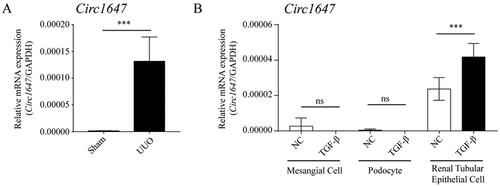
We first evaluated the expression levels of Circ1647 in kidney tissue from CKD mice use qRT-PCR. Our result revealed a significantly higher expression of the novel Circ1647 in CKD kidney tissues compared to sham-operated kidney tissues (As shown in ). Subsequently, we investigated the expression of Circ1647 within the renal parenchyma cell, including mesangial cells, podocytes and renal tubular epithelial cells. Interestingly, our results show that Circ1647 is specifically upregulated only in TGFβ1-stimulated renal tubular epithelial cells (As shown in ). In summary, our study highlights the heightened expression of Circ1647 in both CKD kidney tissues and TGFβ1-stimulated renal tubular epithelial cells.
Figure 1. Knockdown Circ1647 has been shown to attenuate renal fibrosis in a CKD model. (A,B) The level of Circ1647 was detected by qRT-PCR in CKD and the renal parenchyma cells. Data from animal experiments represent the mean ± SEM of six mice per group. Data from cell experiments represent the mean ± SEM of at least three independent experiments.*p < 0.01, **p < 0.005, ***p < 0.001.
3.2. Knockdown Circ1647 could attenuate kidney damage of CKD
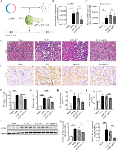
To investigate the impact of the upregulated Circ1647 in CKD kidney tissues on renal damage, we employed electroporation to knock down Circ1647 in CKD. As depicted in , a substantial reduction in the expression level of Circ1647 was observed, with no significant effect on its linear RNA expression, linearSh2d4b. Building upon these findings, we performed HE staining to evaluate the pathological alterations in mouse kidneys after knock down Circ1647 in the UUO model. As showed in , the knockdown of Circ1647 resulted in improved renal tubular dilation and luminal injury. To further assess the impact of Circ1647 knockdown on renal function, we measured the levels of BUN and Scr. While the UUO group and the vehicle group (UUO + EV) exhibited significant increases in both BUN and Scr levels compared to the sham group, Circ1647 knockdown specifically led to a recovery in BUN levels without markedly affecting Scr (As shown in ). Subsequently, we employed immunohistochemistry, immunoblotting, and qRT-PCR (As shown in ) to assess the levels of KIM1, a marker of renal tubular injury. Our results demonstrated that KIM1 predominantly accumulated within the renal tubules, and its expression significantly decreased upon Circ1647 knockdown. Overall, our study concludes that the knockdown of Circ1647 in the UUO model alleviates renal tubular injury.
Figure 2. Knockdown of Circ1647 ameliorates renal tubular injury in UUO-induced mice. (A) Schematic illustrating the depletion strategy of Circ1647 employing specific sgRNA. (B,C) The level of Circ1647/LinearSh2d4b was detected by quantitative RT-PCR (qRT-PCR). (D) Representative micrographs of HE staining. Scale bar represents 50 μm. (E) Immunohistochemistry for KIM1. (F) HE staining of quantitative analysis the percentage of renal tubular necrosis. (G) Percentage of positive area of KIM1 immunohistochemical staining. (H,I) Serum levels of BUN (Blood Urea Nitrogen) and Scr (Serum Creatinine) were measured and quantitatively analyzed to evaluate renal function. (J–L) Detection of RNA and protein levels of KIM1 by qRT-PCR and immunoblotting. Data represent the mean ± SEM for six mice in each group. *p < 0.01, **p < 0.005, ***p < 0.001.
3.3. Knockdown Circ1647 has been shown to attenuate renal fibrosis in a CKD model
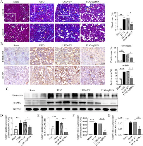
Renal fibrosis represents a crucial pathological factor in the progression of CKD. In this study, we aimed to investigate whether knockdown Circ1647 could inhibit the progression of renal fibrosis in a CKD model. Firstly, we assessed collagen deposition through Masson’s histological staining in different groups of mice. As depicted in , there was a notable escalation in fibrotic deposition following UUO-induced fibrosis compared to the normal group. However, the knockdown of Circ1647 led to a substantial decrease in this fibrotic deposition, while the vehicle group (UUO + EV) showed no significant change. Furthermore, immunohistochemical analysis revealed that the deposition of fibronectin and α-SMA also exhibited corresponding improvements after Circ1647 knockdown (As shown in ). Finally, we evaluated the transcriptional and translational expression levels of c by western blot and qRT-PCR. The results demonstrated a significant decrease in the expression of fibronectin and α-SMA at the RNA and protein levels upon Circ1647 knockdown (As shown in ). In summary, our findings suggest that the knockdown of Circ1647 can notably alleviate the progression of renal fibrosis in the CKD model.
Figure 3. Efficacy of Circ1647 Knockdown in Reducing Renal Fibrosis in UUO-induced mice. (A) Masson’strichrome staining and quantitative analysis of the fibrotic area. Arrows highlight areas of significant collagen deposition. Scale bar represents 50 μm. (B) Immunohistochemistry for fibronectin, α-SMA and percentage of positive area. (C–E) Detection and relative quantification of fibronectin and α-SMA proteins through immunoblotting. (E,F) Quantitative RT-PCR (qRT-PCR) used to assess mRNA levels fibronectin and α-SMA. Data represent the mean ± SEM for six mice ineach group. *p < 0.01, **p < 0.005, ***p < 0.001.
3.4. Circ1647 promotes the synthesis of ECM protein in renal tubular epithelial cells
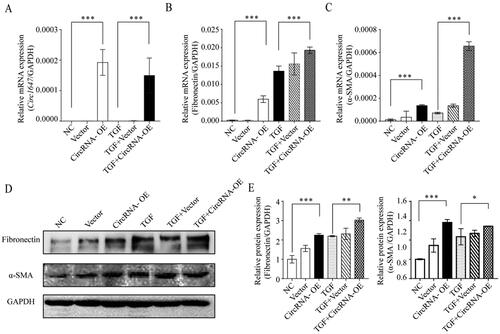
We further examined the overexpression of Circ1647 in renal tubular epithelial cells (TCMK1). Initially, we assessed the overexpression of Circ1647 within the cells, (As shown in ), demonstrating its successful overexpression in TCMK1 cells. Subsequently, we evaluated the expression of fibronectin and α-SMA (As shown in ). We observed an increase in both transcriptional and translational levels of fibronectin and α-SMA upon overexpress Circ1647. Furthermore, it was found that overexpress Circ1647 could enhance the expression of fibronectin and α-SMA at the transcriptional level following stimulation by TGFβ1, but the additive effect at the protein level remained inconspicuous. In conclusion, Circ1647 can facilitate the expression and accumulation of ECM proteins in TCMK1 cells.
Figure 4. Influence of Circ1647 on Extracellular Matrix (ECM) Protein Synthesis in Renal Tubular Epithelial Cells. (A) Detection of overexpressed Circ1647 levels in vivo by qRT-PCR. (B,C) Detection of RNA levels of fibronectin and α-SMA in TCMK1 cell. (D,E) Fibronectin and α-SMA were detected by immunoblotting and relative quantification. Data represent the mean ± SEM for at least three independent experiments. *p < 0.01, **p < 0.005, ***p < 0.001.
3.5. Circ1647 promotes renal fibrosis through the PI3K/AKT signaling pathway
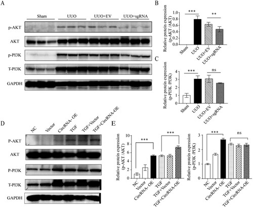
The PI3K/Akt signaling pathway constitutes a significant pathway involved in fibrosis stimulation. In our animal study, targeting Circ1647 for knockdown, shown in , resulted in a marked decrease in phospho-AKT (p-AKT) levels, and an increase in phospho-PI3K (p-PI3K) expression was observed. Conversely, Circ1647 overexpression in renal tubular epithelial cells, illustrated in , significantly heightened the protein expression within the PI3K/AKT pathway. These findings collectively indicate that Circ1647’s role in regulating fibrosis is likely associated with the modulation of the PI3K/AKT pathway.
Figure 5. Circ1647 promotes the abnormal activation of PI3K/AKT signaling in UUO mice. (A–C) Immunoblotting and quantitative analysis for phosphorylated AKT (p-AKT) relative to total AKT andphosphorylated PI3K (p-PI3K) relative to total PI3K in kidney tissues. (D,E) Assessment of p-AKT/AKT and pPI3K/PI3K in TCMK1 cells transfected with Circ1647 to evaluate its impact on the PI3K/AKT signaling pathway, using immunoblotting and quantitative methods. Data from animal experiments represent the mean ± SEM of six mice per group. Data from cell experiments represent the mean ± SEM of at least three independent experiments. *p < 0.01, **p < 0.005, ***p < 0.001.
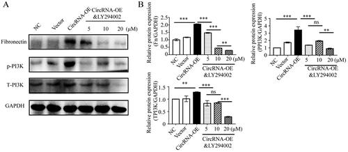
To elucidate whether Circ1647 exerts its effects through the PI3K/AKT pathway, we employed LY294002, a specific PI3K inhibitor. Following Circ1647 overexpression, the addition of varying concentrations of LY294002 led to a decrease in PI3K expression and consequently reduced the cell fibrosis markers, notably fibronectin, as depicted in . These experiments collectively demonstrate that Circ1647 facilitates fibrosis primarily through its action on the PI3K/AKT signaling pathway.
Figure 6. Circ1647 Enhances Renal Tubular Cell Fibrosis via the PI3K Pathway. (A,B) Immunoblotting andquantitative analysis of Fibronectin and the p-PI3K/PI3K ratio in TCMK1 cells. These cells were transfectedwith Circ1647 and then treated with various concentrations of the PI3K inhibitor LY294002 for 24 h, post-transfection. Data represent the mean ± SEM for at least three independent experiments. *p < 0.01, **p < 0.005, ***p < 0.001.
4. Discussion
Recent studies have shown that CircRNAs emerge as a pivotal modulator across both physiological and pathological states [Citation20], particularly in chronic kidney disease (CKD). Its significance in CKD is multifaceted and encompasses vital regulatory functions. The expression of Circ1647 was observed to be upregulated in CKD induced by unilateral ureteral obstruction (UUO). Simultaneously, the study also demonstrated that knockdown of Circ1647 can significantly inhibit the expansion of renal tubules, and inhibit the progression of its fibrosis. These results indicated that Circ1647 may be involved in the development and progression of renal fibrosis. Moreover, in vitro overexpression of Circ1647 has been shown to enhance fibrosis progression via activation of the PI3K/AKT signaling pathway, thereby providing further insights into the role of Circ1647 in the onset and development of renal fibrosis.
Within the realm of CKD, CircRNAs have emerged as crucial regulators across a range of pathological processes, encompassing fibrosis and inflammation [Citation9,Citation21]. CircRNAs regulates the progression of fibrosis in the body through the regulation of gene expression and modulation of signaling pathways. One of the main mechanisms is through the miRNA sponge effect [Citation22]. A specific CircRNA, known as CircRNA_010383, functions as a sponge for miR-135a, effectively delaying the onset of renal fibrosis [Citation23]. Furthermore, CircRNAs can also be regulated through direct interactions with proteins, thereby influencing their activation state and subsequent downstream signaling pathways. This further influences the biological functions associated with fibrosis progression. The upregulation of MYC transcription, mediated by the interaction between CircPTPN14 and FUBP1, contributes to the advancement of renal fibrosis by promoting fibroblast activation and extracellular matrix (ECM) remodeling [Citation9]. These results strongly emphasize the critical involvement of CircRNAs in the context of renal fibrosis. In the present study, it was discovered that Circ1647 possesses the ability to modulate the expression of PI3K/AKT, thus promoting the fibrotic process in CKD. In summary, a specific sgRNA sequence targeting Circ1647 was designed utilizing CRISPR-Cas13 gene editing technology. The results demonstrated that Circ1647 can effectively delay CKD-associated fibrosis by regulating the expression of PI3K/AKT proteins.
RfxCas13d is an RNA-mediated CRISPR-Cas system enzyme that has been successfully employed for targeting CircRNAs in zebrafish embryos [Citation24]. The technology for knocking down RfxCas13d is still in its early stages and currently encounters numerous challenges and limitations, which restrict its applicability for knockdown experiments in mammalian models. In this study, specific sgRNA primers were designed based on the sequence of Circ1647, followed by electroporation to introduce the plasmid into the kidney for knockdown of the target CircRNA. The knockdown efficiency targeting Circ1647 reached up to 50%. Furthermore, in comparison to commonly used siRNA and shRNA methods, the RfxCas13d method employed in this study offers greater flexibility in primer design for CircRNA knockdown without affecting DNA modification. It also demonstrated that dysregulation of the PI3K/AKT signaling pathway is closely associated with the accumulation of extracellular matrix (ECM) and plays a crucial role in the development and progression of fibrosis in various organs and diseases [Citation25]. Research has revealed that reducing the expression of Circ1647 in vitro can retard the progression of renal fibrosis. Conversely, overexpressing Circ1647 in vitro may enhance fibrosis of renal tubular cells. In contrast, in vitro overexpression of Circ1647 has been shown to promote fibrosis in renal tubular cells. Furthermore, introducing the specific PI3K inhibitor LY294002 following Circ1647 overexpression revealed that LY294002 can counteract the fibrosis induced by Circ1647. The observed effects underscore the pivotal role of Circ1647 in renal fibrosis, particularly through its influence on the PI3K/AKT signaling pathway. The inhibition of fibrosis by LY294002 following Circ1647 overexpression points toward a targeted therapeutic potential, though further research is needed to unravel the detailed mechanisms at play.
This study has established that Circ1647 is a key mediator in renal fibrosis, primarily through the PI3K/AKT signaling pathway. The successful inhibition of Circ1647 using RfxCas13d highlights its potential as a therapeutic target in combating renal fibrosis within the context of CKD. These findings not only deepen our understanding of CKD pathogenesis but also open new avenues for therapeutic intervention. By targeting Circ1647, we may develop more effective treatments for CKD, addressing a critical need in current renal disease management.
Authors’ contributions
Yufang Ni and Jianchun Li substantially contributed to the conception and design of the study. Hongmin Zhang, Qianwen Xian, Wenjie Qin and Hongwei Su acquired, analyzed and interpreted the data. Yufang Ni and Jianchun Li drafted the article and provided critical revisions for important intellectual content. Li Wang and Jianchun Li agreed to take responsibility for all aspects of the work, ensuring that any questions regarding the accuracy or integrity of the work are thoroughly investigated and resolved.
Disclosure statement
The authors declare that there are no conflicts of interest.
Data availability statement
Data supporting the findings of this study are available from the corresponding author upon reasonable request.
Additional information
Funding
References
- Lv JC, Zhang LX. Prevalence and disease burden of chronic kidney disease. Adv Exp Med Biol. 2019;1165:1–11. doi:10.1007/978-981-13-8871-2_1.
- Nelson RG, Grams ME, Ballew SH, et al. Development of risk prediction equations for incident chronic kidney disease. JAMA. 2019;322(21):2104–2114. doi:10.1001/jama.2019.17379.
- Liao C, Chen G, Yang Q, et al. Potential therapeutic effect and mechanisms of mesenchymal stem cells-extracellular vesicles in renal fibrosis. Front Cell Dev Biol. 2022;10:824752. doi:10.3389/fcell.2022.824752.
- Huang T, Cao Y, Wang H, et al. Circular RNA YAP1 acts as the sponge of microRNA-21-5p to secure HK-2 cells from ischaemia/reperfusion-induced injury. J Cell Mol Med. 2020;24(8):4707–4715. doi:10.1111/jcmm.15142.
- Kölling M, Seeger H, Haddad G, et al. The circular RNA ciRs-126 predicts survival in critically ill patients with acute kidney injury. Kidney Int Rep. 2018;3(5):1144–1152. doi:10.1016/j.ekir.2018.05.012.
- Xu HP, Ma XY, Yang C. Circular RNA TLK1 promotes Sepsis-Associated acute kidney injury by regulating inflammation and oxidative stress through miR-106a-5p/HMGB1 axis. Front Mol Biosci. 2021;8:660269. doi:10.3389/fmolb.2021.660269.
- Chen Z, Zheng Z, Xie Y, et al. Circular RNA circPPP6R3 upregulates CD44 to promote the progression of clear cell renal cell carcinoma via sponging miR-1238-3p. Cell Death Dis. 2021;13(1):22. doi:10.1038/s41419-021-04462-5.
- Kölling M, Haddad G, Wegmann U, et al. Circular RNAs in urine of kidney transplant patients with acute T cell-mediated allograft rejection. Clin Chem. 2019;65(10):1287–1294. doi:10.1373/clinchem.2019.305854.
- Nie W, Li M, Liu B, et al. A circular RNA, circPTPN14, increases MYC transcription by interacting with FUBP1 and exacerbates renal fibrosis. Cell Mol Life Sci. 2022;79(12):595. doi:10.1007/s00018-022-04603-9.
- Yi L, Ai K, Li H, et al. CircRNA_30032 promotes renal fibrosis in UUO model mice via miRNA-96-5p/HBEGF/KRAS axis. Aging (Albany NY). 2021;13(9):12780–12799. doi:10.18632/aging.202947.
- Cheng X, Ai K, Yi L, et al. The mmu_circRNA_37492/hsa_circ_0012138 function as potential ceRNA to attenuate obstructive renal fibrosis. Cell Death Dis. 2022;13(3):207. doi:10.1038/s41419-022-04612-3.
- Li S, Li X, Xue W, et al. Screening for functional circular RNAs using the CRISPR-Cas13 system. Nat Methods. 2021;18(1):51–59. doi:10.1038/s41592-020-01011-4.
- Konermann S, Lotfy P, Brideau NJ, et al. Transcriptome engineering with RNA-Targeting type VI-D CRISPR effectors. Cell. 2018;173(3):665–676 e14. doi:10.1016/j.cell.2018.02.033.
- Zhang Y, Nguyen TM, Zhang XO, et al. Optimized RNA-targeting CRISPR/Cas13d technology outperforms shRNA in identifying functional circRNAs. Genome Biol. 2021;22(1):41. doi:10.1186/s13059-021-02263-9.
- Tong H, Huang J, Xiao Q, et al. High-fidelity Cas13 variants for targeted RNA degradation with minimal collateral effects. Nat Biotechnol. 2023;41(1):108–119. doi:10.1038/s41587-022-01419-7.
- Li J, Yang J, Zhu B, et al. Tectorigenin protects against unilateral ureteral obstruction by inhibiting Smad3-mediated ferroptosis and fibrosis. Phytother Res. 2022;36(1):475–487. doi:10.1002/ptr.7353.
- Zhu B, Ni Y, Gong Y, et al. Formononetin ameliorates ferroptosis-associated fibrosis in renal tubular epithelial cells and in mice with chronic kidney disease by suppressing the Smad3/ATF3/SLC7A11 signaling. Life Sci. 2023;315:121331. doi:10.1016/j.lfs.2022.121331.
- Peng Z, Guo HY, Li YQ, et al. The Smad3-dependent microRNA let-7i-5p promoted renal fibrosis in mice with unilateral ureteral obstruction. Front Physiol. 2022;13:937878. doi:10.3389/fphys.2022.937878.
- Tan RZ, Li JC, Zhu BW, et al. Neuropeptide Y protects kidney from acute kidney injury by inactivating M1 macrophages via the Y1R-NF-kappaB-Mincle-dependent mechanism. Int J Biol Sci. 2023;19(2):521–536. doi:10.7150/ijbs.80200.
- He AT, Liu J, Li F, et al. Targeting circular RNAs as a therapeutic approach: current strategies and challenges. Signal Transduct Target Ther. 2021;6(1):185. doi:10.1038/s41392-021-00569-5.
- Wang Y, Ding L, Wang R, et al. Circ_0004951 promotes pyroptosis of renal tubular cells via the NLRP3 inflammasome in diabetic kidney disease. Front Med (Lausanne). 2022;9:828240. doi:10.3389/fmed.2022.828240.
- Xue C, Li G, Lu J, et al. Crosstalk between circRNAs and the PI3K/AKT signaling pathway in cancer progression. Signal Transduct Target Ther. 2021;6(1):400. doi:10.1038/s41392-021-00788-w.
- Peng F, Gong W, Li S, et al. circRNA_010383 acts as a sponge for miR-135a, and its downregulated expression contributes to renal fibrosis in diabetic nephropathy. Diabetes. 2021;70(2):603–615. doi:10.2337/db20-0203.
- Li S, Wu H, Chen LL. Screening circular RNAs with functional potential using the RfxCas13d/BSJ-gRNA system. Nat Protoc. 2022;17(9):2085–2107. doi:10.1038/s41596-022-00715-5.
- Zhang Y, Jin D, Kang X, et al. Signaling pathways involved in diabetic renal fibrosis. Front Cell Dev Biol. 2021;9:696542. doi:10.3389/fcell.2021.696542.