
Abstract
Recurrent spontaneous abortions (RSA) affect reproductive health and increase the risk of subsequent abortions. To investigate the role of KISS-1/GPR-54 signaling in RSA progression. Villus tissue was collected from RSA patients, and human trophoblastic HTR-8/SVneo cells were used. KISS-1 and GRP54 levels were detected using RT-qPCR and immunohistochemistry. Western blotting was performed to analyze ZO-1 and ZEB1 levels. Cell proliferation was determined via CCK-8 and cell clone formation assays. Transwell assays were performed to assess cell migration and invasion abilities. KISS-1 was down-regulated in the villus tissues of RSA patients. KISS-1 overexpression dramatically inhibited trophoblast proliferation, migration, and invasion. Mechanistically, ZEB1 expression was down-regulated, whereas ZO-1 expression was up-regulated, after KISS-1 overexpression. GPR54 silencing neutralized the effect of KISS-1 in HTR-8/SVneo cells. Additionally, KISS-1 overexpression inactivated the PI3K/AKT signaling pathway through GRP54. The KISS-1/GPR-54 signaling axis regulates RSA progression by regulating the PI3K/AKT signaling pathway.
1. Introduction
Recurrent spontaneous abortion (RSA) refers to two or more consecutive spontaneous abortions in women of childbearing age [Citation1]. Although the incidence of RSA is less than 5%, it has a significant impact on patients and increases the risk of subsequent abortions [Citation2]. The occurrence of RSA is related to several factors, among which abnormal immune coagulation is key. It is characterized by a positive antiphospholipid antibody, abnormal thrombophilic combination index, vitamin D deficiency, and hyperhomocysteinemia [Citation3–5]. Even if miscarriage-preserving treatment is successful in RSA patients, various pathological pregnancies, such as miscarriage, premature birth, premature rupture of membranes, oligohydramnios, fetal growth restriction, gestational hypertension, intrauterine stillbirth, and gestational diabetes mellitus, are still prone to occur in the later stage, which seriously affects the safety of the mother and fetus [Citation6,Citation7]. Achieving early prediction and reducing maternal and infant damage have always been urgent problems in clinics.
KISS-l is a tumor suppressor discovered by Lee et al. in 1996 in a human melanocyte cell line [Citation8]. The KISS-l gene is located in the lq32-q41 region of chromosomes and includes four exons. In 2001, kisspeptins, products of KISS-l, were found to be the products of different hydrolysis processes of the same protein precursor. Kisspeptins can be further split into kisspeptins-14, −13, −10, etc., which are collectively referred to as kisspeptins [Citation9,Citation10]. G protein-coupled receptor 54 (GPR54, also named KISS1R) is a kisspeptin receptor, belonging to the rhodopsin family of G protein-coupled receptors [Citation11,Citation12]. Previous studies have confirmed that KISS-1 inhibits the metastasis of various human cancers, including breast [Citation13], gastric [Citation14], and colorectal cancer [Citation15]. In 2003, three research groups discovered that KISS-1/GPR54 signaling plays a key role in the initiation of puberty in humans and mice [Citation16–18]. This major discovery has opened a new chapter in the study of mammalian reproductive endocrine regulatory mechanisms. Recently, many studies have shown that the KISS-l/GPR54 system is closely associated with abortion and adverse pregnancy outcomes [Citation19,Citation20]. However, the specific role of KISS-l/GPR54 signaling in RSA remains unclear.
Therefore, this study aimed to explore the function and mechanism of KISS-l/GPR54 signaling in human trophoblastic HTR-8/SVneo cells. We hypothesized that the KISS-l/GPR54 system is a critical factor for the growth and metastasis of HTR-8/SVneo cells, providing a new perspective for the treatment of RSA.
2. Materials and methods
2.1. Participant information
Eighty patients with RSA at the Hangzhou Women’s Hospital were recruited as the RSA group. These patients were aged from 23 to 39 years, with an average of (31.4 ± 2.3) years; The gestational age ranged from 53 to 66 days. All the patients in the RSA group met the 2010 American College of Obstetricians and Gynecologists diagnostic criteria for RSA, and all known causes were excluded. No patients had reproductive tract infections, chromosomal abnormalities, endocrine system diseases, reproductive tract structural abnormalities, or immune system diseases. During the same period, 80 women with normal early pregnancy requiring pregnancy termination were recruited as the control group. The age of the patients ranged from 22 to 37 years, with an average of (31.2 ± 2.5) years. The gestational age ranged from 48 to 62 days. All subjects in the control group showed normal intrauterine early pregnancy and intrauterine embryo development after clinical examination. There were no reproductive tract infections, chromosomal abnormalities, endocrine system diseases, anatomical abnormalities, immune system diseases, or threatened abortions. There were no significant differences in age, gestational age, number of births, or other basic data between the two groups. The clinical characteristics of patients with RSA and healthy controls are shown in . Villus tissues were obtained from the participants and stored in liquid nitrogen. All the participants provided written informed consent. This study was approved by the Ethics Committee of the Hangzhou Women’s Hospital.
Table 1. The clinical characteristics of patients with RSA or normal controls.
2.2. Cell culture and transfection
The human chorionic trophoblast HTR8/Svneo was purchased from the American Type Culture Collection Center (ATCC, USA) and cultured in RPMI1640 medium (containing 10% fetal bovine serum) at 37 °C, 5% CO2, and 98% relative humidity. Additionally, the KISS-1 overexpressing vector (pcDNA 3.1, oe-KISS-1), short hairpin RNA targeting KISS-1 and GPR54 (sh-KISS-1 and sh-GPR54), and their controls (sh-nc and oe-nc) were obtained from Genomeditech (Shanghai, China) and transfected into cells using a TransfectamineTM 5000 Transfection Kit according to the manufacturer’s instructions (AmyJet Scientific, Wuhan, China). The sequences of sh-KISS-1 and sh-GRP54 are as follows:
Sh-KISS-1 #1: 5′-GCACCCATGGAGAATCCTAGA-3′;
Sh-KISS-1 #2: 5′-GGAGAATCCTAGATCTACAGG-3′;sh-GPR54: 5′-GCGATCGACTGGGAGTATAGT-3′.
2.3. RT-qPCR
An RNAiso Plus kit (Takara, Dalian, China) was used for RNA extraction following the manufacturer’s protocol. Subsequently, a Nanodrop2000 ultra-trace spectrophotometer was used to determine the RNA concentration. Then, 1 μg of RNA was used for the reverse transcription to obtain cDNA; the reaction was carried out in accordance with the manufacturer’s instructions. The PCR system included 10 μL of SYBR Green nucleic acid fluorescent dye, 5 μL of cDNA product, and 5 μL of primers; three duplicate wells were established. The reaction conditions were as follows: pre-denaturation at 94 °C for 4 min; amplification: 94 °C for 30 s and 60 °C for 1 min for a total of 40 cycles. Finally, the relative expression was calculated using the 2-△△CT method.
2.4. Immunohistochemistry
After dehydration and embedding, the villus tissues were cut into sections with a thickness of 5 μm. Sections were soaked in methanol containing 0.3% H2O2 for 1 h, rinsed with PBS, and blocked with 10% goat serum for 15 min. Thereafter, the sections were treated with the primary antibodies against KISS-1 (abcam, diluted 1: 1000) or GPR54 (abcam, diluted 1: 1500), overnight at 4 °C. The sections were then washed with PBS for 15 min, followed by incubation with goat anti-rabbit IgG secondary antibody (Nichirei Biosciences Inc., Japan) for 1 h. After rinsing in PBS for 15 min, the sections were washed with streptavidin and incubated with horseradish peroxidase (HRP)-labeled secondary antibody for 30 min. A DAB horseradish peroxidase color development kit (PHYGENE, Shanghai, China) was used to observe immune reaction signals. Sections were observed and photographed under a microscope (Leica, Germany).
2.5. CCK-8 assay
Trophoblasts from each group were seeded in a 96-well plate at a density of 2 × 10⁴ cells/well and incubated for 24, 48, and 72 h. After that, the medium was removed at each time period and 10 μL of CK8 reagent and 90 μL of new medium were added to each well. After incubation at 37 °C for 4 h in the dark, the absorbance was measured at 450 nm using a microplate reader.
2.6. Cell clone formation
Then, the cells were washed with PBS, digested with trypsin, and resuspended in 1 mL of fresh medium after centrifugation. 100 μL of the cell suspension was diluted to obtain a single-cell suspension at a concentration of 1 × 10³ cells/mL. Next, 2 mL of cell suspension was added to each well of a 6-well plate, and three replicate wells were prepared for each treatment group. After 14 days, the medium was discarded and the cells were washed twice with PBS and fixed with 1 mL of 90% ethanol for 30 min. Then, the ethanol was discarded, and the plate was dried and stained with 1 mL of crystal violet for 30 min.
2.7. Determination of cell migration and invasion
Cell migration and invasion assays were performed using Transwell. Matrigel was diluted with DMEM without FBS and spread on the upper layer of the Transwell chamber. Then, this was shaken gently to spread the gel evenly on the membrane and incubated in a 37 °C incubator for 1 h. Next, 200 μL of cell suspensions resuspended in FBS-free medium were added to the upper layer of the Transwell chamber, and the cell density was adjusted to 1 × 10⁶/L. Then, 500 μL of culture medium containing 25% FBS was added to the lower layer of the chamber, and the Transwell was placed it in a 37 °C, 5% CO2 incubator for 24 h. Next, the chamber was taken out and washed with PBS. A wet cotton swab was used to remove the uninvaded cells in the upper chamber. The cells were fixed in 4% paraformaldehyde for 30 min, washed with PBS three times, and stained with hematoxylin for 10 min. After washing with PBS, five fields of view under the microscope were randomly selected and photographed to record the number of invaded cells. To determine cell migration, the upper chamber was not pre-treated with Matrigel. Other procedures were consistent with those used in the invasion experiment.
2.8. Western blotting
The total protein was extracted using RIPA buffer, and the protein content was determined using the BCA method (Beyotime, Shanghai, China). Here, 60 μg of protein was separated with 12% sodium dodecyl sulfate-polyacrylamide gel electrophoresis (SDS-PAGE). After electrophoresis, the proteins were transferred to a nitrocellulose membrane and blocked in 5% skim milk for 1 h. KISS-1 (1:1000), zinc finger E-box binding homeobox 1 (ZEB1) (1:1500), zonula occludes protein-1 (ZO-1) (1:800), PI3K (1:1200), p-PI3K (1:600), AKT (1:800), p-AKT (1:1200), and GAPDH (1:2500) primary antibodies (Abcam, CA, USA) were added and incubated with membranes overnight at 4 °C. The next day, the membrane was washed three times with TBST. Then, HRP-labeled secondary antibody was added and incubated at 37 °C for 1 h. After washing the membranes with TBST, an electrochemiluminescence (ECL) developer (Beyotime) was added, developed in the dark, and photographed using a gel imaging system.
2.9. Statistical analysis
SPSS 21.0 software was used to analyze the experimental data. Measurement data conforming to the normal distribution were expressed as the mean ± SD, and the T-test was used for comparison between the two groups. One-way analysis of variance was used for comparisons among multiple groups, and Tukey’s test was used for further pairwise comparisons. The potential diagnostic value of the KISS in RSA was determined using receiver operating characteristic (ROC) curve analysis. Differences were considered statistically significant at p < 0.05.
3. Results
3.1. KISS-1 was down-regulated in RSA
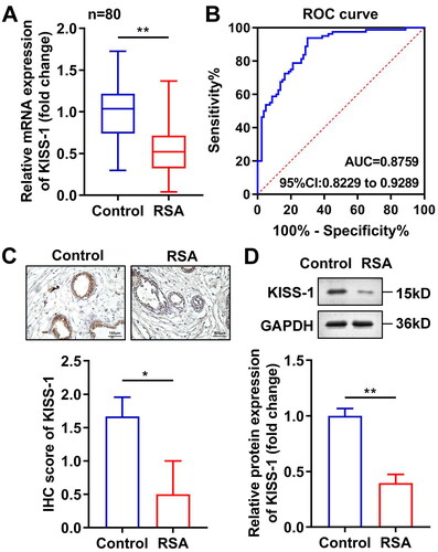
We found that the mRNA levels of KISS-1 were prominently down-regulated in the villus tissue of RSA patients (). The area under the ROC curve (AUC) of KISS-1 was 0.8606, indicating its potential diagnostic value for RSA (). This was further confirmed by the results of the immunohistochemistry () and western blot () assays, which also demonstrated a decrease in KISS levels in the villus tissue of RSA patients.
Figure 1. KISS-1 was down-regulated in RSA. The KISS-1 levels in the villus tissue of the RSA patients were assessed via RT-qPCR (a), immunohistochemistry (C) and western blotting (D) assays. (B) ROC curves of KISS-1 in RSA. **p < 0.01.
3.2. KISS-1 overexpression inhibited the cell proliferation of HTR8/Svneo cells
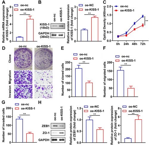
The RT-qPCR and western blotting results showed that oe-KISS-1 transfection dramatically increased KISS-1 levels (). After oe-KISS-1 transfection, the viability of HTR8/Svneo cells was significantly reduced (). In addition, the number of cloned, migrated, and invaded HTR8/Svneo cells was significantly down-regulated after oe-KISS-1 transfection (). Moreover, the ZEB1 protein levels prominently decreased and those of ZO-1 increased after sh-KISS-1 transfection ().
Figure 2. KISS-1 overexpression inhibited the cell growth of HTR8/Svneo cells. The transfection efficiency of oe-KISS-1 was determined via RT-qPCR (A) and western blotting (B) assays. (C) After oe-KISS-1 transfection, the cell viability was tested with a CCK-8 assay. (D) Images of cloned, migrated, and invaded cells. Quantification of cloned (E), migrated (F), and invaded (G) cells. (H) The protein levels of ZO-1 and ZEB1 were calculated via western blotting. **p < 0.01.
3.3. GPR54 silencing inhibited the roles of oe-KISS-1 in HTR8/Svneo cells
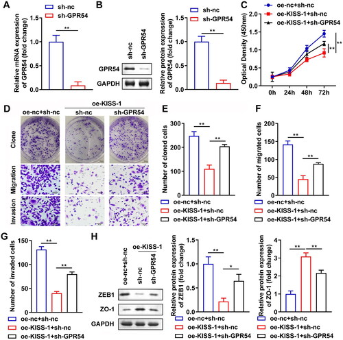
After sh-GPR54 transfection, GPR54 levels were prominently depleted at both the mRNA and protein levels (). Subsequently, sh-GPR54 transfection significantly increased the viability of oe-KISS-1-treated HTR8/Svneo cells (). The number of cloned, migrated, and invaded oe-KISS-1-treated HTR8/Svneo cells prominently increased after sh-GPR54 transfection (). Additionally, the ZEB1 protein levels prominently increased and those of ZO-1 decreased after sh-GPR54 transfection ().
Figure 3. GPR54 silencing neutralized the roles of oe-KISS-1 in HTR8/Svneo cells. The transfection efficiency of sh-GPR54 was determined via RT-qPCR (A) and western blotting (B) assays. (C) After oe-KISS-1 and sh-GPR-54 transfection, the cell viability was tested with a CCK-8 assay. (D) Images of cloned, migrated, and invaded cells. Quantification of cloned (E), migrated (F), and invaded (G) cells. (H) The protein levels of ZO-1 and ZEB1 were calculated via western blotting. **p < 0.01.
3.4. KISS-1 modulated the PI3K/AKT signaling pathway by targeting GPR54
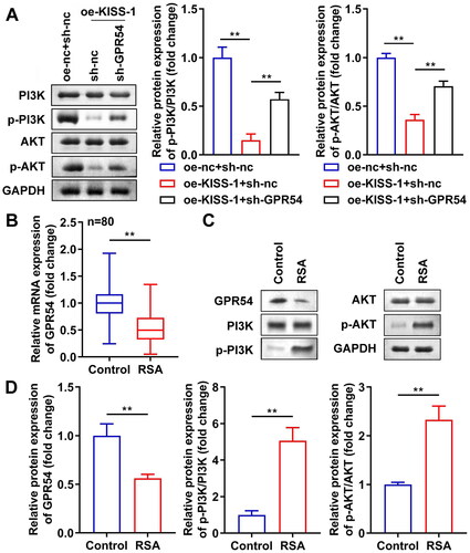
Subsequently, after oe-KISS-1 transfection, p-PI3K and p-AKT protein levels were prominently down-regulated, whereas sh-GPR54 prominently inhibited the role of oe-KISS-1 on p-PI3K and p-AKT protein levels (), indicating that KISS-1 silencing activated the PI3K/AKT signaling pathway by targeting GPR54. In addition, we found that the mRNA levels of GPR54 significantly decreased in RSA patients (). Western blotting results also revealed that the protein levels of GRP54 were significantly decreased, whereas p-PI3K and p-AKT levels were significantly increased in RSA patients ().
Figure 4. KISS-1 modulated the PI3K/AKT signaling pathway by targeting GPR54. (A) The levels of PI3K and AKT were assessed via western blotting. (B) The mRNA levels of GPR54 in the villus tissue of the RSA patients were detected via RT-qPCR assay. (C, D) The protein levels of GPR54, p-PI3K, and p-AKT in the villus tissue of the RSA patients were detected via western blotting. **p < 0.01.
3.5. Inhibition of PI3K/AKT signaling pathway suppressed the growth of sh-KISS-1-transfected HTR8/Svneo cells
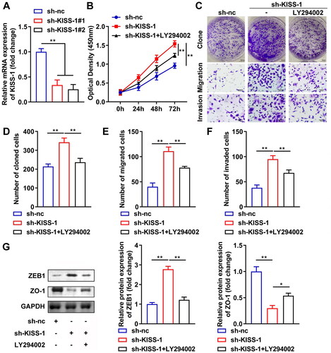
Finally, cells were treated with LY294002, a PI3K/AKT signaling pathway inhibitor. The transfection efficiency of sh-KISS-1 was tested with RT-qPCR. The results showed that sh-KISS-1 significantly decreased KISS-1 levels; thus, sh-KISS-1 #2 was selected for the subsequent experiments (). Subsequently, compared to the sh-nc group, the cell viability () and the number of cloned (), migrated (), and invaded () cells in the sh-KISS-1 group increased prominently. Additionally, ZEB1 protein levels were prominently enhanced, and those of ZO-1 declined (). However, LY294002 treatment reversed the effects of sh-KISS-1.
Figure 5. Inhibition of the PI3K/AKT signaling pathway suppressed the growth of the sh-KISS-1-treated HTR8/Svneo cells. (A) The transfection efficiency of sh-GPR54 was determined via RT-qPCR. (B) After sh-KISS-1 and LY294002 treatment, the cell viability was tested with a CCK-8 assay. (C) Images of cloned, migrated, and invaded cells. Quantification of cloned (D), migrated (E), and invaded (F) cells. (G) The protein levels of ZO-1 and ZEB1 were calculated via western blotting. *p < 0.05, **p < 0.01.
4. Discussion
Here, we demonstrate that KISS1/GPR54 signaling participates in RSA progression. Mechanistically, KISS1 overexpression inhibited the growth, migration, and invasion of HTR8/Svneo cells by targeting the GPR54-mediated PI3K/AKT signaling pathway.
Studies have confirmed that KISS-1/GPR54 signaling plays a central role in mammalian reproductive endocrine regulation [Citation21]. After kisspeptin expressed in the hypothalamus binds to its receptor GPR54, gonadotropin-releasing hormone (GnRH) neurons are activated, which further stimulates the release of luteinizing hormones and regulates reproductive activities [Citation22]. Recently, KISS-1 has been reported to regulate the malignant behavior of tumor cells. Huo et al. [Citation23] have found that KISS-1 is down-regulated in colorectal cancer tissues and identified KISS-1 as a biomarker for colorectal cancer prognosis. Similarly, Chen et al. [Citation24] have demonstrated that KISS-1 overexpression markedly inhibits cell growth and metastasis in the colorectal cancer. Additionally, KISS-1 is most strongly expressed in the placental tissue, and it has been confirmed that KISS-1 inhibits the invasion of trophoblastic cells with an infiltrating ability similar to that of tumor cells [Citation25,Citation26]. In this study, we confirmed that KISS-1 overexpression dramatically inhibited the growth and metastasis of HTR8/Svneo cells. We also showed that GPR54 silencing inhibited the role of oe-KISS-1 in cells, implying that KISS-1 participates in the biological processes of HTR8/Svneo cells by targeting GPR54. Additionally, the transformation of epithelial cells into mesenchymal cells is considered a key regulator of cell growth, invasion, and metastasis [Citation27]. ZO-1, an important component of tight junctions. It combines with the cytoskeleton and signalingg molecules in cells. During EMT, the expression of ZO-1 is down-regulated, and tight junctions are disrupted [Citation28]. ZEB1 induces epithelial mesenchymal transformation [Citation29]. As reported previously, excessive invasion, migration, and growth were the key factors of RSA [Citation4]. Our research also demonstrated that KISS-1 overexpression down-regulated ZEB1 and up-regulated ZO-1, which may be key for KISS-1 regulation of cell migration and invasion. These results indicated a protective effect of KISS-1 against RSA.
Subsequently, through PPI networks, we found that GPR54 was in close contact with the PI3K/AKT signaling pathway. PI3K is an intracellular phosphatidylinositol kinase. When cells are stimulated by growth factors, vascular endothelial factors, or insulin, they become phosphorylated and activated. Activated PI3K induces the downstream protein AKT to bind to its corresponding receptors on the cell membrane surface and phosphorylate AKT (p-AKT). Subsequently, p-AKT is released from the cell membrane into the cytoplasm to transmit signals, thereby promoting cell growth and development [Citation30,Citation31]. Li et al. [Citation32] clarified that blocking the PI3K/AKT signaling pathway is a new fundamental mechanism for RSA treatment in the clinic. Feng et al. [Citation33] showed that BuShen HuoXue decoction promotes the proliferation of decidual stromal cells in RSA by modulating the PI3K/AKT pathway. In our study, we found that KISS overexpression inhibited the PI3K/AKT pathway, which was activated by GPR54 knockdown. In addition, LY294002, a PI3K inhibitor, significantly inhibited cell growth and metastasis following KISS-1 silencing. Our results are similar to those reported by Chen et al. [Citation24] showing that KISS-1 overexpression inhibits the invasiveness of colorectal cancer cells by interacting with the PI3K/AKT pathway.
In summary, our findings suggest that KISS-1/GPR54 signaling plays a critical role in RSA progression and is closely related to the regulation of the PI3K/AKT signaling pathway. These results provide new directions for RSA therapy.
Ethical approval and consent to participate
The study was approved by the Ethics Committee of the Hangzhou Women’s Hospital. Written informed consent was obtained from all patients. All experiments were performed in accordance with relevant guidelines and regulations.
Acknowledgments
Not applicable.
Disclosure statement
No potential conflict of interest was reported by the author(s).
Data availability statement
The datasets used and/or analyzed during the current study are available from the corresponding author on reasonable request.
Additional information
Funding
References
- Li D, Zheng L, Zhao D, et al. The role of immune cells in recurrent spontaneous abortion. Reprod Sci. 2021;28(12):1–8.
- La X, Wang W, Zhang M, et al. Definition and multiple factors of recurrent spontaneous abortion. Adv Exp Med Biol. 2021;1300:231–257.
- Ou H, Yu Q. Efficacy of aspirin, prednisone, and multivitamin triple therapy in treating unexplained recurrent spontaneous abortion: a cohort study. Int J Gynaecol Obstet. 2020;148(1):21–26.
- Lin QD, Qiu LH. Pathogenesis, diagnosis, and treatment of recurrent spontaneous abortion with immune type. Front Med China. 2010;4(3):275–279.
- Tur-Torres MH, Garrido-Gimenez C, Alijotas-Reig J. Genetics of recurrent miscarriage and fetal loss. Best Pract Res Clin Obstet Gynaecol. 2017;42:11–25.
- Liang F, Huo X, Wang W, et al. Association of bisphenol a or bisphenol S exposure with oxidative stress and immune disturbance among unexplained recurrent spontaneous abortion women. Chemosphere . 2020;257:127035.
- Fu J, Li L, Qi L, et al. A randomized controlled trial of etanercept in the treatment of refractory recurrent spontaneous abortion with innate immune disorders. Taiwan J Obstet Gynecol. 2019;58(5):621–625.
- Lee JH, Miele ME, Hicks DJ, et al. KiSS-1, a novel human malignant melanoma metastasis-suppressor gene. J Natl Cancer Inst. 1996;88(23):1731–1737.
- Fratangelo F, Carriero MV, Motti ML. Controversial role of kisspeptins/KiSS-1R signaling system in tumor development. Front Endocrinol (Lausanne). 2018;9:192.
- Kanasaki H, Tumurbaatar T, Tumurgan Z, et al. Mutual interactions between GnRH and kisspeptin in GnRH- and kiss-1-expressing immortalized hypothalamic cell models. Reprod Sci. 2021;28(12):3380–3389.
- Trevisan CM, Montagna E, de Oliveira R, et al. Kisspeptin/GPR54 system: what do We know about its role in human reproduction? Cell Physiol Biochem. 2018;49(4):1259–1276.
- Hu KL, Chang HM, Zhao HC, et al. Potential roles for the kisspeptin/kisspeptin receptor system in implantation and placentation. Hum Reprod Update. 2019;25(3):326–343.
- Guzman S, Brackstone M, Wondisford F, et al. KISS1/KISS1R and breast cancer: metastasis promoter. Semin Reprod Med. 2019;37(4):197–206.
- Kostakis ID, Agrogiannis G, Vaiopoulos AG, et al. KISS1 and KISS1R expression in gastric cancer. J Buon. 2018;23:79–84.
- Zheng Y, Liu Y, Lin Y, et al. MicroRNA-124 and microRNA-378 inhibit the proliferation and invasion of colorectal cancer by upregulating KiSS1. Transl Cancer Res. 2020;9(4):2838–2846.
- de Roux N, Genin E, Carel J, et al. Hypogonadotropic hypogonadism due to loss of function of the KiSS1-derived peptide receptor GPR54. Proc Natl Acad Sci U S A. 2003;100(19):10972–10976.
- Funes S, Hedrick JA, Vassileva G, et al. The KiSS-1 receptor GPR54 is essential for the development of the murine reproductive system. Biochem Biophys Res Commun. 2003;312(4):1357–1363.
- Seminara SB, Messager S, Chatzidaki EE, et al. The GPR54 gene as a regulator of puberty. N Engl J Med. 2003;349(17):1614–1627.
- Smets EM, Deurloo KL, Go AT, et al. Decreased plasma levels of metastin in early pregnancy are associated with small for gestational age neonates. Prenat Diagn. 2008;28(4):299–303.
- Armstrong RA, Reynolds RM, Leask R, et al. Decreased serum levels of kisspeptin in early pregnancy are associated with intra-uterine growth restriction and pre-eclampsia. Prenat Diagn. 2009;29(10):982–985.
- Navarro VM, Tena-Sempere M. The KiSS-1/GPR54 system: putative target for endocrine disruption of reproduction at hypothalamic-pituitary unit? Int J Androl. 2008;31(2):224–232.
- Papaoiconomou E, Msaouel P, Makri A, et al. The role of kisspeptin/GPR54 in the reproductive system. In Vivo. 2011;25(3):343–354.
- Huo X, Zhang L, Li T. Analysis of the association of the expression of KiSS-1 in colorectal cancer tissues with the pathology and prognosis. Oncol Lett. 2018;15(3):3056–3060.
- Chen S, Chen W, Zhang X, et al. Overexpression of KiSS-1 reduces colorectal cancer cell invasion by downregulating MMP-9 via blocking PI3K/akt/NF-kappaB signal pathway. Int J Oncol. 2016;48(4):1391–1398.
- Terao Y, Kumano S, Takatsu Y, et al. Expression of KiSS-1, a metastasis suppressor gene, in trophoblast giant cells of the rat placenta. Biochim Biophys Acta. 2004;1678(2–3):102–110.
- Zhang H, Long Q, Ling L, et al. Elevated expression of KiSS-1 in placenta of preeclampsia and its effect on trophoblast. Reprod Biol. 2011;11(2):99–115.
- Mani SA, Guo W, Liao M, et al. The epithelial-mesenchymal transition generates cells with properties of stem cells. Cell. 2008;133(4):704–715.
- Zhao J, Liang S, Fu W, et al. The LIM domain protein FHL1C interacts with tight junction protein ZO-1 contributing to the epithelial–mesenchymal transition (EMT) of a breast adenocarcinoma cell line. Gene. 2014;542(2):182–189.
- Krebs AM, Mitschke J, Lasierra LM, et al. The EMT-activator Zeb1 is a key factor for cell plasticity and promotes metastasis in pancreatic cancer. Nat Cell Biol. 2017;19(5):518–529.
- Xie Y, Shi X, Sheng K, et al. PI3K/Akt signaling transduction pathway, erythropoiesis and glycolysis in hypoxia (review). Mol Med Rep. 2019;19(2):783–791.
- Ediriweera MK, Tennekoon KH, Samarakoon SR. Role of the PI3K/AKT/mTOR signaling pathway in ovarian cancer: biological and therapeutic significance. Semin Cancer Biol. 2019;59:147–160.
- Li Z, Zhou G, Jiang L, et al. Effect of STOX1 on recurrent spontaneous abortion by regulating trophoblast cell proliferation and migration via the PI3K/AKT signaling pathway. J Cell Biochem. 2018;120(5):8291–8299.
- Feng X, Jiang S, Leung W, et al. BuShen HuoXue decoction promotes decidual stromal cell proliferation via the PI3K/AKT pathway in unexplained recurrent spontaneous abortion. Evid Based Complement Alternat Med. 2020;2020:6868470–6868411.