
ABSTRACT
Purpose: To examine the effects of a biological treatment (adalimumab) on visual function in patients with ankylosing spondylitis and in uveitic patients without macular edema during one-year treatment with adalimumab.
Methods: Sixteen eyes of eight consecutive Caucasian patients treated with adalimumab were followed up using microperimetry (MAIA; CenterVue, Padova, Italy). Five patients had ankylosing spondylitis without uveitis, three patients had panuveitis without macular edema. Macular sensitivity and macular integrity were recorded.
Results: During six-month follow-up, the average threshold did not change significantly (p = .649). Macular integrity was stable (p = .225). The macular sensitivity point analysis showed no significant effects (examination F(3,56) = 0.494 and p = .688; point*examination F(108,2016) = 0.688 and p = .994) during the follow-up.
Conclusions: During one-year follow-up, adalimumab did not affect macular function, unlike the well-established maculopathy induced by hydroxychloroquine. Microperimetry may be considered when following-up macular function in patients undertaking adalimumab.
Biologics are developed and approved to treat systemic inflammatory diseases (e.g. ankylosing spondylitis, juvenile idiopathic arthritis, psoriasis, and inflammatory bowel disease) or to prevent organ transplant rejection.1 Several prospective studies have shown the effectiveness of TNF-α inhibitors (infliximab and adalimumab) in noninfectious uveitis refractory to immunosuppressant and in rheumatologic diseases (e.g ankylosing spondylitis).Citation2–5 TNF-α is a cytokine, which has a major role in regulating the functions of cells involved in the inflammatory process.
Adalimumab is a fully human monoclonal antibody against TNF-α. Previous clinical studies have shown its potential for juvenile uveitis (mainly associated to juvenile idiopathic arthritis) and more recently data have also demonstrated promising results in adults.Citation2,Citation4,Citation5 Adalimumab has been shown to reduce anterior uveitis flares in patients with ankylosing spondylitis and successful adalimumab therapy has been reported for uveitis associated with sarcoidosis, Vogt-Koyanagi-Harada syndrome, birdshot chorioretinopathy, as well as idiopathic noninfectious uveitis.Citation5–7
MicroperimetryCitation8 is a useful tool for the early detection of retinal alteration in patients treated with hydroxychloroquine.Citation9,Citation10 It is a clinical noninvasive tool for assessing macular sensitivity. Microperimetry allows evaluating retinal function by using a fundus-tracking controlled visual field examination. Fundus tracking allows to overcome the eye movements and retinal fixation changes and to obtain precise retina-related sensitivity data.
The aim of the present study was to examine the effect of adalimumab for the retinal function in patients with ankylosing spondylitis without uveitis and in uveitic patients without macular edema by MAIA microperimetry.
MATERIALS AND METHODS
The experiments adhered to the tenets of the Declaration of Helsinki and were approved by the ethics committee of Semmelweis University in Budapest, Hungary (registration number TUKEB 261/2015). Signed informed consent was obtained from each subject after explanation of the nature and possible consequences of the study. Participants were eight subjects (mean age = 49.8 ± 11.4 years at the beginning of the study) with diseases affecting ankylosing spondylitis without uveitis and uveitic patients without macular edema under adalimumab treatment (40 mg every 2 weeks). Five patients had ankylosing spondylitis without uveitis, they were treated by rheumatologist. Two patients had panuveitis with sarcoidosis without macular edema and one patient had idiopathic noninfectious uveitis. Two uveitic patients received methotrexate and one of them received cyclosporine before the adalimumab treatment. All eight patients had no disease activity during the adalimumab treatment. Results from their 16 paired eyes were analyzed.
All subjects underwent microperimetric examinations. The Macular Integrity Assessment System (MAIA; CenterVue, Padova, Italy) was used for the microperimetric measurements. The system is equipped with a scanning laser ophthalmoscope that provides real-time eye-tracking and eye movement compensation. The Expert Protocol was always applied monocularly before the treatment started (baseline), after 1 month (M1), after 3 months (M3), and after 6 months (M6). Four subjects completed 1-year follow-up (M12) at the time of the submission.
Goldman-based standard light stimuli parameters (size III white stimuli) and strategy (threshold 4–2) were used. The maximum luminance of the stimulus (318 cd/m2) allows a stimulus presentation ranging from 0 to 36 decibels (dB). The observer’s task was to press a button to indicate the presence of the light spot whenever it was detected, always keeping central fixation. Visual field locations that required brighter stimuli to reach threshold showed reduced sensitivity, thus, lower dB sensitivity values indicate lower sensitivity. Conversely, higher dB values correspond to dimmer stimuli and represent higher sensitivity. The Expert Protocol was used in order to evaluate thresholds at different macular locations. The test grid consisted of 37 points inside a concentric area of 5° from the central point, meaning that the central area of 10° was examined, with 12 points within each concentric ring (2°, 6°, and 10°) plus the measurement of one central point. Macular integrity index, the numerical value compared to age-adjusted normative data was also evaluated. Besides evaluating sensitivity thresholds and macular integrity, the system allows recording fixation locations 25 times per second. Therefore, fixation stability (P1 and P2) were evaluated.Citation11
Data are expressed as means ± one standard deviation. Two-way ANOVA tests (SPSS, Statistical Package for the Social Sciences, Hong Kong, China) for repeated measure and paired comparisons using Bonferroni post-hoc analyses were performed. We assumed a p value below 0.05 to be statistically significant.
RESULTS
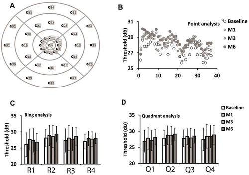
Average threshold varied from 25 to 31 dB (mean = 27.5 ± 2.7) in the first examination (baseline). During 6-months follow-up (M6) there were no significant differences in the average threshold (F(3,59) = 0.551; p = .649), meaning that the means and the standard deviations were statistically similar (M1 = 28.3 ± 3.0; M3 = 28.0 ± 3.0; M6 = 28.6 ± 2.2). shows the means (± one standard deviation) of the average threshold for each of the four consecutive examinations.
FIGURE 1. Average threshold (a) and macular integrity (b) results during the follow-up examinations for all eight subjects. Baseline examination was performed before the beginning of the treatment and the others during a follow-up period (M1 = 1 month, M3 = 3 months, and M6 = 6 months after the treatment). We observed no significant changes (p > .05) during 6-months follow-up
The macular integrity was more variable than the average threshold among the subjects during the baseline examination (mean = 50.8 ± 37.2) ranging from 1 (best performance) to 100 (worst performance). It shows that there were different levels of visual disturbance among the participants. Although macular integrity varied substantially during the baseline examination, its mean became lower (better) during the follow-up examination (M1 = 34.7 ± 28.7; M3 = 37.8 ± 31.3; M6 = 26.9 ± 31.6). However, there was no significant differences during the repeated examinations (F(3,59) = 1.497; p = .225). shows the means (± one standard deviation) of the macular integrity for each of the four consecutive examinations.
In order to verify if the similarities of the global parameters (average threshold and macular integrity) found during the six-month consecutive examinations would be reproduced in a more detailed analysis, we performed regional and local comparisons. As shown in , thresholds were grouped either in rings or in quadrants. In addition, each macular point was analyzed separately. The point analysis shows no significant difference (examination F(3,56) = 0.494 and p = .688; point*examination F(108,2016) = 0.688 and p = .994) during the follow-up (). shows that there were no significant differences during the follow-up in the ring analysis (examination F(3,56) = 0.513 and p = .675; ring*examination F(9,166) = 0.625and p = .775). shows that the quadrant analysis also resulted in stable thresholds during the follow-up since differences were not statistically significant (examination F(3,56) = 0.472 and p = .703; ring*examination F(9,166) = 0.737 and p = .675).
FIGURE 2. Regional and local analysis of eight subjects. Test grid, individual points, and point groups are shown in a diagram (a). It represent the distribution for the right eye. Left eye is represented by the mirror image. Individual (37) points were compared during the follow-up examinations (b) as well as points grouped in rings (c) for R1 (a single foveal point), R2 (points within 1º of eccentricity), R3 (points within 3º of eccentricity); R4 (points within 5º of eccentricity) and in quadrants (d) for Q1 (points placed at the superior quadrant), Q2 (points placed at the inferior quadrant), Q3 (points placed at the temporal quadrant), and Q4 (points placed at the nasal quadrant). Empty symbols represent results from the baseline, symbols with increasing darkness represent M1, M3, and M6 examinations, respectively. We observed no significant changes (p > .05) during 6-months of follow-up for all parameters analyzed
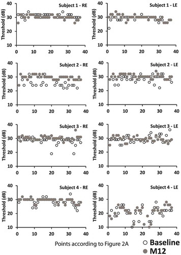
Finally, point-by-point comparison was performed for the eight paired-eyes (four subjects) completing the 12-month follow-up prior to the submission. There was no significant difference between the thresholds recorded in the baseline examination and 1 year after for the four subjects (examination F(1,14) = 0.193 and p = .667; point*examination F(36,504) = 0.430 and p = .999). shows individual results of the baseline thresholds versus the 12-month follow-up (M12) for each of the four subjects completing the follow-up examinations.
FIGURE 3. Point-by-point comparison of eight-paired (four subjects) eyes between baseline (empty symbols) and 12-month follow-up (gray symbols). Despite variability among subjects, we observed no significant changes (p > .05) during 6-months follow-up for all parameters analyzed
DISCUSSION
The present data show no significant differences in the average threshold and in macular integrity during 6-months follow-up of patients undertaking adalimumab.
TNF inhibitors are a group of medications used worldwide in autoimmune disease to treat inflammatory conditions such as rheumatoid arthritis, psoriatic arthritis, juvenile arthritis, inflammatory bowel disease (Crohn’s and ulcerative colitis), ankylosing spondylitis, and psoriasis.Citation3,Citation4 Adalimumab (Humira) is a recombinant, fully human IgG1 monoclonal antibody that binds specifically to tumor necrosis factor (TNF)-alpha, thereby neutralizing the activity of the cytokine. Adalimumab is a useful third-line agent in ophthalmology in the treatment of noninfectious uveitis. It has been approved in ophthalmology for intermediate, posterior, and panuveitis in adults. Adalimumab may be considered first-line therapy for uveitis associated with Behçet’s disease and juvenile idiopathic arthritis.Citation1–3
As the risk of long-term visual impairment is important to recognize any abnormalities of the retina as soon as possible. Microperimetry is a perimetric technique that provides relevant information about retinal sensitivity and can be considered as a screening clinical procedure for the early detection of retinal disturbances. It is especially useful to identify retinal alterations that cannot be detected yet in the funduscopic examination or by optical coherence tomography.Citation12,Citation13
As shown in earlier studies, microperimetry becomes the gold-standard diagnostic technique in the early diagnosis of macular function in disease such as AMDCitation14,Citation15 and retinal toxicity (e.g. hydroxychloroquine maculopathy).Citation9,Citation10,Citation12,Citation13 As previously shown, it provides information concerning the light sensitivity of several macular points with a very good repeatability.Citation11,Citation16
The present results show no retinal impairment associated to adalimumab therapy.
Patients with adalimumab treatment (40 mg every 2 weeks) showed no changes in macular thresholds, fixation stability, and macular integrity during 6- and 12-months follow-up by MAIA microperimetry. Five patients had ankylosing spondylitis without uveitis, they were treated by rheumatologist. Two patients had panuveitis with sarcoidosis without macular edema and one patient had idiopathic noninfectious uveitis. The patients showed no disease activity during the adalimumab treatment.
As shown in earlier studies, the regional analysis, rather than the average macular sensitivity, would be more reliable and more sensitive to establish whether there is or not a sensitivity change.Citation11 In order to verify if the similarities of the global parameters (average threshold and macular integrity) found during the six-month or twelve-month consecutive examinations would be reproduced for a more detailed regional and local analysis. Then each macular point was analyzed separately. Ring analysis and quadrant analysis resulted in stable thresholds during the follow-up period and no significant difference was observed in the point analysis. Fluctuation of the macular sensitivity of the subjects show similarity, no significant changes at all 37 macular points tested.
The present results indicate that a well-controlled adalimumab therapy results in a very low macular sensitivity fluctuation. Although macular integrity varied substantially during the baseline examination, its mean became better during the follow-up examination. This may be related to the fact that adalimumab has documented efficacy in the management of uveitic macular edema.Citation17,Citation18 The other explanation is the learning effect which is an important issue in many psychophysical tests. Several studies showed that the individual experience influences the results of standard-automated perimetryCitation19,Citation20, but the learning effect using microperimetry is not well established. Previously, an improvement of the macular sensitivity in a second examination in healthy subjects was reported and also in AMD patients.Citation11,Citation16 On the other hand, Wong et al. reported no learning effect on the mean sensitivity in glaucoma patients by using MAIA microperimetry.Citation21 In our previous study, a learning effect was seen in fixation stability in the better eye of AMD patients, but there was no learning effect in their worse eye by microperimetryCitation16 or nor in any eye concerning the other parameters, like sensitivity or macular integrity. In our study, all individuals had no previous experience with any type of microperimetry and the mean age of the subjects was 49.8 ± 11.4 years old. The age might be a significant factor for the development of learning effect.
The additional contribution of the present study is that microperimetry can be used to measure and follow up on exact macular function of patients with adalimumab treatment.
Microperimetry is a good tool to evaluate macular function as is done in cases of hydroxychloroquine therapy.
The limitation of our study is the low number of participants, but adalimumab is the third-line therapy in noninfectious uveitis. Further investigations may consider including more subjects to study a homogeneous group of uveitic patients with adalimumab treatment and to be able to compare the results with uveitic patients with macular edema.
In summary, the present data indicate that adalimumab can be used safely in the management of autoimmune disease. Our results demonstrate that this treatment (adalimumab 40 mg every 2 weeks – this is the ordinary dosage in uveitis) did not show negative side-effects on macular function. Microperimetry can be reliably used to follow-up macular sensitivity in patients undertaking adalimumab.
Declaration of interest
The authors report no conflicts of interest. The authors alone are responsible for the content and writing of the paper. No financial support was received for this submission.
References
- Saadoun D, Bodaghi B, Bienvenu B, et al. Biotherapies in inflammatory ocular disorders: -interferons, immunglobulins, monoclonal antibodies. Autoimmunity Rev. 2013;12:774–783. doi:10.1016/j.autrev.2013.02.002.
- Suhler EB, Lowder CY, Goldstein DA, et al. Adalimumab therapy for refractory uveitis: results of a multicentre, open-label, prospective trial. Br J Ophthalmol. 2013;97:481–486. doi:10.1136/bjophthalmol-2012-302292.
- Zannin ME, Birolo C, Gerloni VM, et al. Safety and efficacy of infliximab and adalimumab for refractory uveitis in juvenile idiopathic arthritis. J Rheumatol. 2013;40:74–79. doi:10.3899/jrheum.120583.
- Simonini G, Taddio A, Cattalini M, et al. Superior efficacy of adalimumab in treating childhood refractory chronic uveitis when used as first biologic modifier drug: adalimumab as starting anti-TNF-alpha therapy in childhood chronic uveitis. Pediatr Rheumatol Online J. 2013;11:16. doi:10.1186/1546-0096-11-16.
- Diaz-Lopis M, Salom D, Garcia-de-Vicuna C, et al. Treatment of refractory uveitis with adalimumab: prospective multicenter study of 131 patients. Ophthalmology. 2012;119:1575–1581. doi:10.1016/j.ophtha.2012.02.018.
- Miserocchi E, Modorati G, Di Matteo F, Galli L, Rama P, Bandello F. Visual outcome in ocular sarcoidosis: retrospective evaluation of risk factors. Eur J Ophthalmol. 2011;21:802–810. doi:10.5301/EJO.2011.6417.
- Yang P, Zhong Y, Du L, et al. Development and evaluation of diagnostic criteria for Vogt-Koyanagi-Harada disease. JAMA Ophthalmol. 2018;136:1025–1031. doi:10.1001/jamaophthalmol.2018.2664.
- Shearer RV, Dubois EL. Ocular changes induced by long-term hydroxychloroquine (plaquenil) therapy. Am J Ophthalmol. 1967;64:245–252. doi:10.1016/0002-9394(67)92518-4.
- Molina-Martin A, Pinero DP, Perez-Cambrodi R. Decreased perifoveal sensitivity detected by microperimetry in patients using hydroxychloroquine and without visual field and fundoscopic anomalies. J Ophthalmol. 2015;2015:437271.
- Martinez-Costa L, Victoria Ibanez M, Murcia-Bello C, et al. Use of microperimetry to evaluate hydroxychloroquine and chloroquine retinal toxicity. Can J Ophthalmol. 2013;48:400–405. doi:10.1016/j.jcjo.2013.03.018.
- BarboniMTS, SzepessyZ, VenturaDF, RitchR, HoodDC. Individual test point fluctuations of macular sensitivity in healthy eyes and eyes with age-related macular degeneration measured with microperimetry. Transl Vis Sci Technol. 2018;7:25. doi:10.1167/tvst.7.4.7.
- Markowitz SN, Reyes SV. Microperimetry and clinical practice: an evidence-based review. Can J Ophthalmol. 2013;48:350–357. doi:10.1016/j.jcjo.2012.03.004.
- Rohrschneider K, Bultmann S, Springer C. Use of fundus perimetry (microperimetry) to quantify macular sensitivity. Prog Retin Eye Res. 2008;27:536–548. doi:10.1016/j.preteyeres.2008.07.003.
- Wong EN, Chew AL, Morgan WH. The use of microperimetry to detect functional progression in non-neovascular age-related macular degeneration: A systematic review. Asia Pac J Ophthalmol. 2017;6:70–79.
- Pilotto E, Guidolin F, Convento E, et al. Fundus autofluorescence and microperimetry in progressing geographic atrophy secondary to age-related macular degeneration. Br J Ophthalmol. 2013;97:622–626. doi:10.1136/bjophthalmol-2012-302633.
- Szepessy Z, Barboni MTS, Nagy ZZ, Nemeth J. Retinal sensitivity and fixation stability changes during repeated microperimetry. J Clin Exp Ophthalmol. 2017;8:697. doi:10.4172/2155-9570.
- Muñoz-Gallego A, Barral E, Enríquez E, Tejada P, Barceló A, de Inocencio J. Adalimumab for the treatment of refractory noninfectious paediatric uveitis. Int Ophthalmol. 2017;37:719–725. doi:10.1007/s10792-016-0293-5.
- Thomas AS. Biologics for the treatment of noninfectious uveitis: current concepts and emerging therapeutics. Curr Opin Ophthalmol. 2019;30:138–150. doi:10.1097/ICU.0000000000000562.
- HirasawaK, ShojiN. Learning effect and repeatability of automated kinetic perimetry in healthy participants. Curr Eye Res. 2014;39:928–937. doi:10.3109/02713683.2014.888450.
- Matsuo H, Tomita G, Suzuki Y, Araie M. Learning effect and measurement variability in frequency-doubling technology perimetry in chronic open-angle glaucoma. J Glaucoma. 2002;11:467–473. doi:10.1097/00061198-200212000-00002.
- Wong EN, Morgan WH, Chen FK. Intersession test–retest variability of 10-2 MAIA microperimetry in fixation-threatening glaucoma. Clin Ophthalmol. 2017;11:745–752. doi:10.2147/OPTH.S131371.