
Abstract
Aim: The study of peculiarities of expression of vascular endothelial growth factor (VEGF) in the tissues of the endometrium and myometrium in patients with adenomyosis, associated with chronic pelvic pain syndrome.
Materials and methods: Uterus macro-preparations obtained after hysterectomy in 60 patients with pelvic pain on a background of diffuse adenomyosis II–III degree were used for morphological examination, and macro-preparations received from 30 women with adenomyosis without pain syndrome. The diagnosis of adenomyosis was confirmed by sonography and magnetic resonance imaging. The expression of VEGF in tissues of the endometrium and myometrium was determined using the immune-histochemical method. The significance of differences of the compared parameters was determined using the criterion of Wilcoxon and Mann–Whitney. The differences were considered statistically significant when p < 0.05.
Results: It was found that a higher expression of VEGF is characteristic for patients with pelvic pain associated with adenomyosis, compared to women with adenomyosis and abnormal uterine bleeding, both in epithelial cells of ectopic endometrium (14.7 ± 1.6% against 10.7 ± 1.6%, p < 0.01), and in smooth myocytes of the myometrium (12.6 ± 1.4% against 9.6 ± 1.2%, p < 0.01) and in the stromal cells of the myometrium (10.1 ± 1.9% versus 7.4 ± 1.8%, p < 0.01).
Conclusions: An increased expression of VEGF in the tissues of the uterus is one of the most important pathogenetic mechanisms of algogenesis with adenomyosis, associated syndrome of chronic pelvic pain, compared to the silent form of the disease.
Introduction
Adenomyosis is a uterus disease characterized by the presence of tissue similar to eutopic endometrium, myometrium and smooth muscle hyperplasia. It is a common cause of pelvic pain, abnormal uterine bleeding and abortion in women of reproductive age [Citation1–3]. Some researchers proved the presence of paracrine changes of angiogenesis in adenomyosis, namely, a shift toward angiogenesis the pro-angiogenic components. Peritoneal fluid and the cervical mucus represent the environment, reflecting changes in the status of angiogenesis in adenomyosis [Citation2]. The activity of neovascularization in the endometrium and myometrium of patients with adenomyosis is significantly increased [Citation4]. So, a typical clinical feature of endometriotic lesions is their close vascularization, especially in early lesions. A high density of blood vessels, expansion of vascular structures and an increase in the number of immature vessels are registered. In addition, in the tissues adjacent to the endometrioid heterotopias, an increased vascularization is also noticed [Citation5], so this is why endometriosis/adenomyosis was referred to the group of angiogenic diseases, including solid tumors, rheumatoid arthritis, psoriasis and diabetic retinopathy [Citation6]. The identified imbalance between the activity levels of pro-angiogenic and antiangiogenic factors contributes to the increased proliferative activity of the vessels and growth of ectopic endometrium. Strengthening of the processes of neoangiogenesis, along with the accumulation of heparin-binding growth factors involved in the genesis of uterine bleeding in endometriotic lesions, as well as the development of edema and hypoxia of the myometrium, virtually closes the “vicious circle” with the active adenomyosis [Citation7].
To date, however, the question about the role of angiogenesis in the aspect of pelvic pain caused by adenomyosis remains open.
Typically, neovascularization is explained by angiogenesis and vasculogenesis. Angiogenesis is defined as formation of new micro-vessels from existing ones, which occurs through sprouting of new blood vessels, and intussusception. The start of angiogenesis is tightly regulated by a multi-sequential process, which involves the expression of pro-angiogenic growth factors, matrix degradation by proteases, migration and proliferation of endothelial cells, sprouting and network formation and maturation of new blood vessels. In contrast, intussusception is the internal splitting of blood vessels into two transluminal intussusceptions and the formation of the trunk [Citation7] that allows in extending the endothelium surface area for metabolic exchange and helps to optimize the local geometry of the vascular branching [Citation8].
Vasculogenesis is characterized by the mobilization of tissue resident endothelial progenitor cells or endothelial progenitor cells of bone marrow origin in the blood in response to certain cytokines or tissue ischemia. These circulating endothelial progenitor cells are then recruited to the place of neovascularization, where they are included in the vascular endothelial lining and differentiate in place as endothelial cells [Citation9]. Endothelial progenitor cells are often characterized by the combined expression of different markers on the surface, including CD34, CD133, stem cell antigen-1 and VEGF receptor – VEGFR-2 [Citation10]. Some studies have shown that estradiol causes the proliferation, migration, and mobilization of endothelial progenitor cells [Citation11]. Given the fact that endometriosis/adenomyosis is an estrogen-dependent disease, which means that the hormone-regulated mobilization of endothelial progenitor cells may also play a crucial role in adenomyosis.
VEGF as a mitogen, is the major promoter of angiogenesis and vasculogenesis in pathological and physiological conditions, and is also regarded as a survival factor for endothelial cells [Citation12]. Furthermore, through vascular leakage and leukocytes mobilization, VEGF, as a strong vascular factor that promotes the implementation of mechanisms of inflammation [Citation11].
In a number of scientific works, which have appeared lately, which indicate at a special role of neovascularization in the pathogenesis of pelvic pain in adenomyosis. A close connection of nerve fibers and blood vessels, detected by the dual function of immune localization and acetylcholinesterase histochemical reaction, suggests that neovascularization plays a critical role in the development of chronic pelvic pain [Citation13].
The aim of the study was to investigate the expression features of vascular endothelial growth factor (VEGF) in the tissues of the endometrium and myometrium of patients with adenomyosis, associated with chronic pelvic pain syndrome.
Methods
Macro-preparations of 60 uteruses, obtained after hysterectomy from patients with diffuse adenomyosis of the II–III degrees, accompanied by severe pelvic pain syndrome, were used for morphological studies.
The control group consisted of 30 biopsies of the uterus of patients with a painless form of adenomyosis directed to hysterectomy for the reason of abnormal uterine bleeding. All the patients were operated in the proliferative phase of the cycle.
The diagnosis of adenomyosis was confirmed by ultrasonography (US) and magnetic resonance imaging (MRI). Ultrasound diagnostics was performed using the TOSHIBA APLIO MX scanner (Japan) with a function of the volumetric imaging and directional Doppler transabdominal and transvaginal convex sensors with a frequency of 4.0–7.0 MHz and 5.6–8.0 MHz. A record of echographic images in the form of digital images in two-dimensional mode, photos and videos, was produced on the hard disk of the device. The data were processed with the help of the “Astraia” (Germany) computer program, archiving and processing of ultrasound data. MRI was performed on a Siemens 1.5 T MAGNETOM Avanto magnetic resonance tomograph (Germany).
After hysterectomy, parts of the uterus wall, including endometrium and myometrium, were fixed in neutral buffered 10% formalin (pH 7.4) for 24 h. After dehydration the material was embedded in pure paraffin wax with polymer additives (Richard-Allan Scientific) at a temperature below 60 °C. Sections of 5 μm thickness were obtained on a rotational Microm HM325 microtome with section transfer system STS (Carl Zeiss, Germany). The sections were stained with hematoxylin and eosin.
The expression in tissues of the endometrium and myometrium of VEGF were determined using the immune-histochemical method. An immuno-histochemical (IHC) study was carried out by the Avidin-Biotin-peroxidase method according to standard methods. A specific number of positively stained cells was evaluated using monoclonal antibodies (MABs) against human VEGF (Clone VG1, code No. M7273, “DakoCytomation”, Denmark). A positive control point of known relevant epitopes immunoreactivity when stained for VEGF tissue was angiosarcoma tissue. An omission of primary antibodies served as the negative control point. Nonspecific staining was not detected.
When assessing the VEGF expression, positively stained cells in three fields of vision were counted, and then a percentage of positive cells relative to all cells in stroma or glands were estimated. The calculation was carried out not less than on 1000 cellular elements of the stroma or glands. The estimation of the number of cells was performed in 10 different fields of view of each preparation, at 200 times magnification, corresponding to 0.785 mm2/field of view, with two independent experts involved.
For the purpose of objectification of morphological study, a comprehensive morphometric analysis was performed, using special ImageTool version 3.0. software and Adobe Photoshop CS4 Extended v.11.0.1. The images were made on the Olympus BX51 microscope with a DP70 (Olympus, Japan) digital camera.
Statistical processing of data was performed using the STATISTICA program for Windows 7.0. The differences veracity of the compared parameters were determined using the Wilcoxon and Mann–Whitney criterion. The differences were considered statistically significant when p < 0.05.
Results and discussion
In the study of uterus, macro-preparations obtained from patients with painful and painless forms of adenomyosis, a correlation of the greater part of nerves with blood vessels and appearance of an elevated number of new blood vessels in myometrium was detected.
The analysis of the production sources of VEGF as a key stimulator of neovascularization presented a lot of interest. As the results of IHC studies showed, the main source of VEGF in adenomyosis is epithelial structures of eutopic and ectopic endometrium (), though a significant response to this growth factor was determined also in the myometrium structures.
Figure 1. Immunohistochemistry analysis expression of VEGF in the endometrium and myometrium at adenomyosis. IHC MAT to VEGF, original magnification 80×.
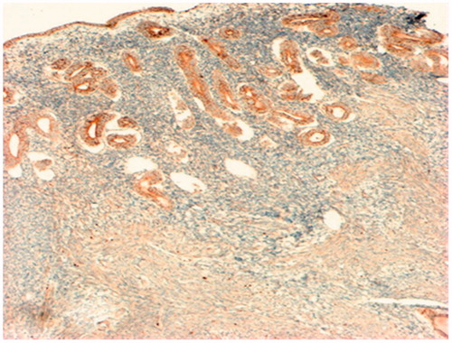
Eutopic epithelial and glandular epithelium was the most significant producer of VEGF in the endometrium. In the stroma of the eutopic endometrium, expression of this growth factor was lower – only 2% of cells were determined by a positive reaction to VEGF.
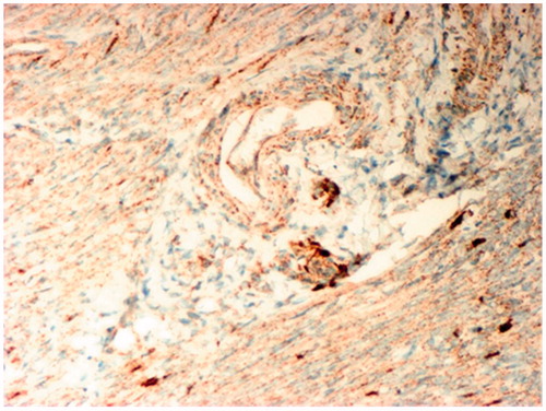
The expression of VEGF in the myometrium of patients with adenomyosis was more extensive in comparison to its expression in the endometrium. Here, the majority of cells had weak or moderate cytoplasmic reaction to VEGF (). Interestingly, in the myometrium, the expression of VEGF was detected in the cytoplasm of smooth myocytes and in the vascular wall, where its producers often served as smooth muscle cells and adventitia. Characteristically, in the perivascular compartment cells secreting VEGF, had an intense reaction (), and even more intense in patients with the painful form of adenomyosis (16.1 ± 1.2% versus 10.9 ± 1.9%).
Figure 2. Immunohistochemistry analysis of VEGF Expression in the myometrium of patients with chronic pelvic pain caused by adenomyosis. A weak reaction to MAT for VEGF in the cytoplasm of smooth myocytes and intense reaction in the cells of the perivascular compartment. IHC MAT to VEGF, original magnification 200×.
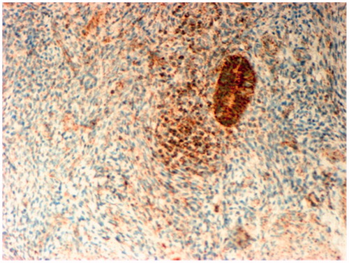
In addition, a severe reaction to VEGF was determined in individual cells between the bundles of smooth myocytes. Moderate expression of VEGF was also detected in the endothelium of thin-walled venous vessels in the myometrium sections of its remodeling (). In addition, a high cytoplasmic expression of VEGF in the glandular epithelium of adenomyosis foci was detected ().
Figure 3. Immunohistochemistry analysis of VEGF Expression in the walls of venous vessels in areas of myometrium remodeling in patients with adenomyosis. IHC MAT to VEGF, original magnification 80×.
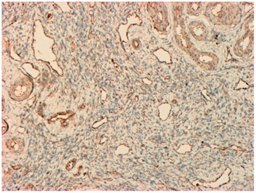
Figure 4. Immunohistochemistry analysis of VEGF Expression in foci of adenomyosis, associated mainly with the epithelial cells. IHC MAT to VEGF, original magnification 80×.
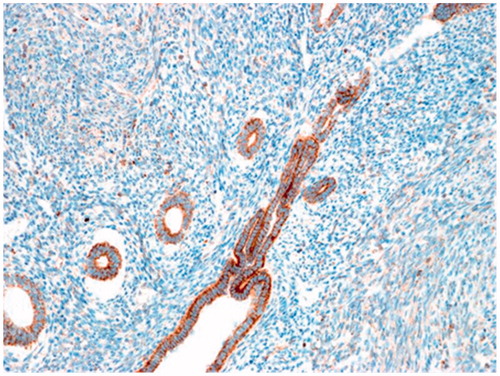
The VEGF expression was detected not only in epithelial cells of uterine glands, but in the stromal cells of the myometrium too. Theoretically, this could be due to the presence of myofibroblasts cells around areas of ectopic endometrial invasion, which are producers of a wide range of growth factors, including VEGF. However, as it turned out, the most pronounced expression of this stimulator of angiogenesis in the stroma of the myometrium is associated mainly with the areas of the remodeling of the myometrium, perivascular regions and with areas of infiltration ().
Figure 5. Immunohistochemistry analysis expression of VEGF in ectopic endometrium and surrounding myometrium stroma with signs of infiltration. IHC MAT to VEGF, original magnification 160×.
It is interesting that in the myometrium the expression of VEGF was detected both in the cytoplasm of smooth myocytes and in the vascular wall, where most often smooth myocytes and cells of the adventitia act as producers of the growth factor. In addition, a severe reaction to VEGF was determined in individual cells between the bundles of smooth myocytes. Moderate expression of VEGF has also been identified in the endothelium of thin-walled venous vessels in the myometrium sections of its remodeling. The cytoplasmic expression of VEGF in the epithelium and foci of adenomyosis was high.
Characteristically, the expression of VEGF in patients with the painful form of adenomyosis was significantly higher than the one in patients with the painless form: in the epithelial cells of ectopic endometrium (14.7 ± 1.6 versus 10.7 ± 1.6%, p < 0.01) in smooth myocytes of the myometrium (12.6 ± 1.4 versus 9.6 ± 1.2%, p < 0.01) in the stromal cells of the myometrium (10.1 ± 1.9 versus 7.4 ± 1.8%, p < 0.01). In the control group, in 3.5% of cases the expression of VEGF were associated only with the epithelium of the uterine glands of endometrioid heterotopias, and in the stromal cells of the myometrium, there was a complete lack of expression of VEGF.
The data obtained on the excessive expression of VEGF in adenomiose coincide with the results of the study [Citation14] where it was shown that it has a diffuse character, both in the myometrium and ectopic endometrium, the relative area of expression of VEGF in adenomyosis is of 11.51 ± 1.33%, which is 57% higher than in the control group, where the rate was equal to 7.32 ± 1.03%. But the obtained [Citation14] data did not concern patients with the painful form of adenomyosis.
According to the literature, VEGF is a key mediator of both angiogenesis and neurogenesis, and is intensely expressed in eutopic and ectopic endometrium of women with adenomyosis [Citation15]. It is also a mitogen for astroglia and Schwann cells in vitro [Citation3,Citation16]. Studies have shown that exogenous VEGF-A increases the number of processes of the sympathetic ganglia in explants [Citation17] and promotes the growth of sympathetic axons from surgically denervated adult rats [Citation18]. VEGF-A promotes the growth of axons, independently from its vascular role, through two receptors: either VEGFR2 [Citation19] or neuropilin-1 (NRP1) [Citation20]. NRP1 and NRP2, originally discovered as neuronal receptors of semaphorins, bind the most common splicing variant of VEGF-A (VEGF165) and form a complex with VEGFR-2 and VEGFR-1 to regulate their signaling [Citation21].
Recent evidence suggests that NRP1 in endothelial cells can also transmit signals independently of VEGF from VEGFR-2 [Citation22,Citation23], and that the loss of NRP1 or NRP2 associated with distinct vascular and neural defects [Citation24]. However, it is still unclear whether these processes happen directly through the angiogenesis induced by nerve growth factor (NGF), or indirectly by induction of the classic angiogenic factors such as VEGF [Citation25].
It is interesting to note that these two pleiotropic factors (VEGF and NGF) and other neurotrophin (NT-3, NT-4/5 and BDNF) expressed ectopic endometrial cells. Recent studies in vitro and in vivo provided the first clues about the mechanisms of pain in endometriosis. In adult female rats with surgical induction of endometriosis, the estrous dynamic changes of VEGF and NGF expression in endometrial cysts occur in parallel with changes in innervation and vascularization of the damaged tissues. It is assumed that the development of innervation in a model of endometriosis occurs through the interaction of perivascular nerve fibers accompanying the sprouting of blood vessels that vascularize the cyst [Citation26–29].
Conclusion
Thus, based on these data, complementing the existing theoretical understanding [Citation3], we can say that one of the important mechanisms of angiogenesis of adenomyosis and pathogenic mechanisms of the formation of chronic pelvic pain caused by adenomyosis is the increased expression of VEGF in eutopic and ectopic endometrium, in areas of remodeling of the myometrium and clusters of nerves and intensification of neovascularization.
Declaration of interest
The authors report no conflict of interest.
References
- Huang TS, Chen YJ, Chou TY, et al. Oestrogen-induced angiogenesis promotes adenomyosis by activating the Slug-VEGF axis in endometrial epithelial cells. J Cell Mol Med 2014;18:1358–71
- Brosens I, Pijnenborg R, Benagiano G. Defective myometrial spiral artery remodeling as a cause of major obstetrical syndromes in endometriosis and adenomyosis. Placenta 2013;34:100–5
- Orazov MR. The role of proliferation and apoptosis in the pathogenesis of pelvic pain in adenomioza. /M. R. Orazov, O. A. Dukhin, E. N. Nosenko//Doctor.Ru. Gynecol Endocrinol 2016;5–7
- Mu Y, Hu X, He J, et al. Serum levels of vascular endothelial growth factor and cancer antigen 125 are related to the prognosis of adenomyosis patients after interventional therapy. Int J Clin Exp Med 2015;8:9549–54
- Laschke MW, Giebels C, Menger MD. Vasculogenesis: a new piece of the endometriosis puzzle. Hum Reprod Update 2011;17:628–36
- Demir R, Yaba A, Huppertz B. Vasculogenesis and angiogenesis in the endometrium during menstrual cycle and implantation. Acta Histochem 2010;112:203–14
- Makanya AN, Hlushchuk R, Djonov VG. Intussusceptive angiogenesis and its role in vascular morphogenesis, patterning, and remodeling. Angiogenesis 2009;12:113–23
- Murasawa S, Asahara T. Endothelial progenitor cells for vasculogenesis. Physiology (Bethesda) 2005;20:36–42
- Bogoslovsky T, Spatz M, Chaudhry A, et al. Stromal-derived factor-1[alpha] correlates with circulating endothelial progenitor cells and with acute lesion volume in stroke patients. Stroke 2011;42:618–25
- Vodolazkaia A, Yesilyurt BT, Kyama CM, et al. Vascular endothelial growth factor pathway in endometriosis: genetic variants and plasma biomarkers. Fertil Steril 2016;105:988–96
- Nagy JA, Benjamin L, Zeng H, et al. Vascular permeability, vascular hyperpermeability and angiogenesis. Angiogenesis 2008;11:109–19
- Barcena de Arellano ML, Arnold J, Lang H, et al. Evidence of neurotrophic events due to peritoneal endometriotic lesions. Cytokine 2013;62:253–61
- Popov JN, Oparina TI, Prokopenko VM, Stepanov MG. Kliniko-patogeneticheskoe obosnovanie kombinirovannogo lechenija giperplasticheskih processov matki. Zhurnal Akusherstva I Zhenskih Boleznej 2010;4:71–5
- Zhang G, Dmitrieva N, Liu Y, et al. Endometriosis as a neurovascular condition: estrous variations in innervation, vascularization, and growth factor content of ectopic endometrial cysts in the rat. Am J Physiol Regul Integr Comp Physiol 2008;294:162–11
- Mani N, Khaibullina A, Krum JM, Rosenstein JM. Astrocyte growth effects of vascular endothelial growth factor (VEGF) application to perinatal neocortical explants: receptor mediation and signal transduction pathways. Exp Neurol 2005;192:394–406
- Kuruvilla R, Zweifel LS, Glebova NO, et al. A neurotrophin signaling cascade coordinates sympathetic neuron development through differential control of TrkA trafficking and retrograde signaling. Cell 2004;118:243–55
- Orazov MR. Expression of vascular endothelial growth factor (VEGF) in tissues of the uterus as one of the mechanisms of allogenes when adenomiose associated with chronic pelvic pain. /M. R. Orazov, V. N. Radzinsky, E. N. Nosenko//Pathol Physiol Exp Ther 2016;40–5
- Long H, Sabatier C, Ma L, et al. VEGF-A and Semaphorin3A: modulators of vascular sympathetic innervation. Dev Biol 2009;334:119–32
- Marko SB, Damon DH. VEGF promotes vascular sympathetic innervation. Am J Physiol Heart Circ Physiol 2008;294:2646–52
- Ruiz de Almodovar C, Fabre PJ, Knevels E, et al. VEGF mediates commissural axon chemoattraction through its receptor Flk1. Neuron 2011;70:966–78
- Erskine L, Reijntjes S, Pratt T, et al. VEGF signaling through neuropilin 1 guides commissural axon crossing at the optic chiasm. Neuron 2011;70:951–65
- Fuh G, Garcia KC, de Vos AM. The interaction of neuropilin-1 with vascular endothelial growth factor and its receptor flt-1. J Biol Chem 2000;275:26690–5
- Wang L, Zeng H, Wang P, et al. Neuropilin-1-mediated vascular permeability factor/vascular endothelial growth factor-dependent endothelial cell migration. J Biol Chem 2003;278:48848–60
- Wang L, Dutta SK, Kojima T, Xu X, et al. Neuropilin-1 modulates p53/caspases axis to promote endothelial cell survival. PLoS One 2007;2:e1161
- Geretti E, Shimizu A, Klagsbrun M. Neuropilin structure governs VEGF and semaphorin binding and regulates angiogenesis. Angiogenesis 2008;11:31–9
- Hansen-Algenstaedt N, Algenstaedt P, Schaefer C, et al. Neural driven angiogenesis by overexpression of nerve growth factor. Histochem Cell Biol 2006;125:637–49
- Benagiano G, Brosens I, Habiba M. Structural and molecular features of the endomyometrium in endometriosis and adenomyosis. Hum Reprod Update 2014;20:386–402
- Giudice LC. Clinical practice. Endometriosis. N Engl J Med 2010; 362:2389–98
- Mackenzie F, Ruhrberg C. Diverse roles for VEGF-A in the nervous system. Development 2012;139:1371–80