
Abstract
Chronic psychological stress has been considered to be a remarkable contributor to diminished ovarian reserve (DOR). However, there is a lack of a psychological stress-induced DOR animal model. We aim to validate the effects of an 8-week chronic unpredictable stress (CUS) paradigm on the ovarian reserve and reproductive hormone secretion of C57BL/6 mice. We found that after an 8-week CUS exposure, the numbers of primordial and preantral follicles and corpus luteum were significantly decreased in CUS model mice. Model mice also presented higher serum follicle-stimulating hormone, corticosterone levels and lower luteinizing hormone, estradiol, testosterone, anti-Müllerian hormone levels compared to those of control mice. Furthermore, we found that FSH receptor and AMH proteins were downregulated in model mouse ovaries. Although a significant litter size difference between the two groups was not found, the ovarian reserve remained significantly lower in the model group 6 weeks after CUS exposure. These results validated the hypothesis that the 8-week CUS paradigm that we adopted could induce the DOR phenotype in C57BL/6 mice and probably had a long-term adverse effect on ovarian reserve. Therefore, our results indicate that we have successfully established an animal model of psychological stress-induced DOR that can be used for further study.
摘要
长期的心理压力被认为是卵巢储备减少(DOR)的重要原因。但是, 缺乏心理压力诱发的DOR动物模型。我们旨在验证8周慢性不可预测压力(CUS)模式对C57BL / 6小鼠卵巢储备和生殖激素分泌的影响。我们发现, 在CUS暴露8周后, CUS模型小鼠的原始卵泡、窦前卵泡以及黄体的数量显著减少。与对照组相比, 模型小鼠的血清卵泡刺激素、皮质酮水平更高, 而黄体生成素、雌二醇、雄激素和抗苗勒管激素水平更低。此外, 我们发现FSH受体和AMH蛋白在模型小鼠卵巢中被下调。尽管两组之间产仔数未发现明显差异, 但暴露于CUS 6周后, 模型组的卵巢储备量显著降低。这些结果证实了以下假设:我们采用的8周CUS模型可以诱导C57BL/6小鼠的DOR模型, 并可能对卵巢储备造成长期不利影响。因此, 我们的结果表明我们已经成功建立了心理应激诱导的DOR动物模型, 可用于进一步研究。
The Chinese abstracts are translated by Prof. Dr. Xiangyan Ruan and her team: Beijing Obstetrics and Gynecology Hospital, Capital Medical University, Beijing 100026, China.
Introduction
Diminished ovarian reserve (DOR) is defined as an intermediate state between normal reproductive physiology and premature ovarian failure and is characterized by a decrease in the number or quality of available oocyte pool. Biomarkers of DOR used in clinical practice include but not limited to lower AMH, higher FSH, FSH/LH and antral follicle count (AFC) [Citation1]. The existing literature indicates that psychogenic factors have emerged as one of the major elements that associated with DOR and impaired female reproduction [Citation2–5]. However, due to the lack of a proper animal model that bears the similar symptoms of DOR, the mechanism of causality is still unclear.
Chronic unpredictable stress (CUS) is one of the most commonly used paradigms to induce anxious-like behavior in animal models and study chronic-stress-associated illness. CUS consists of the continuous exposure of animals to stressful situations or stressful stimuli that mimic the stress of everyday life. This model is widely applied to the study of the neurobiological processes that mediate the effects of chronic stress [Citation6]. There are only a few studies that utilize this type of model to study ovarian biology changes after stress exposure [Citation7–9]. To our knowledge, none of these studies has induced a typical DOR phenotype or explored both the ovarian follicle reserve and serum hormone changes that are associated with DOR.
The present study was undertaken to reveal a DOR phenotype change in C57BL/6 mice using a chronic unpredictable stress paradigm. A series of reproductive hormone changes and follicle development-related factors were also presented to validate the chronic stress damage on ovarian biological functions. A brief probe into the long-term effect of CUS on the ovarian morphology was performed by mating experiments with an ovarian histological analysis.
Materials and methods
Animals
Female C57BL/6 mice undergoing the stress paradigm were aged 6 to 8 weeks. Thirty mice were randomly and equally divided into the model group and the control group. The CUS paradigm experiment was repeated three times. Fifteen-week-old male mice were mated with female mice to evaluate the pregnancy rate. The mating experiment was repeated twice. All of the animal experimental procedures were approved by the Animal Experimental Ethical Committee of Fudan University. The body weight of mice from each group was measured every week. The ovary weight index was calculated using the following formula: ovary weight index = ovarian weight/body weight.
Chronic unpredictable stress (CUS)
The CUS procedure was based on previous reports with a few modifications [Citation10]. Two stressors were administered to each model mouse for eight weeks. To avoid tolerance, the strength of certain stressors was increased each week. Specifically, the stressors were as follows: restraint stress, tail suspension, exhausting swimming, orbital shaking, 45°cage tilt, wet bedding, empty cage, noise stress, flash light, social isolation, social crowding, light-dark shift, food deprivation, and water deprivation. The stressors were randomly scheduled over a one-week period and were repeated throughout the 8 weeks. Food or water deprivation and wet bedding were applied while avoiding the seventh day of each week to prevent the deviation of body weight. Nonstressed control mice were housed in groups without intervention. An example of the weekly CUS schedule is provided in Table 1 in the Supplementary Material. Mice were sacrificed after the 8-week CUS. Sample collection was carried out from 8–10 am when serum corticosterone (CORT) was at the base level of daily fluctuation.
Estrous cycling
Vaginal smears were taken every morning for 3 weeks before sacrifice to evaluate the estrous cycle. The experiment was repeated three times with each replicate containing 6 mice in each group. The estrous cycle stage was defined by a light microscopic analysis of the predominant cell type in vaginal smear samples [Citation11].
Serum hormone assay
Serum hormone was measured by enzyme-linked immunosorbent assay (ELISA) following the manufacturer’s instructions. Commercial ELISA kits were used to assess CORT, LH, FSH, anti-Müllerian hormone (AMH), testosterone (T), and E2.For each assay, there were 6–12 samples in each group with three replicates. Information on the ELISA kits used is listed in Table S2 in the Supplementary Materials.
Ovary serial section and follicle counting
For histological analysis, fixed ovaries were paraffin embedded, serially sectioned and stained for follicle counting based on previous methods [Citation12]. The total number of follicles and corpus luteum per ovary was calculated by adding the counts of every fifth section throughout the entire ovary. The follicles were classified into four stages according to the modified Oktay system [Citation13]. The counting was repeated three times with each replicate containing 6–8 left ovaries from 6–8 mice in each group.
Mating experiments
After 8 weeks of the stress paradigm, ten female mice from each group were selected for breeding to assess their fecundity. Female mice were mated with male C57BL/6 mice at the same age for 1 week at a 2:1 ratio per cage. After the mating period was terminated, the females were serially weighed and examined for signs of pregnancy. Numbers of F1 mice in two groups were counted. After weaning, maternal mice were sacrificed. The ovaries were collected and fixed as above and examined for histological changes. The experiment was repeated two times with each replicate containing 5 mice in each group.
Western blot
Ovaries from both groups were homogenized in lysis buffer. The lysates were boiled for 5 min and subjected to SDS-PAGE. The protein samples were then transferred to a polyvinylidene fluoride (PVDF) membrane and blocked with 5% bovine serum albumin in Tris-buffered saline Tween (TBST) for 1 h. The PVDF membranes were then incubated overnight at 4 °C with primary antibodies against follicle-stimulating hormone receptor (FSHR) (1:1000, PA5-50963, Invitrogen) or AMH (1:100, 103233, Abcam), followed by incubation with a secondary antibody (1:1000, 4414, Cell Signaling Technology) for 1 h at room temperature. Antibody binding was detected by the ECL method using the ECL Western Blotting Substrate (Millipore, WBKLS0500). The bands on the X-ray film were then scanned. The analysis was repeated two times with each replicate containing 5 mice from each group.
Immunohistochemistry
Ovarian sections were deparaffinized in xylene, re-hydrated in a graded ethanol series, and boiled in 10 mM sodium citrate for antigen retrieval. After incubation in a 0.3% H2O2 solution and blocking with 10% normal goat serum in 0.1% Triton X-100, sections were incubated overnight with anti-FSH receptor antibody (Abcam, ab150557) or anti-AMH antibody (Abcam, ab24542) at 4 °C. After washing, the sections were incubated for 2 h with HRP-conjugated horse anti-rabbit IgG at room temperature, visualized with DAB, dehydrated in ethanol, and mounted with coverslips for observation. The analysis was repeated three times with each replicate containing 6 slices of 6 ovaries from 6 mice in each group.
Statistical analysis
At least three replicates were performed for each treatment except for the mating experiments and post-labor ovarian follicle count. All data are presented as the mean value ± standard error (SEM). SPSS 22.0 software was used for statistical analysis. A comparison of the distributions of the continuous variables between two groups was analyzed using Student’s t-test. The comparison of body weight changes between two groups was made using repeated measurements analysis of variance. p values of less than .05 were considered statistically significant.
Results
Effects of CUS exposure on mice stress biomarkers and ovarian function
Body weight (), ovarian weight index () and serum level of CORT () were monitored to control for the efficacy of stressors. In the model group subjected to the 8-week protocol of CUS, the mean body weight was significantly decreased during the 8-week protocol (p < .001). There was also a significant increase of serum CORT (p < .001) and a significant decrease of the ovary weight index (p < .001) in model group.
Figure 1. The validity of CUS to induce stress in mice and impair reproductive function. (A) Ovary weight index. (B) Mean serum CORT concentrations. (C) Number of follicles at different stages. (D) Mean cycle length of different stages of estrous cycle in the final two weeks before sacrifice. (E-J) Serum reproductive hormone concentrations. M = CUS model group; C = control group; primordial: primordial follicle; primary: primary follicle; secondary: secondary follicle; antral: antral follicle; atretic: atretic follicle; *: p < .05; **: p < .01; ***: p < .001.
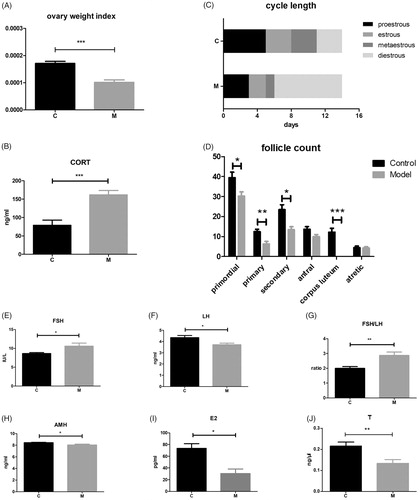
Table 1. Comparison of weekly body weight in model and control mice.
Mice from the model group exhibited a significant increase of the mean length of the diestrous phase of the estrous cycle (). There was also a decrease in the days of the estrous phase compared to the control group. The numbers of primordial follicles (p < .05) and preantral follicles (primary follicle, p < .01; secondary follicle, p < .05) were significantly lower in the model group. There was also a significant decrease in the number of corpus luteum in model group (p < .05), which proved the ovulation inhibiting effect of CUS on mice ovaries ().
Moreover, consistent with the morphology changes in the ovaries, CUS also induced disturbances in sex hormone secretion (). Serum FSH (p < .05), FSH/LH (p < .01) in the model group was significantly increased, while LH (p < .05), AMH (p < .05), E2 (p < .05) and T (p < .01) was significantly decreased than those of the control group.
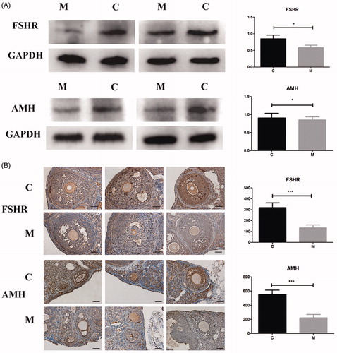
CUS suppresses AMH and FSH receptor (FSHR) protein expression in mouse ovaries
Immunohistochemistry and western blotting were employed to evaluate the expression of several markers of folliculogenesis. As shown in , the model group showed significantly lower FSHR and AMH protein expression in model mouse ovaries. FSHR is a transmembrane receptor that interacts with FSH to regulate follicle development. AMH is expressed in the granulosa cells of the preantral and small antral follicles in ovaries. As shown in the immunohistochemical analysis, FSH was expressed less in antral follicles, and AMH was expressed less in preantral and small antral follicles in the model group compared to that in the control group.
Figure 2. Protein expression analysis of FSHR and AMH in the ovaries. (A) Western blot images of FSHR and AMH in the two groups. (B) Immunoreactivity staining results showing FSHR and AMH protein expression in antral follicles and preantral follicles. Magnification: ×400. Scale bar = 12.5 μm.
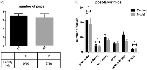
CUS does not significantly affect mice fecundity, but has a long-term effect on follicle depletion
After the 8-week treatment, model mice exhibited a slightly lower fertility rate. However, the number of pups was similar between the two groups (). After 21 days of pregnancy and 21 days of lactation, follicle depletion effects were still found in the model group. () The numbers of primordial follicles, primary follicles and antral follicles in the model group were still significantly lower than those in the control group. There was also an increase in atretic follicles in the model group.
Figure 3. Fecundity of mice in both groups and long-term stress effect in mice. (A) Litter size and fertility rate of both groups. (B) The number of follicles at different stages in postlabor mice.
Discussion
Clinical studies have suggested various links between accelerated follicle decline and psychological disorders [Citation14]. However, direct evidence of the depletion of the resting pool of follicles can be reliably provided only through an assessment of the total number of follicles in whole ovaries after oophorectomy. This highlights the importance of developing an animal model of DOR induced by chronic psychological stress. In this study, we adopted an 8-week CUS paradigm on C57BL/6 mice to mimic the various stressors women experience every day and valid the direct effects of stress on female mice reproduction.
As the most widely used inbred strain of genetically modified mice, C57BL/6 mice seem to be less vulnerable to stress than other mouse strains [Citation15,Citation16]. Therefore, we observed the effect of CUS on mice after a series of different stress exposure time (data not given). And we observed significant reproductive biological changes under the 8-week stress paradigm, longer than the typical 4-week paradigm; and chose this stress exposure time to be the suitable condition to induce DOR. Body weight gain and serum CORT level are other biomarkers of chronic stress [Citation17–19]. We observed a distinct disturbed body weight gain during the CUS process and increased serum CORT level after the 8-week CUS treatment.
In previous animal studies, researchers found that a 30-day CUS regimen could reduce secondary and antral follicle depletion in mice [Citation7]. However, they did not observe primordial or primary follicle changes. In this study, using the 8-week CUS paradigm, we demonstrated that the numbers of primordial follicles and the stages of preantral follicles were all significantly decreased.
Previous clinical studies have revealed that women under high daily stress exhibit higher serum FSH level and lower LH and E2 levels as well as higher odds of anovulation [Citation2,Citation20]. Using the CUS animal model, we found similar higher FSH and lower LH and E2 changes in model mice serum, which is in accordance with these clinical studies. Downregulated FSHR protein expression confirmed damaged hormone responsiveness in model mice. AMH is one of the most often used markers of the ovarian reserve, it has also been revealed to be related to early follicle depletion [Citation21–22]. Another hormone with emerging importance is T, the precursor of E2. Both AMH and T have been revealed to be effective in improving the functional ovarian reserve in females with DOR [Citation23–28]. Our study found the same decreased serum T and AMH levels in CUS model mice. These results further verified that our 8-week CUS paradigm can cause the DOR phenotype in mice.
We did not observe significantly damaged fertility in the mating experiment. However, despite the fertility rate, a follicle depletion effect still existed in model mice 6 weeks after the stress paradigm. This result suggests the possibility of long-term adverse effects of chronic stress on the ovarian reserve.
In light of these results, our study proves that an 8-week CUS paradigm can induce the diminished ovarian reserve phenotype in C57BL/6 mice. CUS may also exert a long-term detrimental effect on DOR in mice. The stressed DOR animal model provides further opportunities to study the mechanisms, complications and treatment of DOR induced by psychological factors.
Supplementary_Material-revised_2019.04.02.docx
Download MS Word (22.2 KB)Disclosure statement
No potential conflict of interest was reported by the authors.
Additional information
Funding
References
- Cohen J, Chabbert-Buffet N, Darai E. Diminished ovarian reserve, premature ovarian failure, poor ovarian responder—a plea for universal definitions. J Assist Reprod Genet. 2015;32:1709–1712.
- Pal L, Bevilacqua K, Santoro NF. Chronic psychosocial stressors are detrimental to ovarian reserve: a study of infertile women. J Psychosomat Obstet Gynecol. 2010;31:130–138.
- Williams KE, Marsh WK, Rasgon NL. Mood disorders and fertility in women: a critical review of the literature and implications for future research. Human Reproduction Update. 2007;13:607–616.
- Kaplan JR, Manuck SB. Ovarian dysfunction, stress, and disease: a primate continuum. ILAR Journal. 2004;45:89–115.
- Harlow BL, Wise LA, Otto MW, et al. Depression and its influence on reproductive endocrine and menstrual cycle markers associated with perimenopause: the Harvard Study of Moods and Cycles. Arch Gen Psychiatry. 2003;60:29.
- Willner P. The chronic mild stress (CMS) model of depression: history, evaluation and usage. Neurobiol Stress. 2017;6:78–93.
- Wu LM, Liu YS, Tong XH, et al. Inhibition of follicular development induced by chronic unpredictable stress is associated with growth and differentiation factor 9 and gonadotropin in mice. Biol Reprod. 2012;86:121.
- Wu LM, Hu MH, Tong XH, et al. Chronic unpredictable stress decreases expression of brain-derived neurotrophic factor (BDNF) in mouse ovaries: relationship to oocytes developmental potential. PLoS One. 2012;7:8.
- Gao Y, Chen F, Kong Q-Q, et al. Stresses on female mice impair oocyte developmental potential. Reprod Sci. 2016;23:1148–1157.
- Monteiro S, Roque S, de Sa-Calcada D, et al. An efficient chronic unpredictable stress protocol to induce stress-related responses in C57BL/6 mice. Front Psychiatry. 2015;6:6.
- Turner HE, Vivien T. Principles and practice of endocrinology and metabolism. 3rd edition on CD-ROM. Clin Endocrinol. 2003;59:655–655.
- Bolon B, Bucci TJ, Warbritton AR, et al. Differential follicle counts as a screen for chemically induced ovarian toxicity in mice: results from continuous breeding bioassays. Fundam Appl Toxicol. 1997;39:1–10.
- Oktay K, Schenken RS, Nelson JF. Proliferating cell nuclear antigen marks the initiation of follicular growth in the rat. Biol Reprod. 1995;53:295–301.
- Bleil ME, Adler NE, Pasch LA, et al. Depressive symptomatology, psychological stress, and ovarian reserve: a role for psychological factors in ovarian aging? Menopause-J N Am Menopause Soc. 2012;19:1176–1185.
- Razzoli M, Carboni L, Andreoli M, et al. Different susceptibility to social defeat stress of BalbC and C57BL6/J mice. Behav Brain Res. 2011;216:100–108.
- Anisman H, Hayley S, Kelly O, et al. Psychogenic, neurogenic, and systemic stressor effects on plasma corticosterone and behavior: mouse strain-dependent outcomes. Behav Neurosci. 2001;115:443–454.
- Nollet M, Le Guisquet AM, Belzung C. Models of depression: unpredictable chronic mild stress in mice. Curr Protoc Pharmacol. 2013;61:5.65.1–5.65.17.
- Yeon JJ, Hoon LD, Kang SS. Effects of chronic restraint stress on body weight, food intake, and hypothalamic gene expressions in mice. Endocrinol Metab. 2013;28:288–296.
- Gong S, Miao YL, Jiao GZ, et al. Dynamics and correlation of serum cortisol and corticosterone under different physiological or stressful conditions in mice. PLoS One. 2015;10:e0117503.
- Schliep KC, Mumford SL, Vladutiu CJ, et al. Perceived stress, reproductive hormones, and ovulatory function a prospective cohort study. Epidemiology. 2015;26:177–184.
- La Marca A, Sighinolfi G, Radi D, et al. Anti-Mullerian hormone (AMH) as a predictive marker in assisted reproductive technology (ART). Hum Reprod Update. 2010;16:113–130.
- Depmann M, Broer SL, van der Schouw YT, et al. Can we predict age at natural menopause using ovarian reserve tests or mother's age at menopause? A systematic literature review. Menopause. 2016;23:224–232.
- Durlinger AL, Kramer P, Karels B, et al. Control of primordial follicle recruitment by anti-Mullerian hormone in the mouse ovary. Endocrinology. 1999;140:5789–5796.
- Gleicher N, Barad DH. Dehydroepiandrosterone (DHEA) supplementation in diminished ovarian reserve (DOR). Reprod Biol Endocrinol. 2010;21:360–365.
- Gleicher N, Weghofer A, Barad DH. The role of androgens in follicle maturation and ovulation induction: friend or foe of infertility treatment? Reprod Biol Endocrinol. 2011;9:116.
- Lu Q, Shen H, Li Y, et al. Low testosterone levels in women with diminished ovarian reserve impair embryo implantation rate: a retrospective case-control study. J Assist Reprod Genet. 2014;31:485–491.
- Sen A, Hammes SR. Granulosa cell-specific androgen receptors are critical regulators of ovarian development and function. Mol Endocrinol. 2010;24:1393–1403.
- Gleicher N, Kim A, Weghofer A, et al. Hypoandrogenism in association with diminished functional ovarian reserve. Hum Reprod. 2013;28:1084–1091.