
Abstract
Objective
To assess the impacts of Platelet-Rich Plasma(PRP) and Granulocyte Colony-Stimulating Factor(G-CSF) on a rat model with induced ovarian follicular damage caused by cyclophosphamide(Cy).
Materials and Methods
Forty-two Sprague–Dawley rats were randomly allocated into seven distinct groups as; Group 1(control): NaCl intraperitoneal (IP) injection was administered on days D1, D7, and D14. Group 2(Cy):Cy IP injection on D1 + NaCl IP injection on D7 and D14 were administered. Group 3(PRP): PRP IP injection on D1,D7 and D14 were administered. Group 4(Cy + PRP):Cy IP injection on D1 and PRP IP injection on D1, D7 and D14 were administered. Group 5(G-CSF): G-CSF IP injection on D1, D7 and D14 were administered. Group 6(Cy + G-CSF):Cy IP injection on D1+ G-CSF IP injection on D1, D7 and D14 were administered. Group 7(Cy + PRP + G-CSF):Cy IP injection on D1+ PRP IP injection on D1,D7 and D14+ G-CSF IP injection on D1,D7 and D14 were administered. Follicular number, histological scores of AMH and INSL3 stained follicles at different stages of follicular development, and serum Anti-Müllerian hormone(AMH) were evaluated.
Results
The primary, secondary, and antral follicle intensity scores for AMH-positive staining were most prominent in Groups 3 and 5. There was no significant difference between groups 4, 6 and 7 compared to group 1 in terms of follicule counts and AMH staining. The intensity scores of AMH-positive staining follicles were notably reduced in group 2 compared to groups 4, 6, and 7, with a significant difference (p < .01). Among the groups, group 2 exhibited the least intense antral follicle staining for INSL3, displaying a significant difference(p < .01) compared to the remaining groups.
Conclusions
Autologous PRP and G-CSF might protect ovarian function in the face of ovarian damage caused by Cy-induced effects.
Introduction
The ovaries’ development and function are crucial to a fertility function [Citation1]. Premature ovarian insufficiency (POI) is characterized by the halt of ovarian activity in women who are under the age of 40 [Citation2]. It is portrayed by primary/secondary amenorrhea, infertility, estrogen deficiency and elevated gonadotrophin levels [Citation3]. The pathologic mechanism of POI is not completely understood. There are multifactorial contributors such as autoimmune damage, environmental factors and a multitude of genetic deficiencies have been identified [Citation4]. In the past few decades, significant therapeutic progress has been achieved in oncology which has improved the survival rates of children and young adults. However, the excellent success results are achieved with compromise on reproductive function and subsequent POI [Citation5].
Cyclophosphamide is an alkylating agent widely used in many cancer treatment protocols and autoimmune disorders. Cyclophosphamide (Cy) use leads to accelerated loss of ovarian reserve follicules, disruption of folliculogenesis, and malfunctioning of steroid synthesis [Citation6]. Dysfunction of the ovary and consequent POI in rats which is treated with Cy is related to destruction of the granulosa cells [Citation7]. Cy requires metabolic activation with the end metabolite being phosphoramide mustard which is responsible for the toxic effect [Citation8]. The cumulative toxicity of Cy is generated by its production of oxidative stress, induction of apoptosis, and increased inflammatory effects. Additionally, Cy is thought to provoke POI via its damaging activation of dormant primordial follicles [Citation9].
Several agents in the last decade have been studied to ameloriate the damage caused by Cy treatment. One agent is PRP (Platelet Rich Plasma), a derivative of whole blood after the blood is centrifuged and separated. The centrifugation procedure generates the elimination of red blood cells and the production of an enriched plasma containing higher concentration of growth factors. Upon activation; the alpha granules of platelets contribute to release of many growth factors such as VEGF (Vascular Endothelial Growth Factor), PDGF (Platelet Derived Growth Factor) and TGF-β (Transforming Growth Factor Beta) [Citation10]. These growth factors present in PRP have been demonstrated to have a significant role. These growth factors promote angiogenesis and the regeneration of tissues [Citation11,Citation12]. The main advantage of utilizing PRP is the autologous nature; accordingly, there are no risks associated with an immune reaction nor microorganism transmission from other donors [Citation13]. The efficacy of PRP has been demonstrated in the rat POI model induced by Cy [Citation14].
Another substance used to alleviate ovarian damage is G-CSF (Granulocyte colony-stimulating factor), a cytokine growth factor family member. It is extensively employed for mobilizing stem cells, as it triggers the movement of hematopoietic stem cells from the bone marrow to the bloodstream [Citation15]. G-CSF is a hematopeitic growth factor. It regulates the mobilization, proliferation, and differentiation of bone marrow cells which are responsible for fighting infection and promoting healing. G-CSF has been utilized in treating congenital neutropenia or in cases of neutropenia induced by chemotherapy. Beyond its ability to promote proliferation and differentiation of bone marrow cells. G-CSF plays a role in anti-inflammatory and anti-apoptotic cell function. Research has shown that G-CSF may reduce follicular degeneration particularly after cisplatin chemotherapy [Citation16].
Antral Follicule Count (AFC) and the established biochemichal including estradiol, antimullerian hormone (AMH) and inhibin B markers, mainly derived from granulosa cells, are widely used to predict and evaluate ovarian function. Insulin-like factor 3 (INSL3) is a relatively recent peptide hormone identified in women. INSL3 stands out from the rest of the circulating hormones due to its ability to mirror the activity of theca cells within recruited antral follicles. Research findings indicate a positive correlation between the level of INSL3 and the count of antral follicles (AFC) [Citation17,Citation18].
In this study, our aim was to evaluate the possible positive effects of PRP and G-CSF on an experimental rat model with Cy-induced ovarian follicular damage by evaluating the different stages of follicullar number (primordial, secondary, antral and atretic follicle counts), histological scores of AMH and INSL3 stained follicules at different stages of follicullar development and serum Anti-Müllerian hormone (AMH).
Material and methods
Animals
Forty-two female Sprague–Dawley rats weighing 200–250 g; were utilized in the experiments. The animals were accommodated in the Experimental Animal Laboratory of Bezmialem University. The rats were maintained under a 12-h light-dark cycle and had unrestricted access to food and water. The experimental procedure received approval from the Institutional Animal Care and Use Committee of Bezmialem Vakif University. (2021/141//24.05.2021). All steps of the procedures were conducted in adherence to the guidelines outlined in the National Academy of Sciences Guide for the Care and Use of Laboratory Animals. (1996).
Experimental design
The animals were randomly allocated into seven groups by one investigator blindly, each with six rats ().
Figure 1. The Time frame of the experiments. The animals were divided into seven groups containing six rats in each group.
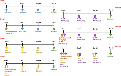
Group 1(control): On days 1 (D1), 7 (D7), and 14 (D14), a single intraperitoneal (IP) injection of 0.9% sodium chloride solution (1 mL/kg) was administered.
Group 2(Cy): On day 1 (D1), a single intraperitoneal (IP) injection of Cy (75 mg/kg) was administered. On days 7 (D7) and 14 (D14), single IP injections of 0.9% sodium chloride solution (1 mL/kg) were given. The selection of drug doses was determined using the parameters established in a previous study published by our group [Citation14].
Group 3(PRP): IP injections of PRP (200 μl) were administered as single doses on days 1 (D1), 7 (D7), and 14 (D14).
Group 4(Cy + PRP): A single intraperitoneal (IP) injection of Cy (75 mg/kg) was given on day 1 (D1), while PRP (200 μl) IP injections were administered as single doses on days 1 (D1), 7 (D7), and 14 (D14).
Group 5 (G-CSF): IP injections of G-CSF (100 IU/kg, Leucostim 30 MIU; Dem ilaç, Istanbul, Turkey) were given as single doses on days 1 (D1), 7 (D7), and 14 (D14). The selection of drug doses was based on parameters established in a previously published study. [Citation15].
Group 6 (Cy + G-CSF): A single intraperitoneal (IP) injection of Cy (75 mg/kg) was administered on day 1 (D1), along with IP injections of G-CSF (100 IU/kg, Leucostim 30 MIU; Dem ilaç, Istanbul, Turkey) given as single doses on days 1 (D1), 7 (D7), and 14 (D14).
Group 7 (Cy + PRP + G-CSF): On day 1 (D1), a single intraperitoneal (IP) injection of Cy (75 mg/kg) was administered, along with IP injections of PRP (200 μl) as single doses on days 1 (D1), 7 (D7), and 14 (D14). Additionally, IP injections of G-CSF (100 IU/kg, Leucostim 30 MIU; Dem ilaç, Istanbul, Turkey) were given on days 1 (D1), 7 (D7), and 14 (D14).
Preparing platelet-rich plasma
Six mature male Sprague–Dawley rats other than those used in the experiments were utilized to prepare PRP. The right ventricle was punctured to collect whole blood. The collected samples were transferred into test tubes containing 3.2% sodium citrate (Merck, Darmstadt, Germany). Afterwards centrifugation was performed at 400 × g for 10 min. Following the removal of the buffy coat centrifugation was repeated at 800 × g for 10 min in another tube. The upper 2/3 of the supernatant containing platelet-poor plasma was discarded. The remaining lower layer (1/3) was used for further procedures.
Sample collection
On day 21 (D21), all the animals were weighed and then anesthetized through intramuscular injection of 50 mg/kg ketamine hydrochloride (Ketalar; Eczacibasi Warner-Lambert Ilac Sanayi, Levent, Istanbul, Turkey). Blood samples were collected to measure AMH levels once the rats were immobilized on a surgical table. A ventral midline incision was used to enter the abdomen under sterile conditions. The ovaries were removed for histomorphometric and immunohistochemical analysis.
Serum AMH concentrations
Serum AMH levels were assessed using the USCN Life Science Enzyme-Linked Immunosorbent Assay. The assay’s range spanned from 0.31 to 20 ng/mL, with a minimum detectable dose of less than 0.078 ng/mL.
Histological analysis and ovarian follicle count
The histopathologic examination was performed by same histologist blinded to the groups. The rats’ ovaries were carefully removed and then immersed in 10% neutral formalin for 72 h to ensure fixation. Following fixation, the ovaries underwent a thorough water rinse and were subjected to a gradual dehydration process using ascending concentrations of alcohol (ranging from 70% to 100%). Subsequently, the specimens were immersed in paraffin and left overnight at 60 °C. Sections measuring 5 µm in thickness were obtained using the paraffin blocks and mounted onto slides. Paraffin sections were stained with Hematoxylin & Eosin (H&E) for histomorphometric analysis. A photomicroscope (ZEISS Axio Zoom.V16, Germany) was utilized to count number of primary, secondary, antral and atretic follicles using the techniques described by Tilly et al. [Citation19]. Follicle counts were performed on 10 serial sections taken at 50 micrometer intervals from each sample by the same histologist.
Immunohistochemistry of AMH and INSL3
Following deparaffinization and rehydration, the sections were exposed to a solution containing 3% hydrogen peroxide in methanol for 10 min to inhibit the activity of endogenous enzymes. Following the hydrogen peroxide treatment, the sections were washed and then subjected to microwave irradiation at 200 W using a citrate buffer (pH 6.1) for 20 min to facilitate antigen retrieval. Subsequentially phosphate buffered saline (PBS) wash was performed. Incubation in blocking solution for 10 min and then incubation in mouse anti-Mullerian hormone (AMH) antibody (1:20, GeneTex, Cat: GTX42794) and Insulin-like 3 (INSL3) antibody (1:50, Novus Biologicals, Cat: NBP3-04435) at 4 °C overnight was undertaken. After antigen retrieval, secondary antibody staining was carried out using the Histostain®-Plus 3rd Gen IHC Detection Kit (Cat: 85-9073, Invitrogen, CA, USA), following the manufacturer’s instructions. After washing, the sections were exposed to streptavidin–peroxidase (ready-to-use) for a duration of 10 min at room temperature, followed by incubation with 3,3′-diaminobenzidine (DAB) for 5 min. Finally, the slides were counterstained using Mayer’s hematoxylin and subsequently covered with a mounting medium. In the each section, AMH positive follicles were counted and evaluated the staining intensity (0 to 3 as follows: 0, no staining; 1, weak staining; 2, moderate staining; and 3, strong staining) semi-quantitatively. INSL3 intensity of folicular theca layer was evaluated semi-quantitatively as AMH scoring. For AMH and INSL3 immunohistochemistry staining, 3 serial sections taken from each sample at 100 micrometer intervals were evaluated by the same histologist.
Statistical analysis
A sample size and power calculation determined that sufficient statistical power required six rats for each group (power = 0.80, type 1 error = 0.05 and type 2 error = 0.20). The power calculation was based on primordial follicle count to estimate a difference of 5.56 units at a 95% confidence level (α = 0.05 significance level,80% power) based on a previously published study [Citation16]. Statistical analysis was performed using GraphPad Prism 6.0 software (GraphPad Software, Inc., La Jolla, CA, USA). Data were analyzed by One-Way ANOVA followed by Tukey test. All data were presented as mean ± standard error. p values were regarded as significant when it was lower than 0.05. (*p < .05, ** p < .01, *** p < .001, **** p < .0001).
Results
The animals were randomly allocated into seven groups, each with six rats (). and –Citation4 display the serum AMH concentrations, follicle counts (including primordial, primary, secondary, atretic, and antral), as well as the counts and intensity scores of primary, secondary, and antral AMH positive staining follicles. The antral INSL3 positive staining follicle intensity scores for all groups are also presented. Statistically significant differences were found in terms of all parameters between six groups (p < .05).
Figure 2. (a) Number of follicles. Primordial, primary, secondary, antral, and atretic follicle counts of ovaries in the all groups. ++ p < .01, +++ p < .001 ++++ p < .0001, compared to group 1; * p < .05, ** p < .01, *** p < .001, **** p < .0001, compared to group 2.2. (b) Serum concentrations of AMH.
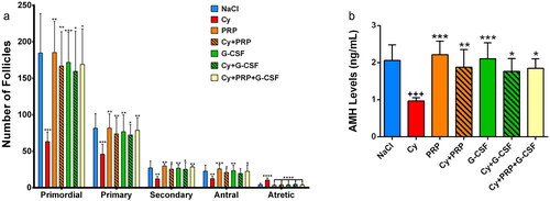
Figure 3. (a) Primary, secondary, and antral AMH positive staining follicle counts in the all groups. ++ p < .01, +++ p < .001, ++++ p < .0001, compared to group 1; * p < .05, ** p < .01, *** p < .001, **** p < .0001, compared to group 2. (b) Primary, secondary and antral AMH positive staining follicle intensity score in the all groups. ++++ p < .0001, compared to group 1; **** p < .0001, compared to group 2. 3c) Immunohistochemistry of AMH in the ovary. In the upper panel, AMH positive stained primary follicules are seen in the ovary sections. Densely stained primary follicles were observed in NaCl, PRP and G-CSF groups. In Cy group, primary follicles were stained weakly. Intensity of AMH was increased and stained moderately in Cy + PRP, Cy + G-CSF and Cy + PRP + G-CSF groups. (Bar: 50 μm) In the Middle panel, AMH positive stained secondary follicules are seen in the ovary sections. Intensity of AMH was stained strongly in PRP and G-CSF groups. In Cy group, secondary follicles were stained weakly. In NaCl, Cy + PRP, Cy + G-CSF and Cy + PRP + G-CSF groups, intensity of AMH was stained moderately. (Bar: 100 μm) In the bottom panel, AMH positive stained antral follicules are seen in the ovary sections. Intensity of AMH was stained strongly in PRP and G-CSF groups. In Cy group, secondary follicles were stained weakly. In NaCl, Cy + PRP, Cy + G-CSF and Cy + PRP + G-CSF groups, intensity of AMH was stained moderately. (Bar: 200 μm).
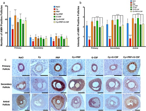
Table 1. Comparison of the primary, secondary and antral AMH-positive staining follicle counts, AMH-positive staining follicle intensity, antral INSL3 positive staining follicle intensity and primordial, primary, secondary, antral, atretic follicle counts of all groups.
The mean serum AMH concentrations were 2.05 ± 0.42, 0.96 ± 0.21, 2.21 ± 0.36, 1.87 ± 0.47, 2.1 ± 0.43, 1.76 ± 0.34 and 1.84 ± 0.26 ng/ml in groups 1, 2, 3, 4, 5, 6 and 7 respectively (p < .0001) ( and ). Group 2 has the lowest serum AMH levels. The serum AMH concentrations exhibited significant increases in groups 4, 6, and 7 compared to group 2 (p < .01, p = .02, p = .02, respectively). No significant differences were observed between groups 4, 6, and 7 compared to group 1 (p = .97, p = .85, p = .97, respectively).
Significant differences were observed in the counts of primordial, primary, secondary, antral, and atretic follicles among all the groups. Group 2 exhibited the lowest counts of primordial, primary, and secondary follicles and the highest count of atretic follicles among all the groups. The number of atretic follicles was statistically significantly lower in groups 4,6 and 7 compared to group 2 (p < .01). There was no significant difference between group 4,6,7 compared to group 1. The counts of primordial, primary, secondary, and antral follicles were notably more significant in group 4 compared to group 2, with significant differences observed (p < .01, p < .01, p = .02, and p = .02, respectively). Compared to group 2, the primordial, primary, and secondary follicle counts were significantly elevated in group 6 (p = .02, p = .02, and p = .03, respectively). No statistically significant differences were observed between groups 4 and 6, 3 and 5, 4 and 7, and 6 and 7 about the primordial, primary, antral, and atretic follicle counts.
Primary, secondary and antral AMH positive staining follicle counts were the most intense in group 3 and least in group 2. A statistically significant distinction was observed in the AMH positive staining when comparing the primary, secondary, and antral follicle counts across the groups. (p < .0001, p = .0003 and p < .0001 respectively) (). In group 2, the AMH-positive staining of both primary and secondary follicle counts was notably lower compared to group 4, group 6, and group 7; this difference was statistically significant. (p < .01, p = .02; p < .01, p = .03; p < .01, p < .01 respectively). No statistically significant differences were observed in AMH-positive staining when comparing the primary, secondary, and antral follicle counts between groups 1 and 3, groups 1 and 5, and groups 3 and 5. AMH positive staining of primary and secondary follicle count was no significant difference in group 1 compared to group 4, group 6 and group7. (p = .25, p = .8; p = .052, p = .36; p = .8, p = .95 respectively).
The primary, secondary, and antral AMH-positive staining follicle intensity score was lowest in group 2.
The primary, secondary, and antral AMH-positive staining follicle intensity score was highest in both group 3 and group 5. The intensity scores of AMH-positive staining for primary, secondary, and antral follicles were notably lower in group 2 compared to group 4, group 6, and group 7. This difference in intensity scores was statistically significant. (p < .01 for all). Primary, secondary, and antral AMH positive staining follicle intensity scores of Cy + PRP applied to group 4 and Cy + G-CSF applied to group 6; no statistically significant difference was found when compared to group 7 in which both were involved (p = .99).
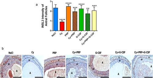
Antral INSL-3 positive staining follicle intensity score in all groups are shown in and . Group 2 had the lowest antral INSL3 positive staining follicle intensity score compared to the other groups (p < .01). Antral INSL-3 positive staining follicle intensity score was significantly higher in group 4,6,7 compared with group 2 (p < .01). There was no significant difference between groups 3 vs. 5, 4 vs. 7, and 6 vs. 7 in antral INSL-3 positive staining follicle intensity score. Also there was no significant difference between group 1 compared to group 4,6 and 7 (p = .45, p = .08, p = .85).
Figure 4. (a) INSL3 intensity of antral follicles. ++++ p < .0001, compared to group 1; ** p < .01, *** p < .001, **** p < .0001, compared to group 2. (b) Immunohistochemical analysis of INSL3 in the antral follicles of ovaries. Immunoreactivity of INSL3 was detected as brown staining. Strong INSL3 staining was observed in the follicular theca and granulosa cell layer in NaCl, PRP and G-CSF groups. Weakly INSL3 staining was detected both follicular theca and granulosa cell layer in Cy group. Moderate INSL3 staining was observed in Cy + PRP, Cy + G-CSF and Cy + PRP + G-CSF groups. A, antrum of antral follicle; tc, follicular theca cell layer; gc, granulosa cell layer. All figures include same magnification. Bar: 200 μm.
Discussion
The global overall prevalence of POI among women is 3.5%[Citation20]. Chemotherapy-induced Primary Ovarian Insufficiency (POI), also known as iatrogenic POI, is a frequently observed outcome of cancer treatments.
Chemotherapy drugs exhibit diverse mechanisms of ovarian toxicity and affect ovarian follicles at different stages of development. Alkylating agents like Cyclophosphamide (Cy) lead to the gradual reduction of ovarian reserve by affecting primordial follicles through cumulative impact [Citation21]. Germ cells are deposited as primordial follicles in the human ovary and they may be activated under physiological conditions [Citation22]. Stimulating the activation of these primordial follicles could potentially hold the solution for effectively managing women with Primary Ovarian Insufficiency (POI). Cy, one of the most reported toxic agents leading to ovarian failure, does not depend on the cell cycle and has the potential to deter ovarian function in females at any age [Citation23]. Deghgani et al. showed that the number of follicles at different developmental stages, especially follicles in the preantral stage, were significantly diminished due to Cy induced POI [Citation24].
In this study, we focused mainly on the potential protective effects of PRP and GCSF on ovarian function against Cy-induced ovarian damage. Cyclophosphamide was used in our animal model for inducing ovarian damage. Our data demonstrated the catastrophic damage caused by Cy in the ovaries. However, the hormonal, histopathological, and immunohistochemical methods used in our study clearly demonstrated the possible ameliorative effects of PRP and GCSF against ovarian damage induced by Cy. Following the administration of PRP and G-CSF, a significant increase was observed in the serum levels of Anti-Müllerian Hormone (AMH) and in the counts of primordial, primary, secondary, and antral follicles. Additionally, the number of follicles displaying positive staining for AMH substantially rose, while the count of atretic follicles notably decreased.
Additionally INSL-3 staining scores were increased compared to Cy treatment. INSL3 was previously considered to be a male-specific hormone secreted by testicular Leydig cells [Citation25]. Later it was discovered to be secreted in theca interna cells of females [Citation26]. Zhu et al. demonstrated that serum INSL3 decreased in patients with POI and therefore suggested that INSL3 may be of use as an auspicious theca-cell specific marker for POI [Citation18]. Furthermore the former study illustrated that INSL3 was negatively correlated with FSH levels and positively correlated with AMH, inhibin B, antral follicle count. Likewise, in our study, we demonstarted that INSL3 staining scores were correlated with serum AMH level and AMH positive staining follicle count and related to Cy induced ovarian damage. As another observation from our experimental study; INSL3 staining scores improved with administration of PRP and G-CSF as did the serum AMH level and AMH positive staining follicle count.
Our findings align with previous studies investigating substances for administering of PRP to counteract gonadotoxic damage due to Cy exposure. The constituting growth factors in platelet rich plasma may contribute to cell growth, proliferation, differentiation, angiogenesis and the formation of the extracellular matrix, although the regenerative mechanisms of PRP remain undetermined. It has been hypothesized, yet not definite, that ovarian stem cells are present in the ovaries of adult human and rats [Citation27, Citation28]. Upon injecting PRP into the ovaries, the growth factor ligands engage with receptors, thereby influencing cell differentiation. We have previously demonstrated the effects of PRP on Cy induced ovarian damage [Citation14]. In our previous study, we clearly demonstrated that the addition of PRP in the presence or absence of cyclophosphamide showed a significant increase in primordial, primary, and antral follicle count and a significant increase in primary, secondary and antral AMH-positive staining follicle intensity score [Citation14]. Similarly, Dehghani et al. also illustrated that PRP administration could increase the activation of follicles in POI [Citation24]. It has been postulated that intraovarian PRP therapy would augment ovarian function by both increasing dormant follicles and inducing the differentiation of potential ovarian stem cells into new young oocytes.
Our findings also align with previous studies investigating substances for administering of GCSF to counteract gonadotoxic damage due to Cy exposure. In a recent article by Huang and colleaugues the effects of combined administration of G-CSF-mobilized PBMCs (peripheral blood mononuclear cells) and PRP to rat ovaries damaged by Cy was examined [Citation29]. They reported that the agents individually had the ability to restore rat ovarian function, but their combination significantly accelerated the restoration process synegistically. Conversely, in this current study, our results could not demonstrate the additional beneficial effect of the combination of PRP with G-CSF compared to only-PRP or only- G-CSF treatment.
The strengths of our study are that we demonstrated ovarian restoration by follicule counts and immunostaining with multiple markers and INSL-3 as a possible new marker for POI, for administering of PRP and G-CSF to counteract gonadotoxic damage due to Cy exposure. Our study’s main limitation is that it is an experimental animal study with relatively small sample size. We believe that it will be instructive in terms of supporting it with clinical studies and with a larger sample size. The future clinical trials on humans ought to be designed to determine the optimum dosage, schedule, and duration of PRP and G-CSF treatment.
In conclusion our study provides evidence that autologous PRP and G-CSF could protect ovarian function against ovarian damage induced by Cy. Nevertheless, findings derived from experimental animal models might not directly translate to accurate human outcomes. Further research is necessary to explore the impact of Platelet-Rich Plasma (PRP) and Granulocyte Colony-Stimulating Factor (G-CSF) on women with Primary Ovarian Insufficiency (POI).
Ethical approval
The experimental protocol was approved by the Institutional Animal Care and Use Committee of Bezmialem University (2021/141//24.05.2021). All procedures were carried out in accordance with the National Academy of Science’s Guide for Care and Use of Laboratory Animals (1996).
Informed consent Statement
Our study was an experimental animal study thus ‘informed consent’ was N/A.
Disclosure statement
No potential conflict of interest was reported by the author(s).
Data availability statement
The data sets used and/or analyzed during the current study are available from the corresponding author on reasonable request.
Correction Statement
This article has been corrected with minor changes. These changes do not impact the academic content of the article.
Additional information
Funding
References
- Kawamura K, Ishizuka B, Hsueh AJ. Rug-free in-vitro activation of follicles for infertility treatment in poor ovarian response patients with decreased ovarian reserve. Reprod Biomed Online. 2020;40(2):1–8. doi:10.1016/j.rbmo.2019.09.007.
- Luo X, Xu J, Zhao R, et al. The role of inactivated NF-κB in premature ovarian failure. Am J Pathol. 2022;192(3):468–483. Mar 1 doi:10.1016/j.ajpath.2021.12.005.
- Wang H, Chen H, Qin Y, et al. Risks associated with premature ovarian failure in han chinese women. Reprod Biomed Online. 2015;30(4):401–407. doi:10.1016/j.rbmo.2014.12.013.
- Busnelli A, Vitagliano A, Mensi L, et al. Fertility in female cancer survivors: a systematic review and meta-analysis. Reprod Biomed Online. 2020;41(1):96–112. doi:10.1016/j.rbmo.2020.02.008.
- Spears N, Lopes F, Stefansdottir A, et al. Ovarian damage from chemotherapy and current approaches to its protection. Hum Reprod Update. 2019;25(6):673–693. Nov 5 doi:10.1093/humupd/dmz027.
- Pascuali N, Scotti L, Di Pietro M, et al. Ceramide-1-phosphate has protective properties against cyclophosphamide-induced ovarian damage in a mice model of premature ovarian failure. Hum Reprod. 2018;33(5):844–859. doi:10.1093/humrep/dey045.
- Hassanpour A, Yousefian S, Askaripour M, et al. Ovarian protection in cyclophosphamide-treated mice by fennel. Toxicol Rep. 2017;4:160–164. doi:10.1016/j.toxrep.2017.03.002.
- Barberino RS, Silva RL, Palheta Junior RC, et al. Protective effects of antioxidants on Cyclophosphamide-Induced ovarian toxicity. Biopreserv Biobank. 2022;21(2):121–141. doi:10.1089/bio.2021.0159.
- Chang EM, Lim E, Yoon S, et al. Cisplatin induces overactivation of the dormant primordial follicle through PTEN/AKT/FOXO3a pathway which leads to loss of ovarian reserve in mice. PLoS One. 2015 2;10(12):e0144245. doi:10.1371/journal.pone.0144245.
- Seckin S, Ramadan H, Mouanness M, et al. Ovarian response to intraovarian platelet-rich plasma (PRP) administration: hypotheses and potential mechanisms of action. J Assist Reprod Genet. 2022;39(1):37–61. doi:10.1007/s10815-021-02385-w.
- Bakacak M, Bostanci MS, İnanc F, et al. Protective effect of platelet rich plasma on experimental ischemia/reperfusion injury in rat ovary. Gynecol Obstet Invest. 2016;81(3):225–231. doi:10.1159/000440617.
- Pintat J, Silvestre A, Magalon G, et al. Intra-articular injection of mesenchymal stem cells and platelet-rich plasma to treat patellofemoral osteoarthritis: preliminary results of a Long-Term pilot study. J Vasc Interv Radiol. 2017;28(12):1708–1713. doi:10.1016/j.jvir.2017.08.004.
- Le ADK, Enweze L, DeBaun MR, et al. Current clinical recommendations for use of platelet-rich plasma. Curr Rev Musculoskelet Med. 2018;11(4):624–634. doi:10.1007/s12178-018-9527-7.
- Ozcan P, Takmaz T, Tok OE, et al. The protective effect of platelet-rich plasma administrated on ovarian function in female rats with Cy-induced ovarian damage. J Assist Reprod Genet. 2020;37(4):865–873. Apr doi:10.1007/s10815-020-01689-7.
- Bostanci MS, Budak Ö, Çakiroğlu H, et al. The effectiveness of granulocyte Colony-Stimulating factor (G-CSF) against experimental ischemia–reperfusion injury in rat ovaries and its effect on in vitro fertilization outcomes. Reprod Sci. 2022;30(5):1660–1667. Novdoi:10.1007/s43032-022-01132-5.
- Akdemir A, Zeybek B, Akman L, et al. Granulocyte-colony stimulating factor decreases the extent of ovarian damage caused by cisplatin in an experimental rat model. J Gynecol Oncol. 2014;25(4):328–333. Oct 1 doi:10.3802/jgo.2014.25.4.328.
- Ivell R, Anand-Ivell R. Insulin-like peptide 3 (INSL3) is a major regulator of female reproductive physiology. Hum Reprod Update. 2018;24(6):639–651. doi:10.1093/humupd/dmy029.
- Zhu C, Luo W, Li Z, et al. New theca-cell marker insulin-like factor 3 is associated with premature ovarian insufficiency. Fertil Steril. 2021;115(2):455–462. Feb 1 doi:10.1016/j.fertnstert.2020.08.005.
- Tilly JL. Ovarian follicle counts—not as simple as 1, 2, 3. Reprod Biol Endocrinol. 2003;1(1):11. doi:10.1186/1477-7827-1-11.
- Li M, Zhu Y, Wei J, et al. The global prevalence of premature ovarian insufficiency: a systematic review and meta-analysis. Climacteric. 2022;14:1–8.
- Bedoschi G, Navarro PA, Oktay K. Chemotherapy-induced damage to ovary: mechanisms and clinical impact. Future Oncol. 2016;12(20):2333–2344. doi:10.2217/fon-2016-0176.
- Terren C, Munaut C. Molecular basis associated with the control of primordial follicle activation during transplantation of cryopreserved ovarian tissue. Reprod Sci. 2021;28(5):1257–1266. May doi:10.1007/s43032-020-00318-z.
- Cosgrove CM, Salani R. Ovarian effects of radiation and cytotoxic chemotherapy damage. Best Pract Res Clin Obstet Gynaecol. 2019;55:37–48. doi:10.1016/j.bpobgyn.2018.07.008.
- Dehghani F, Aboutalebi H, Esmaeilpour T, et al. Effect of platelet-rich plasma (PRP) on ovarian structures in cyclophosphamide-induced ovarian failure in female rats: a stereological study. Toxicol Mech Methods. 2018;28(9):653–659. doi:10.1080/15376516.2018.1491662.
- Pusch WO, Balvers MA, Ivell RI. Molecular cloning and expression of the relaxin-like factor from the mouse testis. Endocrinology. 1996;137(7):3009–3013. doi:10.1210/endo.137.7.8770925.
- Bamberger AM, Ivell R, Balvers M, et al. Relaxin-like factor (RLF): a new specific marker for leydig cells in the ovary. Int J Gynecol Pathol. 1999;18(2):163–168. doi:10.1097/00004347-199904000-00011.
- Telfer EE, Anderson RA. The existence and potential of germline stem cells in the adult mammalian ovary. Climacteric. 2019;22(1):22–26. doi:10.1080/13697137.2018.1543264.
- Johnson J, Canning J, Kaneko T, et al. Germline stem cells and follicular renewal in the postnatal mammalian ovary. Nature. 2004;428(6979):145–150. doi:10.1038/nature02316.
- Huang Q, Liu B, Jiang R, et al. G-CSF-mobilized peripheral blood mononuclear cells combined with platelet-rich plasma accelerate restoration of ovarian function in cyclophosphamide-induced POI rats. Biol Reprod. 2019;101(1):91–101. doi:10.1093/biolre/ioz077.