
Abstract
Small molecule drugs play a major role in the study of human platelets. Effective action of a drug requires it to bind to one or more targets within the platelet (target engagement). However, although in vitro assays with isolated proteins can be used to determine drug affinity to these targets, additional factors affect target engagement and its consequences in an intact platelet, including plasma membrane permeability, intracellular metabolism or compartmentalization, and level of target expression. Mechanistic interpretation of the effect of drugs on platelet activity requires comprehensive investigation of drug binding in the proper cellular context, i.e. in intact platelets. The Cellular Thermal Shift Assay (CETSA) is a valuable method to investigate target engagement within complex cellular environments. The assay is based on the principle that drug binding to a target protein increases that protein’s thermal stability. In this technical report, we describe the application of CETSA to platelets. We highlight CETSA as a quick and informative technique for confirming the direct binding of drugs to platelet protein targets, providing a platform for understanding the mechanism of action of drugs in platelets, and which will be a valuable tool for investigating platelet signaling and function.
Plain Language Summary
Platelets control blood clotting in health and disease. Small molecule drugs are often used to study human platelets. Here, describe how Cellular Thermal Shift Assay (CETSA) can be used in platelets to investigate the binding between these drugs and their targets inside platelets. This technique can be used to increase our understanding of how existing and future drugs work in platelets.
Introduction
Platelets are key mediators of hemostasis, thrombosis and inflammation.Citation1 As platelets lack a nucleus, small molecule drugs have been crucial for investigating human platelet function and regulation.Citation2 Although genetically modified mouse strains have also been immensely useful in platelet research, differences between murine and human platelets limit the ability to extrapolate between species.Citation3 CRISPR-edited human megakaryocytesCitation4 and platelet-like particles derived from them are likely to have an increasing contribution to future platelet research but are not yet widely used. Therefore, drugs are likely to remain a key tool in human platelet research.
Effective action of a drug requires it to bind to one or more targets within the platelet (target engagement). In vitro assays with isolated proteins can be used to determine drug affinity to the target but these are often within a physiologically inaccurate buffer.Citation5 In addition, drugs must first cross the plasma membrane to bind to intracellular targets, and may be metabolized within a cell, with the consequence that a concentration giving high target engagement in vitro may not give similarly high target engagement when applied to intact platelets. Moreover, specific cellular factors can have a major effect on drug binding and consequent effects, meaning that action in platelets cannot always be extrapolated from action in cell lines. These factors include expression level of the target and related proteins, accessibility of the target in a cellular context, and expression of multidrug influx/efflux transporters. Mechanistic interpretation of the effect of drugs on platelet activity requires comprehensive investigation of drug binding in the proper cellular context, i.e. in intact platelets.
The Cellular Thermal Shift Assay (CETSA) is a valuable method to investigate target engagement within complex cellular environments.Citation6 First developed in 2013 by Molina et al.Citation7 CETSA is now an established and widely used approach to evaluate drug–target interactions in a wide variety of cell types. The assay is based on the principle that drug binding to a target protein increases that protein’s thermal stability.Citation8 In thermal shift assays, a purified protein is subjected to increasing temperatures that lead to protein denaturation and aggregation. An aggregation curve is used to calculate a characteristic melting temperature (Tagg). Drug binding and protein stabilization shifts this aggregation curve to higher temperatures (ΔTagg). CETSA is based on the same principle but can be performed on cell lysates and intact cells.Citation9 Moreover, CETSA can also be used at a fixed temperature to explore the effects of compound concentration on target occupancy in an experiment referred to as isothermal dose–response (ITDR) CETSA.Citation10 However, although there are a small number of reports of CETSA being applied to platelet lysates,Citation11,Citation12 we are not aware of any reports of CETSA being used to investigate target engagement in intact platelets.
In this technical report, we describe the application of CETSA to platelets.
Materials and methods
Reagents and antibodies
Reagents and antibodies were sourced as follows: Complete Mini protease inhibitors, EDTA free (Roche); NP-40 detergent, calcium ionophore A23187 and rabbit anti-CD41 (Abcam); rabbit anti-phospho ERK1/2, anti-ERK1/2, rabbit anti-MKK-6, rabbit anti-phospho p38 (Thr180/Tyr182) and rabbit anti-p38 (Cell Signalling Technologies); mouse anti-Talin (Sigma); Gossypetin (Cayman Chemicals); SB239063 (Tocris); ulixertininb (MedChemExpress); mouse anti-SHP-1 (BD Transduction Laboratories); FITC conjugated anti-CD41a (Invitrogen); and PE-conjugated anti-P-selectin (BD Pharminogen).
Washed platelet preparation
Washed platelets were isolated from blood drawn from healthy drug-free volunteers with approval from the Human Biology Research Ethics Committee, University of Cambridge, as previously described.Citation13 We anticipate that any similar washed platelet procedure would yield comparable results. Platelets were adjusted to a concentration of 1 × 10⁹/mL (for CETSA) or 1 × 10⁸/mL (for flow cytometry).
Cellular Thermal Shift Assay (CETSA)
Washed platelets were pre-incubated for 30 min with drugs as indicated in the Results and Discussion or corresponding volume of DMSO at a maximum concentration of 0.1%. 100 µL of treated platelets were aliquoted into PCR tubes (Qiagen) and heated in a TC-3000 G thermal cycler (Techne) at the indicated temperature range for 8 minutes, although optimal incubation times may need to be determined for different cell types. IDTR CETSA was also performed at 8 min at a set temperature following platelet treatment with a concentration range of drug (0.1 nM to 50 µM). Following hearting the platelets were equilibrated to room temperature for 3 min then mixed with an equal volume of lysis buffer (50 mM Tris, 150 mM NaCl, 1% NP40, pH8.5) containing protease inhibitors and centrifuged at 17 000g for 30 min at 4°C. The supernatant was carefully aspirated and subjected to Western blotting under reducing conditions.
Western blotting
Supernatants from lysed centrifuged platelets were loaded onto 12% SDS-PAGE gels and transferred onto 0.2 micron nitrocellulose membranes using the Trans-Blot Turbo System (BioRad). After blocking with 5% Blotto nonfat dry milk (Santa Cruz) in TBST, the membranes were probed with primary antibodies at a dilution of 1:1000 at 4°C overnight, washed x3 with TBST and incubated with Alexa-Fluor secondary antibody for 1 h at room temperature (1:10,000 in TBST). After repeated washing, bands were visualized using a LI-COR Odyssey (LI-COR Biosciences) and mean fluorescence of each band quantified using FIJI. CETSA band intensities were normalized to 100%, with protein remaining in supernatant at 40°C set to 100% and subsequent bands expressed as a percentage thereof. We chose 40°C as a starting temperature for platelet aggregation curves so that the protocol can be equally applied whether platelet experiments were performed at room temperature (as we have done here) or at 37°C. For ITDR CETSA, protein remaining in the supernatant at 50 µM was set to 100% for normalization. Tagg values (temperature at 50% aggregation) were calculated using a sigmoidal 4 parameter concentration–response equation (GraphPad Prism). No parameters were constrained in the curve-fitting.
Flow cytometry
Washed platelets were incubated with the Ca2+ ionophore A23187 (10 µg/ml final concentration), then incubated with PE-conjugated anti-P-selectin antibody (to measure platelet granule secretion) and FITC–conjugated anti-CD41 antibody (to positively identify platelets) for 2 min (1:20 dilution in HEPES buffer) prior to analysis by flow cytometry (BD Accuri C6; BD Biosciences). For each experiment, 10 000 platelets were collected for analysis.
Results and discussion
CETSA is based on the principle that many proteins denature and aggregate when exposed to increasing temperature. Protein aggregates are insoluble and therefore the protein moves from soluble to insoluble fractions. By quantifying the amount of remaining soluble protein at each temperature, the melting curve of a particular protein can be generated and a point where 50% of the protein remains soluble identified (Tagg). Stabilization of a protein, either by the presence of a binding ligand results in a discernible (usually positive) shift in Tagg.Citation14
The process for performing CETSA on platelets is illustrated in .
Figure 1. CETSA of platelet proteins. (A) schematic of the CETSA process; see main text for further details of each step. (B) Thermal shift curves for the indicated proteins were derived from Western blotting of the soluble protein fraction following heating to the indicated temperature. For each protein represented, N ≥ 4 platelet preparations from independent donors (mean ±S.E.M.) (C) Representative Western blots of the proteins screened.
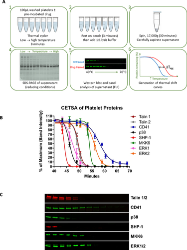
(1) Washed platelets are incubated with drugs of interest at the desired concentration. Aliquots of treated platelets are then heated to different temperatures for a short time (8 min). For this, we use a programmable thermal cycler. The original CETSA protocols heated samples for 3 min,Citation15 but this can be varied to accurately capture aggregation profiles. For platelets, we found 8 min to be an optimal incubation time. (2) Immediately following heating, the aliquots are equilibrated to room temperature (3 min). The platelets are rapidly lysed by addition of detergent-containing lysis buffer and (3) immediately centrifuged to separate soluble and insoluble components. Different lysis techniques/buffers can be applied to CETSA to avoid resolubilizing protein aggregates or to promote precipitation of partly denatured material. For platelets we achieved this using a Tris buffer with 1% NP40, which worked well for the proteins we have studied so far, although this may require optimisation for different proteins. (4) The soluble fraction (supernatant) is aspirated and separated by SDS-PAGE. (5) The protein of interest is then detected by Western blotting. (6) A thermal aggregation curve can be constructed by expressing the soluble protein present at each temperature as a percentage of the soluble protein present in a sample without denaturation (e.g., at 40°C). Tagg is the temperature at which 50% of the protein remains in the soluble fraction.
To illustrate the potential application of CETSA to a variety of platelet proteins, we have generated thermal aggregation curves for the platelet membrane protein CD41a (αIIb), the structural proteins Talin-1 and −2, and the cytosolic signaling proteins SHP-1, ERK1/2, p38 and MKK6. A wide range of Tagg were observed ( & ), with CD41a proving most stable (Tagg 57.4°C; −0.7/+0.7 [95% Cl] respectively) and SHP-1 least (Tagg 44.0°C; −0.4/+0.4 95% Cl respectively) and validate the optimization of the experimental approach. Representative images of the Western blots used to calculate Tagg values are shown in , and Tagg values for all proteins tested listed in .
Table I. Tagg values with 95% confidence intervals of initial platelet proteins tested.
Effect of heating platelets
As the CETSA protocol involves heating intact platelets, it is useful to consider what effects this may have that could affect interpretation of CETSA data. Previous studies have shown that platelets exposed to temperatures above 42°C lose their discoid shape and become irregularly swollen. Their granules are transported to the cell center where they fuse together. Such heat-altered platelets do not aggregate in response to ADP or thrombin and will not retract clots.Citation16 This is likely to be caused by many factors, including aggregation of CD41a (Supplemental ). However, despite heat-induced forward and side scatter changes observed by flow cytometry, surface P-selectin expression did not increase prior to its aggregation, suggesting that the incrementally heated platelets did not rapidly activate.
Effect of platelet activation
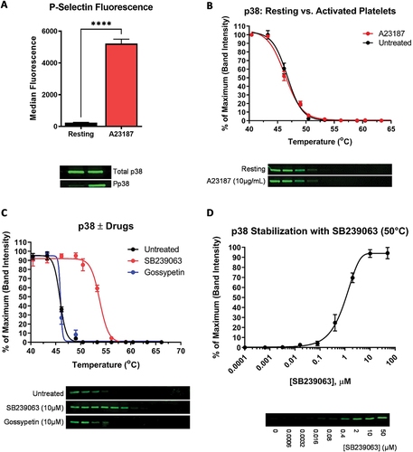
Protein thermal stability can be affected by post-translational modificationsCitation17 such as protein phosphorylation, which in turn might confound interpretation of drug-induced shifts in Tagg. When optimizing CETSA for a new platelet protein, we recommend investigating whether platelet activation affects the protein’s thermal aggregation curve. For example, the kinase p38 is phosphorylated during platelet activation (), which might in principle affect its thermal aggregation curve. However, despite its small 38kDa size, CETSA analysis of p38 revealed no change in Tagg upon phosphorylation (), indicating that the phosphorylation status of this protein is unlikely to conflict with drug-induced Tagg shifts. Our findings concur with an extensive study performed in 2020 by Potel et al. who investigated the thermal stabilities following phosphorylation of over 7000 phosphoproteins and peptides.Citation18 They concluded that 98.7% of these showed no significant change in melting behavior and that cases in which phosphorylation results in a decrease in thermal stability constitute the exception rather than the rule.
Figure 2. Activation and CETSA of p38. Platelets were activated using the Ca2+ ionophore, A23187. (A) Platelet activation was confirmed by P-selection surface expression (as a marker of α-granule secretion) and p38 phosphorylation (Pp38 = phospho-p38). (B) CETSA was conducted on A23187-treated and unstimulated platelets. Thermal stability curves showing soluble p38 at each temperature, and representative Western blots, are shown. (C) CETSA of p38 pre-incubated with the p38-specific inhibitor SB239063 and the MKK6 inhibitor Gossypetin (both 10 µM). The untreated sample contains an equivalent volume of the solvent, DMSO. (D) ITDR CETSA of p38 vs. SB239063 at 50°C. For all panels, values shown are the means ±S.E.M (N = 4).
Effect of pharmacological agents
The main purpose of CETSA is to determine whether a pharmacological agent has bound to a target within platelets. To demonstrate this, we incubated washed platelets with SB239063, a potent, cell-permeable, reversible and selective competitive inhibitor which targets the ATP binding site of p38 MAPK. 10 µM SB239063 resulted in a clear stabilization of this MAP kinase, with a Tagg shift from 46.4°C -/+0.3 [95% Cl] (untreated) to 53.8°C −0.33/+0.34 95% Cl in the presence of the drug. In contrast, 10 µM gossypetin (an inhibitor of the p38-activating protein MKK6) did not induce an isothermal shift (), which is as expected as gossypetin is not believed to bind directly to p38. SB239063 has reported in vitro IC50 of 44 nM against the α and β isoforms.Citation19 To achieve ITDR CETSA, platelets were heated at 50°C with increasing concentrations of SB239063 which yielded a higher stabilization IC50 of 1.27 µM () and required 10 µM for maximum effect (Similarly, SB203580, a compound closely related to SB239063, also has an in vitro IC50 of 44 nM, rising to 0.2–1 µM for p38 signaling in intact platelets.Citation20). The assessment of drug specificity and affinity to a target through biochemical means often fail to adequately mimic the complex setting of proteins within the cells, and thus exhibit significantly different behaviors than they would as isolated proteins. ITDR CETSA can be used to identify the concentration of a drug necessary to give extensive target engagement in a platelet.
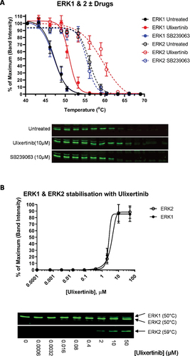
The extracellular signal-regulated kinases (ERKs) including ERK1 and ERK2, are another group of the MAP kinase family and often activated in response to growth factor stimulation.Citation21 The protein sequences of ERK1 and ERK2 in humans are 84% identical, with ERK1 approximately 2kDa larger than human ERK2. Their role and regulation are also largely similar, contributing to intracellular signaling by phosphorylating a common subset of substrates.Citation22 Despite these similarities, ERK1 and ERK2 have very distinct aggregation curves in platelets, with a Tagg of 46.9°C −0.55/+0.54 [95% Cl] and 55.4°C −0.4/+0.5, respectively (). Though slightly lower in value, our findings concur with those of Labraud et al. who also observed the thermal stability profiles of ERK2 to be more stable than ERK1 in HCT116 cells, reporting a Tagg of 51–53°C and 58–60°C, respectively.Citation10 Ulixertinib, a potent, selective, cell-permeable inhibitor of ERK1/2, stabilized both ERK1 and ERK2. In contrast, the p38 inhibitor SB239063 had no effect. These data again demonstrate how CETSA can be used to investigate selective drug binding in platelets. ITDR CETSA of ERK1 and 2 revealed that similar stabilization of these kinases by Ulixertinib (). Ulixertinib has an in vitro IC50 value of <0.3 nM, but a Tagg IC50 value in platelets of 2.48 and 3.54 µM for ERK1 and ERK2, respectively.
Figure 3. CETSA of ERK1 & ERK2. (A) CETSA of ERK1 and ERK2 pre-incubated with the ERK1/2-specific inhibitor Ulixertinib or the p38 inhibitor SB239063 (10 µM). The untreated sample contains an equivalent volume of the solvent, DMSO. Representative blots are shown. (B) ITDR CETSA of ERK1 and ERK2 vs. Ulixertinib at 50°C and 59°C, respectively. Platelets were treated with the indicated concentrations of Ulixertinib prior to heating. For all panels, values shown are the means ±S.E.M (N = 4).
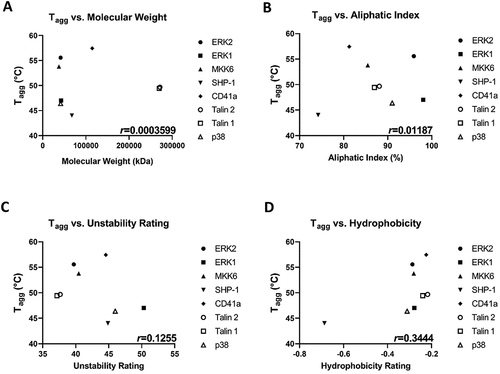
The discrepancy between the Tagg measurements between homologous ERK1 and ERK2 highlights the fact that size is not everything. As illustrated in , a protein’s Tagg is not governed by its molecular weight. There are several measures and determinants of protein stability, which can be thought of as a net balance of forces that determine whether a protein will be its native folded conformation (protein structure and atomic/group interactions, such as hydrophobic, electrostatic, hydrogen bonding, van der Waals and disulfide bonds) or a denatured unfolded or extended state (dominated by entropic and nonentropic free energies). The net stability of proteins is quite small and is the difference between these two large opposing forces. Predicting the stability of a protein is potentially useful for industrial applications; improving a protein’s half-life or robustness at elevated temperatures can often be the deciding factor for its commercialization.Citation23 Several computational tools for predicting protein stability changes have been developed, with each tool employing different algorithms and features that may produce conflicting prediction results making it difficult for users to decide upon the correct design approach.Citation24 We have attempted to correlate the results of our initial CETSA screen of various platelet proteins () against molecular weight (kDa), aliphatic index (the relative volume of a protein occupied by its aliphatic side chains), dipeptide composition-based Instability IndexCitation25 and hydrophobicity. Although we acknowledge that this preliminary analysis encompasses only a few proteins, we find that the best correlation (Pearson correlation coefficient) of protein stability lies with its hydrophobicity (r = 0.344; ). Hydrophobicity is thought to be a key mediator of the folding of proteins. Hydrophobic residues occur preferentially in the core of a protein structure, whereas polar residues tend to occur at the surface.Citation26 The hydrophobicity density of a protein is determined through calculation of the radii of the hydrophobic ellipsoid of a protein using the Eisenberg hydrophobicity scale. Although our data concur with the proposition that an increase in the calculated hydrophobic density of the protein correlates with an increase of its mid-point transition temperature,Citation27 which could be used to help predict the stability and hence Tagg prior to CETSA of any particular protein, the stability of different proteins may be differentially affected by factors such as a particular detergent for example. We also appreciate that in the complex cellular environment, hydrophobicity may not be the sole determinant. Proteins that exist as dimers or multimers (such as CD41 on the platelet surface) may well be inherently stabilized by their binding partner(s) and so we recommend that researchers empirically determine the Tagg for their protein of interest at the start of a new CETSA project.
Figure 4. Tagg moderately correlates with protein hydrophobicity. Tagg of all platelet proteins tested in are plotted against (A) molecular weight; (B) Aliphatic index; (C) Unstability rating; and (D) Hydrophobicity. The Pearson correlation coefficient (r) is shown on each plot. Each protein represented is the mean value from N ≥ 4 separate donors.
In conclusion, although significant progress has been made in understanding platelet signaling in the last decade, there is much that remains poorly understood. Small molecule drugs are likely to retain their major role in platelet studies in the future. Since its introduction in 2013, CETSA has become a popular tool to demonstrate physical target engagement. Here, we have used a “traditional” CETSA based on Western band quantification. This approach can be time consuming, but faster, high-throughput CETSA techniques are becoming increasingly available. CETSA therefore has increasing potential as a quick and informative technique for confirming the direct binding of drugs to platelet protein targets. This provides a platform for understanding the mechanism of action of drugs in platelets, which will be a valuable tool for investigating platelet signaling and function.
Author contributions
Matthew Harper designed the research and helped write the manuscript, and Joanna-Marie Howes designed and performed the research, and wrote the manuscript.
Disclosure statement
No potential conflict of interest was reported by the author(s).
Additional information
Funding
References
- Ho-Tin-Noe B, Boulaftali Y, Camerer E. Platelets and vascular integrity: how platelets prevent bleeding in inflammation. Blood. 2018;131(3):277–8. doi:10.1182/blood-2017-06-742676.
- Patrono C. Role of clinical pharmacology in the development of antiplatelet drugs. Clin Ther. 2014;36(12):2096–111. doi:10.1016/j.clinthera.2014.10.012.
- Balkenhol J, Kaltdorf KV, Mammadova-Bach E, Braun A, Nieswandt B, Dittrich M, Dandekar T. Comparison of the central human and mouse platelet signaling cascade by systems biological analysis. BMC Genomics. 2020;21(1):897. doi:10.1186/s12864-020-07215-4.
- Montenont E, Bhatlekar S, Jacob S, Kosaka Y, Manne BK, Lee O, Parra-Izquierdo I, Tugolukova E, Tolley ND, Rondina MT. et al. CRISPR-edited megakaryocytes for rapid screening of platelet gene functions. Blood Adv. 2021;5(9):2362–74. doi:10.1182/bloodadvances.2020004112.
- Rowlands H, Tschapalda K, Blackett C, Ivanov D, Plant D, Shaw J, Thomas A, Packer M, Arnold L, Holdgate GA. High throughput screening of 0.5 million compounds against CRAF using alpha CETSAⓇ. SLAS Discov. 2023;28(3):102–10. doi:10.1016/j.slasd.2023.01.006.
- Owens AE, Iannotti MJ, Sanchez TW, Voss T, Kapoor A, Hall MD, Marugan JJ, Michael S, Southall N, Henderson MJ. High-throughput cellular thermal shift assay using acoustic transfer of protein lysates. ACS Chem Biol. 2022;17(2):322–30. doi:10.1021/acschembio.1c00760.
- Martinez Molina D, Jafari R, Ignatushchenko M, Seki T, Larsson EA, Dan C, Sreekumar L, Cao Y, Nordlund P. Monitoring drug target engagement in cells and tissues using the cellular thermal shift assay. Science. 2013;341(6141):84–7. doi:10.1126/science.1233606.
- Celej MS, Montich GG, Fidelio GD. Protein stability induced by ligand binding correlates with changes in protein flexibility. Protein Sci. 2003;12(7):1496–506. doi:10.1110/ps.0240003.
- Henderson MJ, Holbert MA, Simeonov A, Kallal LA. High-throughput cellular thermal shift assays in research and drug discovery. SLAS Discov. 2020;25(2):137–47. doi:10.1177/2472555219877183.
- Lebraud H, Surova O, Courtin A, O’Reilly M, Valenzano CR, Nordlund P, Heightman TD. Quantitation of ERK1/2 inhibitor cellular target occupancies with a reversible slow off-rate probe. Chem Sci. 2018;9(45):8608–18. doi:10.1039/c8sc02754d.
- Chen Y, Zhao Y, Bajor DL, Wang Z, Selfridge JE. A facile and sensitive method of quantifying glutaminase binding to its inhibitor CB-839 in tissues. J Genet Genomics. 2020;47(7):389–95. doi:10.1016/j.jgg.2020.06.001.
- Lim YT, Prabhu N, Dai L, Go KD, Chen D, Sreekumar L, Egeblad L, Eriksson S, Chen L, Veerappan S. et al. An efficient proteome-wide strategy for discovery and characterization of cellular nucleotide-protein interactions. PLOS ONE. 2018;13(12):e0208273. doi:10.1371/journal.pone.0208273.
- Wei H, Davies JE, Harper MT. 2-aminoethoxydiphenylborate (2-APB) inhibits release of phosphatidylserine-exposing extracellular vesicles from platelets. Cell Death Discov. 2020;6(1):10. doi:10.1038/s41420-020-0244-9.
- Delport A, Hewer R. A superior loading control for the cellular thermal shift assay. Sci Rep. 2022;12(1):6672. doi:10.1038/s41598-022-10653-7.
- Sanchez TW, Ronzetti MH, Owens AE, Antony M, Voss T, Wallgren E, Talley D, Balakrishnan K, Leyes Porello SE, Rai G. et al. Real-time cellular thermal shift assay to monitor target engagement. ACS Chem Biol. 2022;17(9):2471–82. doi:10.1021/acschembio.2c00334.
- White JG. Effects of heat on platelet structure and function. Blood. 1968;32(2):324–35. doi:10.1182/blood.V32.2.324.324.
- Lee JM, Hammaren HM, Savitski MM, Baek SH. Control of protein stability by post-translational modifications. Nat Commun. 2023;14(1):201. doi:10.1038/s41467-023-35795-8.
- Potel CM, Kurzawa N, Becher I, Typas A, Mateus A, Savitski MM. Impact of phosphorylation on thermal stability of proteins. Nat Methods. 2021;18(7):757–9. doi:10.1038/s41592-021-01177-5.
- Kuliopulos A, Mohanlal R, Covic L. Effect of selective inhibition of the p38 MAP kinase pathway on platelet aggregation. Thromb Haemost. 2004;92(12):1387–93. doi:10.1160/TH04-03-0187.
- Saklatvala J, Rawlinson L, Waller RJ, Sarsfield S, Lee JC, Morton LF, Barnes MJ, Farndale RW. Role for p38 mitogen-activated protein kinase in platelet aggregation caused by collagen or a thromboxane analogue. J Biol Chem. 1996;271(12):6586–9. doi:10.1074/jbc.271.12.6586.
- Roux PP, Blenis J. ERK and p38 MAPK-activated protein kinases: a family of protein kinases with diverse biological functions. Microbiol Mol Biol Rev. 2004;68(2):320–44. doi:10.1128/MMBR.68.2.320-344.2004.
- Vantaggiato C, Formentini I, Bondanza A, Bonini C, Naldini L, Brambilla R. ERK1 and ERK2 mitogen-activated protein kinases affect Ras-dependent cell signaling differentially. J Biol. 2006;5(5):14. doi:10.1186/jbiol38.
- Huang P, Chu SKS, Frizzo HN, Connolly MP, Caster RW, Siegel JB. Evaluating protein engineering thermostability prediction tools using an independently generated dataset. ACS Omega. 2020;5(12):6487–93. doi:10.1021/acsomega.9b04105.
- Chen CW, Lin MH, Liao CC, Chang HP, Chu YW. iStable 2.0: predicting protein thermal stability changes by integrating various characteristic modules. Comput Struct Biotechnol J. 2020;18:622–30. doi:10.1016/j.csbj.2020.02.021.
- Gamage DG, Gunaratne A, Periyannan GR, Russell TG. Applicability of instability index for in vitro protein stability prediction. Protein Pept Lett. 2019;26(5):339–47. doi:10.2174/0929866526666190228144219.
- Moelbert S, Emberly E, Tang C. Correlation between sequence hydrophobicity and surface-exposure pattern of database proteins. Protein Sci. 2004;13(3):752–62. doi:10.1110/ps.03431704.
- Mozo-Villarias A, Cedano J, Querol E. Hydrophobicity density profiles to predict thermal stability enhancement in proteins. Protein J. 2006;25(7–8):529–35. doi:10.1007/s10930-006-9039-y.