
 ?Mathematical formulae have been encoded as MathML and are displayed in this HTML version using MathJax in order to improve their display. Uncheck the box to turn MathJax off. This feature requires Javascript. Click on a formula to zoom.
Abstract
Purpose: Cohorts allowing joint epidemiological and biological analyses are essential for radiation risk assessment. The French Hemangioma Cohort (FHC), studied within the European project EpiRadBio, is one of the rare cohorts suitable for studying the effect of low dose radiation exposure (<100 mGy at organs), with a long-term follow-up. This highly homogeneous cohort consists of healthy individuals belonging to a normal population, except for the presence of skin hemangioma (age at exposure: between 6 months and 3 years of age). Published epidemiological studies have demonstrated that the risk of developing cancer is three times higher in the exposed individuals than in the general population. Here, we present the biobanking of samples (nucleated blood cells, cytogenetic slides of T and B lymphocytes) from the FHC and a primary feasibility study of biomarker analysis focusing on mean telomere length (MTL). Telomeres act as an internal clock, regulating the lifetime of the cell by their shortening during cell division. MTL is thus a biomarker of age. Many in vitro studies have linked MTL and radiosensitivity. The FHC will make it possible to discriminate between the effects of aging and radiation on this biomarker.
Conclusion: The establishment of a biobank of essentially healthy individuals (369 in total), exposed 40–70 years before, during their early childhood, is a logistical challenge. Even among those who previously participated to a self-questionnaire based study, the response rate was only 30%. The first biomarker to be studied was the MTL to discriminate age effects from those of radiation exposure. MTL showed significant variation within age groups (4–11 kb) in both the exposed and non-exposed groups. MTL within the limited age window (i.e. 40–73 year) examined, showed age-dependent changes of 46 bp/year, consistent with the age-dependent decline of 41 bp/year previously reported. We observed no significant changes in MTL according to the average active bone marrow dose. However, we were able to demonstrate that exposure to radiation causes the loss of cells with, on average, shorter telomeres, by applying a model in which both the heterogeneity of the individual dose received at the bone marrow and the heterogeneity of the intercellular distribution of MTL were taken into account.
Introduction
Exposure to ionizing radiation presents a health risk to humans. Epidemiological studies of atomic bomb survivors have demonstrated the correlation between high radiation doses and dose rates (above 100 mSv) and the appearance of solid cancers and leukemia. The effect of low doses (below 100 mSv), based on a linear non-threshold (LNT) extrapolation of cancer risk, is still a matter of debate and the subject of intense study. Recent epidemiological studies suggest that cancer risk after exposure to doses comparable to the dose limits for occupationally exposed workers may be larger than previously assumed (Brenner et al. Citation2003; Abbott Citation2015; Richardson et al. Citation2015).
Cohorts that allow joint epidemiological and biological analyses are essential for direct radiation risk assessment and the study of radiation-induced cancers which require accurate information, especially dosimetry, age at exposure, and post-irradiation long-term follow-up. To be informative, such cohorts need to fulfill several criteria:
They have to include a detailed and individual whole body dosimetric reconstruction. This criterion has to exclude environmental exposure and is probably only possible in using portable dosimeter for occupational cohorts or with access to medical records for medical cohorts.
If a cohort is consisting of patients who received radiotherapy, these patients have to have not received other carcinogens, and the cohort has to include unexposed subjects treated without irradiation for the same disease which had motivated the radiotherapy.
The cohort needs to permit dose response analysis, and therefore to include subjects who received a wide range of radiation dose.
Cohort members have to be followed up for a very long time (>30 years) in a way that guaranties the absence of bias, what cannot be achieved via a simple questionnaire or medical contact, but only by cross linkage with data bases of national hospitals or/and the medical insurance.
The French Hemangioma Cohort (FHC) is exceptional as it fulfils all necessary characteristics for low-dose studies and is therefore one of the rare cohorts suitable for studying the effect of low-dose exposure (<100 mGy) to ionizing radiation on individual cancer risk. It allows joint epidemiological and biological analyses to be performed for radiation risk assessment and the study of radiation-induced diseases, due to accurate dosimetry calculations (i.e. the dose received by all major organs, considering the size of the baby/child during treatment), the access to radiotherapy medical records and a long-term post-irradiation follow-up (Dondon et al. Citation2004). The cohort contains patients who received radiotherapy from different sources (226Ra, X-rays, 32P, 90Y or 90Sr), as well as untreated individuals and those who received cryotherapy and serve as internal controls. The estimation of doses received by all major organs during radiotherapy was performed between 1985 and 1997 by the Unit 605 of INSERM and the Department of Radiophysics at the Institute Gustave Roussy (IGR, Villejuif, France), using a specially developed software that integrated the individual medical records for every child (Ligot et al. Citation1998; Shamsaldin et al. Citation2000).
Moreover, health-related information of the donors has been updated with the help of the SNIIRAM (Système National d’Information Inter-régimes de l’Assurance Maladie [National Inter-Plan Health Insurance Information System]). The SNIIRAM assembles nationwide information of the national health insurance system.
The study of cancer incidence in the FHC was started in 2005. The dose-effect relationship for the dose of radiation received at the cancer site was analyzed and revealed a higher cancer risk than expected for very low local doses (<100 mSv) (Haddy et al. Citation2010, Citation2011). Epidemiological analyses demonstrated a 3-fold higher risk of developing cancer (especially skin, breast, and thyroid cancer) (Dondon et al. Citation2004). Also within the Swedish Hemangioma Cohort an increase of breast cancer risk was described resulting from a radiation-induced genomic instability (Eidemuller et al. Citation2009, Citation2011, Citation2015).
A biobank of the FHC could make it possible to estimate the cancer risk after low-dose exposure to ionizing radiation by integrating molecular biology and epidemiology on individuals exposed 40–70 years ago.
Telomeres play a crucial role in regulating cellular lifespan and aging (Harley, Citation1991; Wright & Shay, Citation1992; Vaziri et al. Citation1994), and are the guardians of genomic stability and integrity in cells (Raynaud et al. Citation2008). Telomere length in somatic proliferative tissues naturally declines with each cell division until they fall below their size ‘threshold’ and become critically short or dysfunctional (Hayflick & Moorhead Citation1961; Hayflick Citation1985) and are sensed as DNA damage (Allsopp & Harley Citation1995). The damage signal stops the cells from further division and they enter a stage of permanent growth arrest called ‘replicative senescence.’ Progressive loss of telomeric DNA limits the number of cell divisions, and therefore limits the lifespan of normal cells as a tumor suppressor mechanism (Raynaud et al. Citation2008).
Reduced telomere length is associated with numerous chronic diseases that are generally considered to be diseases of aging, such as diabetes, cancer, and heart diseases (Price et al. Citation2012; Guzzardi et al. Citation2015; Needham et al. Citation2015; Zhang et al. Citation2015, Citation2016). Abnormal loss of telomeres may contribute to the late side-effects seen in long-term cancer survivors as several studies have shown that cancer patients have shorter telomeres in their blood cells after radiotherapy, even long after treatment has stopped [for review, see Shim et al. (Citation2014)]. It is currently unclear how radiotherapy causes alterations in telomere dynamics, length, structure, and function, and what mechanisms underlie radiation-induced telomere shortening in normal human cells (Li et al. Citation2012). There is evidence that there is a correlation between short telomeres and cancer incidence (solid tumors) or mortality (Svenson & Roos Citation2009; Willeit et al. Citation2010).
G0 human lymphocytes are highly radiosensitive and play an important role in immunity. Thus, telomere length in lymphocytes may be an accurate prognostic indicator of long-term disease, progression, survival, and the evaluation of the efficacy of radiotherapy. Several studies have shown that telomere length in blood cells can even serve as a marker for risks of solid malignancies (Svenson et al. Citation2009). Telomere length in tumor cells and, healthy tissues, such as peripheral blood lymphocytes and epithelial cells, may carry valuable information for future cancer treatment strategies (Svenson & Roos Citation2009). In addition, correlations between radiation-induced long-term diseases and short telomeres in peripheral blood lymphocytes have been previously described for cancer (M’Kacher et al. Citation2003, Citation2007, Citation2010) and cardiovascular disease (Girinsky et al. Citation2014; M’Kacher et al. Citation2015) in a cohort of Hodgkin lymphoma patients.
The age-dependent decrease of telomere length has been well-studied over the last 20 years (Vaziri et al. Citation1993), but their behavior in radiobiology is still not entirely understood and appears to depend on the sample, the received dose, and the type of radiation [for review: Shim et al. (Citation2014)].
We first describe the challenge of creating the biobank of the FHC followed by the results of the first biological (feasibility) study of the biobank. It consisted of analyzing the mean telomere length in mononucleated blood cells, in function of age, from exposed and non-exposed donors (both healthy and without cancer or cardiovascular diseases). The aim of the study was to identify the effect of exposure to low doses of ionizing radiation on mean telomere length, 40–60 years after treatment.
Materials and methods
Study population
The French Hemangioma Cohort (FHC) was established in the 1980s and was followed up by the group U1018/INSERM. The cohort includes 8335 individuals treated as children for a hemangioma at the Institute Gustave Roussy (IGR, Villejuif, France) between 1940 and 1973. Among the 8335 individuals of the cohort, 5744 were treated by radiotherapy and 7800 were under the age of 15 years when treated (among them, 5473 by radiotherapy). Since the major reason for the treatment was esthetical, 80% of the FHC patients were women. A first mortality study was published in 2004 (Dondon et al. Citation2004). Authorization to contact members of the FHC was obtained by INSERM in 2000. In 2014, a specific Council of State decree (Décret en Conseil d’Etat) was obtained, that authorized linkage with the French National Hospital Database and the French National Medical Insurance (Decree no. 2014-96, 3 Feb 2014). In the frame of the FP7-Euratom project EpiRadBio, two new authorizations were obtained to collect biological samples (blood): CCTIRS (Agreement no. 11.700, 11 Dec 2011) and CNIL (Agreement no. 912.039, 10 Jan 2012).
The dose-response relationship was evidenced for thyroid and breast cancers and for melanomas. The observed risks were higher than expected for very low local doses (<100 mSv) (Haddy et al. Citation2010, Citation2011).
Dosimetry
Radiotherapy procedures
The radiotherapy technique depended on the surface and thickness of the hemangioma (Sancho & Beyer Citation1975) and evolved during the treatment period. Different types of radiation were used: Radium 226 and artificial beta emitters, including phosphorus 32, strontium 90, and yttrium 90, as well as X-rays of various energies (Haddy et al. Citation2010).
Individual dose reconstruction
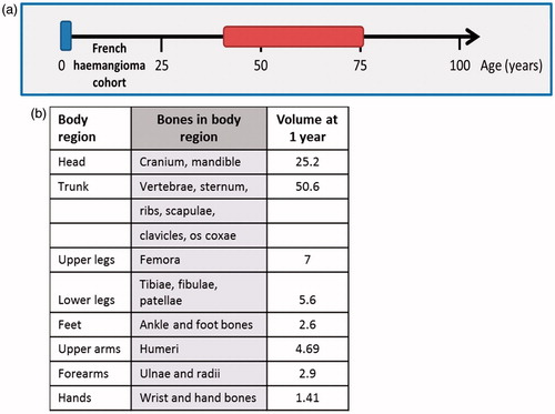
The distribution and proportion of bone marrow per body region is dependent on age and changes during one’s lifetime. This is especially true for babies and children during their physical development (e.g. see ). The FHC is unique for its highly accurate dosimetry, taking into account the age, hence the size of the body, and relative body proportions (distance of body regions from the radiation source).
Individual dose reconstructions were performed for each patient treated by radiotherapy, based on technical radiation therapy records. Whole body dose reconstructions were performed using a software developed and validated by U1018/INSERM in collaboration with the Medical Physics department of Gustave Roussy: Dos_EG software (Diallo et al. Citation1996) for X-rays and ICTA software package (Ligot et al. Citation1998; Shamsaldin et al. Citation2000) for the other techniques. Dose estimations take into account the type of radiation, the surface of the applicators (cm2), their size, shape, and location, and the delivered treatment dose, as well as the size of the child at the respective age (Haddy et al. Citation2010). This method makes it possible to estimate the dose received at every major organ. The age-weighted average radiation dose received to the active bone marrow was estimated by applying age-dependent coefficients determined by Cristy (Citation1981) to bone marrow compartments.
Dosimetry data demonstrated that the dose received by the bone marrow was highly heterogeneous. The mean dose at the bone marrow did not consider hotspots with locally higher doses during treatment. The estimation of the dose was highly accurate through the use of phantoms based on approximately 20 anatomical points (Cristy Citation1981) and donor body size at the time of treatment; importantly, the ratio of head size to that of the body changes considerably with age. Six different dose groups were classified for exposed individuals depending on the total dose received by the bone marrow ().
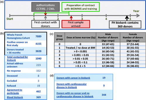
Figure 1. Establishment of the FHC Biobank. (a) Milestones of the administrative process are shown over the time scale, including the authorization required from the French research ministry (CCTIRS, Agreement no. 11.700, 11 Dec 2011) and the National Commission on Informatics and Liberty (CNIL, Agreement no. 912.039, 10 Jan 2012), and major steps during the process of patient acquisition and progress in biobanking are shown underneath. (b) Black square: Follow-up and general information about the FHC. Blue square: Description of participation and the outcome of the establishment of the FHC biobank. (c) Categorization of exposed donors in the FHC biobank into dose groups based on the mean dose received by the bone marrow. Gender and age-distribution in the FHC biobank within the different dose groups. (d) Detailed information about the cancer cases (19 new cancer cases present among the 369 donors of the FHC biobank) and donors with cardiovascular diseases (nine in total, among them three with cancer) within the FHC biobank.
Biobanking
The collection of blood samples from donors living throughout France was performed by the private company Biomnis (Ivry-sur-Seine, France), which specializes in the treatment (transportation, processing, and culture) of blood samples. The biobank contains cytogenetic slides of nucleated blood cells and metaphase spreads for T and B lymphocytes, as well as isolated nucleated blood cells frozen in liquid nitrogen under conditions (10% DMSO in serum) that allow future cell culture and the performance of DNA/RNA extraction and FACS analyses. The population was enriched for B-lymphocytes by including 12-O-tetradodecanoyl-phorbol-13-acetate (TPA; Sigma; Ref: P8139; 0.3 μg/ml final concentration) during cell culture, whereas the division of T-lymphocytes was induced by using phytohemagglutinin (PHA; AppliTech; Ref: EKAMTB-100; all in one medium), as recommended by the manufacturer.
Supplementary information on confounding factors was provided by every donor in a questionnaire, including height and weight, type of work, smoking and alcohol consumption, number of pregnancies (for women), presence of cancer or a benign tumor, radiological procedures during their lifetime, chronic diseases, and skin type and phototype. The blood lead concentration at the time of blood donation was also determined.
Telomere quantification
Mean telomere length was determined for healthy donors (without cancer or cardiovascular diseases) of the dose group 0 (non-exposed) and dose groups 2–6 (exposed donors that received ionizing radiation at the bone marrow). The number of donors and the gender and age-distribution within each dose category are listed in . There were more women than men in all dose groups (including the control).
Preparation of metaphase chromosome and cytogenetic slides for in situ hybridization was performed as previously described (Dutrillaux et al. Citation1981). Slides were prepared by Biomnis using an automated system for the uniform spreading of metaphases and nuclei and stored at −20 °C until use.
We determined mean telomere length in nuclei of mononucleated blood cells scored on cytogenetic slides of T-lymphocyte metaphases using Q-FISH (Quantitative Fluorescent in situ hybridization). For Q-FISH, telomeres and centromeres were stained with a Cy3-labeled peptide nucleic acid (PNA; an artificially synthesized polymer similar to DNA) probe specific for TTAGGG of telomeres and a fluorescein isothiocyanate (FITC)-labeled PNA probe specific for centromere sequences (both from Panagene, Daejon, South Korea), as described in Pottier et al. (Citation2013). After washing with phosphate buffered saline (PBS), the slides were counterstained with 4′,6-diamidino-2-phenylindole (DAPI, a nuclear and chromosome counterstain) and mounted with p-phenylenediamine, antifade solution (PPD). Image acquisition, analysis, and quantification were performed using Metacyte software (version 3.9.1, MetaSystems, Newton, MA, U.S.A.). Quantification was performed by grayscale analysis (Q-FISH) (Piqueret-Stephan et al. Citation2016).
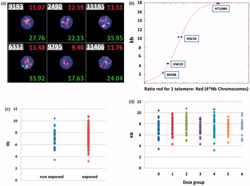
We first obtained the relative mean telomere length by quantifying the entire telomere signal (Cy3, red signal) of all chromosomes within the individual nuclei (). Images of approximately 10,000 nuclei were captured for each donor and the relative mean telomere length per donor was calculated using the overall average fluorescent signals for all nuclei. We then translated the fluorescent signal (relative telomere length) into the mean telomere length in kb using a standard curve based on four cell lines with known telomere lengths.
The cell lines used have telomeres of different mean lengths: REMB (non-Burkitt lymphoblastoid cell line) with the shortest telomeres of 2.3 kb; two HeLa wild type cell lines, HW19 and HW18, with telomeres of 4.36 and 10.32 kb, respectively; and HT1080 (human fibrosarcoma) with the longest telomeres of 17.82 kb (). We quantified and confirmed the telomere lengths by Southern blot analysis (data not shown). One large batch of cytogenetic slides for every cell line was prepared for the entire duration of the project. In each experiment, we included and stained two slides per cell line in parallel. This allowed (A) comparison of the different experiments, and (B) translation of the relative telomere length based on fluorescent signal into telomere length in kb.
Chromosomal aberrations
Following uniform staining, image acquisition, analysis, and quantification were performed using Isis software (MetaSystems, Newton, MA, U.S.A.). Chromosomal aberrations were identified based on the detection of centromeric regions and telomeric sequences against the chromosomal morphology (M’Kacher et al. Citation2014). The detection of dicentric chromosomes was performed semi-automatically using DCscore software (Metasystems). This software automatically identifies and counts dicentric chromosomes in a high throughput mode. At least 1000 metaphases of T-lymphocytes per donor were analyzed.
Statistics and normalization
For higher accuracy, every donor was analyzed in triplicate by staining three different slides, and each hybridized in a separate experiment. Images of 10,000 nuclei were captured per slide. Normalization between experiments and the translation of relative telomere length (fluorescence intensity) into kb was performed using the four different cell lines with known telomere lengths (REMB: 2.3 kb, two HeLa lines: 4.36 and 10.32 kb, and HT1080: 17.82 kb; determined by Southern blot).
Only donors with an inter-experimental variation <15% for the triplicates (variation from the mean value of telomere length between the experiments for each donor) were used to plot the results.
Averaging approach
The impact of the mean radiation dose delivered to the bone marrow on age-dependent telomere shortening was investigated using the following linear regression model:
(1)
(1)
where MTL is the mean telomere length in kb, Dose the mean absorbed dose to the active bone marrow, Age the current Age of the donor, and MTL0, αRad, αAge the parameters of the model estimated by a least squares criterion.
Model (1) can be interpreted as an immediate effect model since it allows a dose-dependent intercept, namely MTL0 + αRad×Dose, and a common age shortening rate αAge for the exposed and unexposed donors.
Functional data analysis approach
The previous averaging approach only partially exploits the abundant information provided by the Q-FISH technique since intra-individual variability is not considered, either for exposure to ionizing radiation or telomere length.
The individual distribution of telomere lengths and bone marrow radiation dose, illustrated in , suggests that these data are derived from processes that can most naturally be described as functional via probability density functions.
Note that a density f can equivalently be represented as a cumulative distribution function (cdf) F such as F′ = f, a quantile function (qf) Q = F−1 or a quantile-density function (dqf), which is the derivative of the quantile function .
The major advantage of the ‘quantile’ point of view is ‘quantile synchronization’ (Zhang & Müller Citation2011) across all densities, especially in the presence of large horizontal shifts due to heterogeneity of the data values.
Functional data analysis (FDA) (Ramsay & Silverman Citation2005) is concerned with the statistical analysis of such data and has the same general goal as multivariate statistical analyses by providing a functional version for dimension reduction, regression, etc. Thus, we generalized model (1) according to functional data regression models:
(2)
(2)
(3)
(3)
where QTL is the quantile function of the telomere length distribution, Age is the current age of the donor, Dose (resp QDose) is the mean active bone marrow dose (resp. bone marrow quantile function). Finally, β0(t), βAge(t) and βDose(t) (resp βDose(t,s)) are the parameters of the model. Note that model (3) differs from model (2) in that both the individual dose and telomere distribution heterogeneity are taken into account.
These functional parameters were estimated by penalized spline functions using the REFUND package of R software (R Foundation for Statistical Computing, Citation2011).
We applied the inverse transformation Ψ−1 to visualize the impact of age and radiation on the Kb scale of the telomere length distribution as described in (Petersen & Müller Citation2016), to back into the space of densities:
where:
and θTL=
Results
Establishment of the FHC biobank
We established a biobank for the FHC blood samples through a collaboration between INSERM (U1018) and the CEA (Radiation and Oncology Laboratory, named now PROCyTOX) during the European project, EpiRadBio, in close cooperation with, and under the supervision of, a medical doctor (onco-dermatologist at the Institute Gustave Roussy, Villejuif, France).
It took one-and-a-half years to obtain the necessary authorizations, including the application to the French Research Ministry (CCTIRS) and the National Commission on Informatics and Liberty (CNIL) (). We then contacted the potential donors to obtain their agreement to participate. The engagement of the private company (Biomnis, Ivry-sur-Seine, France), which specializes in the transportation, processing, and analysis of blood samples, allowed the consistent transport of blood samples under appropriate conditions within a maximum of 1 day, even for donors living outside the local vicinity (outside of the region of Ile de France) and offered to each donors the possibility to get the blood punction in a biology lab (or hospitals, private laboratories, home nurse, etc. for donors living outside the Paris region) of their choice. This ‘easiness’ is very important in order to limit the attrition between the number of donors who agree to give blood and those doing it. After finalizing the contract and providing training for specific sample preparation with Biomnis (on site as well as in our own laboratories), the blood-drawing kits were shipped out two and a half years after the start of the project.
The first blood samples arrived 32 months after the start of the project (). We tried to contact 1040 subjects of the FHC as potential donors, selected on criteria of active bone marrow dose (0.001–1 Gy) among cohort members without cancer at the time of the first self-administrated questionnaire (2000–2006) and who had been treated with radiotherapy before the age of 3 years. Of the 1040 contacted subjects, 462 gave their agreement to participate (). The address of the current residence was not valid for 223 potential donors (21% of all contacted individuals), 332 (32%) did not answer the invitation, 19 (1.89%) refused to participate, seven (0.7%) were excluded from the study, and three (0.3%) died since the previous update of the FHC. Of the 462 individuals who initially accepted to participate in the study, not all provided blood samples. The biobank consists of samples from 369 donors (), 5 years after the start of the project.
Among the 369 donors of the FHC biobank (), a total of 19 new cancers were identified via questionnaires completed by each participating donor, by cytogenetic analysis performed in the present study, and the linkage with the database of the national health insurance information system (SNIIRAM). These cancer cases, as well as donors with cardiovascular diseases (nine in total, among them three with cancer, ) were excluded from further analysis of telomere length or cytogenetic anomalies and were not compared to other donors.
The biobank was prepared on the site of Biomnis using a specific anonymization code to ensure the privacy of donors. A scheme of the biobank is shown as Supplementary data in Figure 6. Blood was collected into two ethylenediaminetetraacetic acid (EDTA) tubes with a maximum volume of 4 ml, two Lithium-Heparin tubes of 4 ml, and one Lithium-Heparin tube of 2 ml for each donor. The value of the FHC biobank rests on the homogeneous preparation of high quality cytogenetic slides for B- and T-lymphocytes, essential for all types of hybridization techniques, such as fluorescent in situ hybridization (FISH), and the availability of living cells for future cell culture experiments. A description of the biobank and contact points are available in the bulletin AIR2 and the infrastructure database AIR2D2.
Characterization of the biobank and dose categories
Only individuals who received radiotherapy before the age of 3 years were contacted (, blue square), along with non-exposed controls. At the time of the blood donation, the donors had been between 40 and 73 years old (, red square), and 63% of them where women ().
Figure 2. Characterization of the FHC Biobank. (a) Visualization of the age range at the time of blood donation (red) relative to the age at the time of exposure during treatment for hemangioma (blue). (b) Allocation and proportion per body region of bone marrow in a 1-year-old child.
Exposed donors were classified according to six groups of average active bone marrow dose (). Dose group 1 contains individuals that received radiotherapy but with extremely low and non-evaluable (<0.001 mGy) doses at the bone marrow. This dose group was not included in the study since we concentrated on the direct effect of ionizing radiation and not on the abscopal effect.
We adjusted the number of donors to 50–60 per dose group (). We were only able to recruit 30–40 donors for dose groups 5 and 6, who received higher mean doses to the bone marrow, because they are less well-represented in the cohort than members of the other dose groups.
Quantification of telomere length (kb)
We determined the mean telomere length in nuclei of mononucleated blood cells scored on cytogenetic slides of T-lymphocyte metaphases using Q-FISH (quantitative FISH).
The mean telomere length of every donor is based on FISH experiments performed in triplicate. We observed a non-significant increase in mean telomere length in the exposed groups compared to the non-exposed groups (, Kruskal-Wallis ANOVA p-value =0.2). This effect was more visible, including fluctuations, when we plotted the different dose categories individually versus non-exposed donors (). This approach represents the classical method to analyze the effect of exposure to ionizing radiation on telomere length used in most publications.
Figure 3. Overall mean Telomere Length in non-exposed vs. exposed Individuals. (a) Images of individual nuclei of T-lymphocytes used to measure mean telomere length by Q-FISH and quantification of the entire telomere signal (Cy3, red signal). (b) Calibration curve using the four different cell lines (REMB, HW19, HW18, HT1080) of known telomere lengths used to compare different experiments and translate the relative telomere length by fluorescent signal into telomere length in kb. (c) Distribution of mean telomere length in T-lymphocytes in exposed vs. non-exposed donors and (d) within the different dose groups.
However, it does not consider the major factor that naturally influences telomere length: the age of the donor. We therefore performed a multivariate regression based on age and bone marrow radiation dose to adjust the radiation-effect relationship on aged-dependent telomere shortening.
Age-weighted average radiation dose received by the bone marrow
We first plotted the curve for the age-dependent decline of mean telomere length of non-exposed () and exposed () donors relative to the curve established by Vaziri et al. in 1993, who also used human blood cells. The FHC biobank contains donors within a small age-window (40–73 years old) relative to those of Vaziri and colleagues, who included donors of all ages starting from new-born babies up to centenarians. In contrast to our study using the Q-FISH technique, Vaziri quantified telomere length by Southern blot analysis. Both curves show the same telomere length for the respective ages in both studies, as well as a comparable age-dependent decline of telomere length: 41 bp per year for Vaziri and 46 bp for the FHC (p-value <0.001).
Figure 4. Mean Telomere Length in non-exposed vs. exposed Donors. Mean telomere length as a function of age in non-exposed (a) and exposed (b) donors versus the curve of Vaziri et al. (Citation1993). (c) Table showing the results of model 1. (d) Visualization of mean telomere length as a function of the dose and the age for all donors. The estimated surface of model 1 (middle) is represented with its confidence bands (lower and upper). (e) Color-coded plot of mean telomere length (red represents long telomeres and blue short telomeres) against the age of the donor and the mean dose received by the bone marrow.
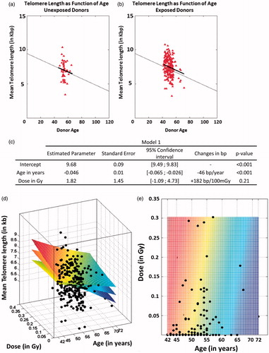
We investigated the impact of the mean dose to the bone marrow on mean telomere length using model 1 (). The dose slope obtained by the model 1 indicates, for a fixed age, a no significant increase of the mean telomere length of 182 bp per 100 mGy received (p = 0.21).
We created a three-dimensional visualization of the correlation of mean telomere length depending on the age of the donor and the mean dose received by the bone marrow to better illustrate the results of model 1 (). Thus, at a fixed mean dose to the bone marrow, the age-dependent decrease in mean telomere length is clearly visible. This is particularly evident for the non-exposed donors.
We performed more exhaustive statistical analyses to determine whether telomere length increased or cells with short telomeres were simply lost due to exposure, resulting in longer overall mean telomere length.
Dose and inter-individual distribution of telomere length
We refined our statistical analysis by considering two parameters in more detail: (1) We included not only the mean dose, but also the entire dose distribution received by the bone marrow during treatment. illustrates the bone marrow dose distributions of five different donors and shows how the treatment varied among the individuals who received different mean doses, as well as those in the same dose category (two different dose distributions leading to the same mean dose). Considering solely the mean received dose fails to address so called ‘hot-spots’, parts of the body that received very high local doses; (2) We also considered the inter-cellular distribution of mean telomere length. Variations of telomere length between the different cells within one donor are missed if only the overall mean telomere length is analyzed.
Figure 5. Mean telomere length as a function of age and the mean dose received by the bone marrow. (a) Examples of dose-distribution quantile functions after radiotherapy for skin hemangioma (mean doses in legend). (b) Distribution of intra-individual telomere length in all donors of the FHC biobank (red = exposed donors, green = non-exposed donors). (c) Predicted telomere length distribution for unexposed individuals of various ages using model 2. (d) Telomere length distribution at 50 years of age as a function of the mean dose received. (e) Correlation of telomere and dose quantile distribution (red indicates an increase in the frequency of larger telomeres; blue indicates an increase in the frequency of shorter telomeres). (f) Dose effect as a projection of the surface coefficient (model 3).
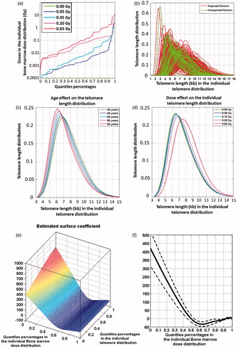
The intercellular distribution of individual telomere length differed noticeably between exposed and non-exposed donors of the FHC biobank ( non-exposed, green; exposed, red). Exposed donors tended to have a higher proportion of cells with longer telomeres than non-exposed donors (especially by kb). This was confirmed by an extension of the classical inferential t-test to the functional domain (Ramsay & Silverman Citation2005), which found a significant difference between exposed and non-exposed quantile functions (p-value =0.0254). More precisely, the quantiles above the median in the exposed telomere distribution were higher than the corresponding quantiles in the non-exposed telomere distribution.
summarizes the results of the fit of models 2 and 3. With the functional approach, the impact of radiation doses became significant (p < 0.001), after adjusting for age.
Table 1. Summary of results obtained for models 2 and 3 fit.
The impact of age and the mean dose in the fit of model 2 is illustrated in . Age is still significantly associated (p < 0.001) with telomere shortening, whereas the dose effect is significantly associated with the distribution of telomere lengths, including the highest range of cell telomere length (p < 0.001).
The correlation of telomere length distribution and dose distribution by applying quantiles in percentages using model 3 is presented in . The red coloration represents an increase in the proportion of larger telomeres, whereas blue represents a higher proportion of shorter telomeres.
As described in Material and methods, model 3 offers the possibility to include all of the dose distribution information rather than simply the mean dose delivered to the bone marrow.
Thus, model 3 makes it possible to model the correlation between telomere length and the radiation dose quantiles. The statistical performance of model 3 outperforms that of model 2 with a significantly lower (more than 4 points) Akaike information criterion: 91756 vs. 92721 (see also ).
The dose-effect, according to model 3, can be visualized by projecting the surface coefficient as illustrated in .
We observe that the doses below the median dose (quantile 50%) are associated with a positive weight, whereas the doses above the median dose are associated with a negative weight. Furthermore, the weights associated with the lowest doses are approximately 10 times greater than those for the highest doses (hundreds for positive weights versus tens for the negative weights).
The effect of the dose distribution on the telomere quantile values is then obtained as a weighted mean of the surface projection with all the ordered doses in the individual dose distribution. According to this model, radio-induced telomere shortening results from a dose distribution with a large gradient of doses for a given age.
Discussion
In France, children were treated for skin hemangioma during early childhood with radiotherapy from 1940–1973. Epidemiological analysis of this cohort has demonstrated a 3-fold higher risk of developing cancer. We established a biobank of blood samples of the FHC with a follow up of at least 40 years (), including cytogenetic slides of T- and B-lymphocytes, and isolated living nucleated blood cells.
We launched an exposed/non-exposed study focusing on healthy donors (at the time of biosampling). We excluded donors with cancer, because cancer therapy is often combined with cytotoxic treatments (chemo- or/and radiotherapy), and cardiovascular disease. We focused our analysis on mean telomere length as potential biomarker since changes in telomere length may help to better estimate the risk of developing cancer and personalize the follow-up needed after radiotherapy and/or cancer screening. There are only a few studies analyzing the long-term effect of low doses of ionizing radiation on telomeres. Ilyenko et al. (Citation2011) described telomere shortening in blood lymphocytes up to 20 years after exposure to low-dose ionizing irradiation in Chernobyl workers. However, they did not consider the age of the donors, although telomere length is strongly correlated with age. Short telomeres have been reported to be particularly radiosensitive at high doses (radiotherapy) (Mirjolet et al. Citation2015). The long-term effect of radiation exposure (dose categories 0–0.005 Gy, 0.005–0.5 Gy, 0.5–1 Gy and ≥1 Gy; bone marrow dose) on T-lymphocyte telomere length was studied in atomic bomb survivors describing longer telomeres at low doses and a decreasing trend of telomere length at doses higher than 0.5 Gy (Yoshida et al. Citation2016).
The most critical step in our study has been the creation of the biobank of blood samples from donors belonging to the FHC. The combination of information obtained from the SNIIRAM database and the questionnaire accompanying the blood donation for the FHC biobank proved to be necessary for the identification of cancer cases, as some were not included in the SNIIRAM data, mostly due to their recent appearance (question of updating the system). In contrast, the SNIIRAM data indicated cancer cases that were not declared in the questionnaire (missing information in the declaration of donors). A crosscheck of the cytogenetic analyses should confirm the genotoxic treatment and permit the identification of cancer cases not declared in the questionnaire and not yet included in the SNIIRAM database.
Information about members of the FHC is not static. The updating of personal data, including health and mortality data, as well as simple address changes, is essential for long-term follow-up. It enables subsequent studies, such as biological analyses and the development of biobanks, and the possibility of multiple sample collections over a long period of time (also molecular follow-up studies). This is the major strength of the FHC, which is unique in this respect. The FHC is smaller (8300 members) than other hemangioma cohorts, such as the Swedish cohorts (Stockholm and Goteborg) with a total of more than 26,000 individuals, which allow accurate epidemiological studies of cancer risk (Karlsson et al. Citation1998; Lundell et al. Citation1999; Preston et al. Citation2002; Eidemuller et al. Citation2015). However, the possibility to remain in contact with the FHC members has already allowed their integration into several studies (development of the FHC biobank within EpiRadBio; cognitive and cerebrovascular risk analysis in CEREBRAD). The FHC represents a living and relatively young cohort, including many members between 45 and 60 years old. It would be possible to obtain additional biological samples. A new sampling campaign every 5–10 years may offer the opportunity of individual follow-up with very precise dosimetry.
Radiation dose reconstruction is a considerable challenge (Bouville et al. Citation2015). The FHC is unique for the accuracy of the dosimetry techniques applied. The data for the Swedish program covers only Ra226 and P32, and does not include children below 6 months. In contrast, the ICTA software, developed at the IGR, can be used for all body sizes and ages and is applicable to other radio-nuclides (plates of Sr90 and P32 and Y90 needles). It more accurately considers the morphology and anatomy of the patient and supports more penetrating radiation (gamma Ra226). Most model calculations are based on in vitro experiments. More in vivo studies are still needed, as well as the identification of good biomarkers, for the development of efficient models to estimate the cancer risk after exposure to low doses of ionizing radiation. There are only a few simultaneous low-dose epidemiological and molecular biology studies. The FHC is one such cohort.
We studied several aspects of the effect of radiation treatment for skin hemangioma on telomere length, considering not only the mean values, but also the distribution of telomere lengths and doses received by the bone marrow. Nuclei on cytogenetic slides of T-lymphocyte metaphases from non-exposed donors of the FHC showed comparable values and a similar age-dependent decline of mean telomere length (, approximately 46 bp per year, p-value <0.001) to those of a non-exposed population and data already published by Vaziri et al. (Citation1993). This result represents the first validation of the Q-FISH method for telomere quantification in kb. We choose to use the Vaziri data as a reference because they performed Southern blots for their estimates as we did for our calibration curve to estimate telomere length in kb based on fluorescence intensity. The age-range from 41–73 years within the FHC is smaller than that of Vaziri, which ranged from new-born babies to adults of more than 100 years. However, the slope of the curve for non-exposed donors was consistent with that reported by Vaziri et al. (Citation1993). Exposed donors showed the same age-dependent decrease in telomere length ().
There are at least are three possible mechanisms for the described observations:
Telomere shortening may accelerate after exposure. This hypothesis is unlikely considering that the slope for the age-dependent decrease in mean telomere length for exposed donors of the FHC was the same as that of non-exposed donors.
There is an immediate effect after exposure, resulting in the presence of cells with long mean telomere lengths.
There is a combination of both telomere shortening and lengthening, including a direct effect of exposure and an effect on the age-dependent decrease in telomere length per year. However, as discussed for the first possibility, the similar decrease in telomere length per year observed for exposed and non-exposed donors of the FHC makes this unlikely, favoring the second possibility.
In support of the second hypothesis we demonstrated a shift towards a higher proportion of cells with longer telomeres after exposure of bone marrow to higher mean doses based on accurate model calculations (model 2), considering the intra-individual and intercellular distribution of telomere lengths.
illustrate the highly complex and more precise correlation of the dose and telomere length distributions by also considering the age of the donor.
Starting from the lowest doses, there is telomere elongation that is reduced as the median dose is reached followed by shortening for the highest 50% of doses, with a peak around the 70% quantile.
Above the 5–10% most elevated doses, which represent the hot-spots during radiation treatment, this reduction appears to stop, which may indicate a significant loss of cells with shorter telomeres, leading to an overall increase of mean telomere length for the individual.
This finding is consistent with a previous study (M’Kacher et al. Citation2007) on telomere shortening in the context of radiation radiotherapy involving high doses of several tens of Gy as model 3 predicts a clear reduction in telomere length due to the high dose gradient and high hotspot doses.
Dose-dependent model calculations () demonstrated that the loss of cells with short telomeres occurred above a certain dose threshold, suggesting increased sensitivity of cells with shorter telomeres to ionizing radiation. Comparable observations have been reported for a study on the long-term effect of radiation exposure on T-lymphocyte telomere length in atomic bomb survivors (Yoshida et al. Citation2016). This non-linearity is not restricted to this biomarker as the question of linear/non-linear radiation-induced biological responses has arisen in many biological systems (Averbeck Citation2009). These results confirm those of previous studies showing increased in vitro radiosensitivity of cells with shorter telomeres (McIlrath et al. Citation2001; Castella et al. Citation2007) and increasing radiation sensitivity as telomeres shorten during cell proliferation (Soler et al. Citation2009). A decrease of the fraction of cells with short telomeres has been observed in cancer patients after radiation therapy (Maeda et al. Citation2013). Moreover, shorter telomeres result in organismal hyper-radiosensitivity (Goytisolo et al. Citation2000; Wong et al. Citation2000).
In future studies, we would like to perform a follow-up of exposed donors without cancer and/or cardiovascular diseases and compare them with those having these diseases (these donors will need to be newly recruited). This study would address whether the intracellular polymorphism of telomere length, specifically short telomeres, is associated with the risk of developing radiation-induced cancer, as previously suggested (Murnane & Sabatier Citation2004; Shim et al. Citation2014; Sishc et al. Citation2015).
In summary, during EpiRadBio, we created the first biobank of hemangioma patients. We demonstrated a strong correlation between telomere length and age, and provided evidence that exposure to low doses of ionizing radiation directly affects cells with, on average, shorter telomeres, which can be detected long after exposure (decades). The study can now be extended to include the analysis of the heterogeneity of telomere length within individual cells and its potential association with the occurrence of cancer. Additionally, telomere features and other cytogenetic analyses, such as genomic instability, may also be related to other diseases, such as cardiovascular or cognitive diseases.
We plan to enlarge and extend the FHC biobank in the future, including blood samples for the isolation of living nucleated blood cells and immediate cell culture for cytogenetic preparations. This will include not only recruiting new donors, but also contacting donors for whom there are already samples in the biobank for multiple sample analyses over a longer period (at 5-year intervals, if possible). As presented here, the distribution of telomere lengths, as well as mean telomere length, vary inter-individually. A comparison of the slopes determined for every individual donor over a period of 15 years could help to refine our understanding of the age-dependent decrease of telomere length.
Tribute to Bill Morgan
The establishment of the French Hemangioma Biobank and the described exposed/non-exposed study was perfomed within the European project EpiRadBio, financed under FP7 EURATOM FISSION. The main aim of the project was to combine epidemiology and radiobiology to assess cancer risk in the breast, lung, thyroid, and digestive tract after exposure to ionizing radiation with cumulative equivalent doses of approximately 100 mSv or below. One of the key objectives of EpiRadBio was to perform feasibility studies of telomere length measurement with blood samples from members of radio-epidemiological cohorts.
Bill Morgan joined EpiRadBio as an external expert in the advisory board and enthusiastically promoted the consortium thanks to his wide expertise in the field of radiobiology and dose/dose-rate studies (Brooks Citation2015; Limoli et al. Citation2016). The establishment of the FHC Biobank and the study on telomere length within the exposed/non-exposed study were consistently supported by Bill Morgan. He highly contributed to the success of EpiRadBio through his advice and interest.
Notes on Contributors
Monika Frenzel completed her PhD in Physical Biochemistry, Radiation Biology followed by a post-doctoral position at the CEA (Fontenay aux Roses, France) in cytogenetics, and cohort studies in the laboratory of radiobiology and oncology (LRO). She manages and coordinates the contribution of the LRO in the EpiRadBio project.
Michelle Ricoul, research engineer at the CEA, is head of the Platform of Radiobiology and Oncology Cytogenetic (PROCyTox). She is an expert in biological dosimetry and molecular cytogenetics. She performed all the quantifications of telomeres on interphase nuclei in the EpiRadBio cohort.
Mohamed Amine Benadjaoud, PhD in Biostatistics and MSc in medical physics, researcher at the Institute for Radiological Protection and Nuclear Safety (IRSN). His main activities include the development of multiscale risk models through radiobiological data integration. He involved in the EpiRadBio project in the modelling of radio-induced telomere length response.
Marion Bellamy obtained her second master degree in cellular and genetic engineering. She has wide expertise in molecular biology, biochemistry, cell biology, imaging and cytogenetics as well as cohort and epidemiological studies. She is one of the main actors in the development of the FHC biobank and the biological studies conducted.
Aude Lenain, engineer, made her degree in biochemistry with a specialisation in biomedical research. She gained wide experiences in molecular biology, biochemistry, cell biology, imaging, histology, animal experiments and cytogenetics. Within EpiRadBio, she contributes in the development of the biobank and the biological studies.
Nadia Haddy, PhD in public health, Researcher at Radiation and Cancer Team of the Research Center in Epidemiology and Population Health (CESP). Her main fields of interest are to develop the molecular epidemiology in the team and specially to research predictive biomarkers of the occurrence of iatrogenic diseases.
Ibrahima Diallo, PhD in Medical Physics, leads the Radiation Dosimetry group of the Cancer and Radiations Team of the Research Center in Epidemiology and Population Health (CESP). His main areas of expertise are Radiation Therapy dose reconstructions for epidemiology and the development of radiotherapy patient phantoms and dosimetry software.
Christine Mateus, Dermatologist with a specialization on oncology, titular in Dermatology Unit at Gustave-Roussy, since 2006. She gained her medical degree at the Paris XI University in 2000 and received her French Board Certification in Dermatology in 1999. She obtained in 2007 her French Board Complementary Certification in Oncology.
Florent de Vathaire, PhD in Biostatistics and in Ecology, leads the Radiation and Cancer Team of the Research Center in Epidemiology and Population Health (CESP). He has initiated the French Hemangioma Cohort and the French Childhood Cancer Survivor Study (FCCSS). His main areas of expertise are radiation epidemiology, cancer survivorship and thyroid cancer epidemiology.
Laure Sabatier, PhD in Human Genetics, Research Director at CEA. Her main research interests are the understanding of the role of chromosomal instability in the bypass of senescence process, cellular immortalization and transformation of primary human cells after irradiation. She supervises the biobanking activities and biomarkers studies of this cohort.
frenzel_et_al_revised2_supplem_fig6.pptx
Download MS Power Point (509.9 KB)Acknowledgements
We gratefully thank Annie Vernique for her valuable contribution to the biobanking. We are in debt to Radhia M’kacher and Elena Giulotto (University of Pavia) for the cell lines used for the calibration curves. We would also like to thank the hemangioma patients for allowing this study to be conducted. In addition, we are grateful to the professionals at Biomnis for the chromosome preparations and the biobanking of the blood samples. The creation of the FHC biobank with Biomnis as subcontractors was organized by the LRO (CEA). All telomere measurements and cytogenetic analyses were performed at the LRO, whereas the preparation of authorizations associated with the establishment of the biobank and the statistical analysis and model calculations have been performed by the Radiation Epidemiology Group, INSERM U1018 at INSERM.
Disclosure statement
The authors report no conflicts of interest. The authors alone are responsible for the content and writing of the paper.
Additional information
Funding
References
- Abbott A. 2015. Researchers pin down risks of low-dose radiation. Nature. 523:17–18.
- Allsopp RC, Harley CB. 1995. Evidence for a critical telomere length in senescent human fibroblasts. Exp Cell Res. 219:130–136.
- Averbeck D. 2009. Does scientific evidence support a change from the LNT model for low-dose radiation risk extrapolation? Health Phys. 97:493–504.
- Bouville A, Toohey RE, Boice JD Jr, Beck HL, Dauer LT, Eckerman KF, Hagemeyer D, Leggett RW, Mumma MT, Napier B, et al. 2015. Dose reconstruction for the Million Worker study: status and guidelines. Health Phys. 108:206–220.
- Brenner DJ, Doll R, Goodhead DT, Hall EJ, Land CE, Little JB, Lubin JH, Preston DL, Preston RJ, Puskin JS, et al. 2003. Cancer risks attributable to low doses of ionizing radiation: assessing what we really know. Proc Natl Acad Sci U S A. 100:13761–13766.
- Brooks AL. 2015. Dr William F. Morgan 1952–2015. Int J Radiat Biol. 91:913.
- Castella M, Puerto S, Creus A, Marcos R, Surralles J. 2007. Telomere length modulates human radiation sensitivity in vitro. Toxicol Lett. 172:29–36.
- Cristy M. 1981. Active bone marrow distribution as a function of age in humans. Phys Med Biol. 26:389–400.
- Diallo I, Lamon A, Shamsaldin A, Grimaud E, de Vathaire F, Chavaudra J. 1996. Estimation of the radiation dose delivered to any point outside the target volume per patient treated with external beam radiotherapy. Radiother Oncol. 38:269–271.
- Dondon MG, de Vathaire F, Shamsaldin A, Doyon F, Diallo I, Ligot L, Paoletti C, Labbe M, Abbas M, Chavaudra J, et al. 2004. Cancer mortality after radiotherapy for a skin hemangioma during childhood. Radiother Oncol. 72:87–93.
- Dutrillaux B, Viegas Pequignot E, Aurias A, Prod’homme M, Sportes M, Prieur M. 1981. Tentative estimate of the risk of chromosomal disease due to radiation-induced translocations in man. Mutat Res. 82:191–200.
- Eidemuller M, Holmberg E, Jacob P, Lundell M, Karlsson P. 2009. Breast cancer risk among Swedish hemangioma patients and possible consequences of radiation-induced genomic instability. Mutat Res. 669:48–55.
- Eidemuller M, Holmberg E, Jacob P, Lundell M, Karlsson P. 2011. Breast cancer risk after radiation treatment at infancy: potential consequences of radiation-induced genomic instability. Radiat Prot Dosimetry. 143:375–379.
- Eidemuller M, Holmberg E, Jacob P, Lundell M, Karlsson P. 2015. Breast cancer risk and possible mechanisms of radiation-induced genomic instability in the Swedish hemangioma cohort after reanalyzed dosimetry. Mutat Res. 775:1–9.
- Girinsky T, M’Kacher R, Lessard N, Koscielny S, Elfassy E, Raoux F, Carde P, Santos MD, Margainaud JP, Sabatier L, et al. 2014. Prospective coronary heart disease screening in asymptomatic Hodgkin lymphoma patients using coronary computed tomography angiography: results and risk factor analysis. Int J Radiat Oncol Biol Phys. 89:59–66.
- Goytisolo FA, Samper E, Martin-Caballero J, Finnon P, Herrera E, Flores JM, Bouffler SD, Blasco MA. 2000. Short telomeres result in organismal hypersensitivity to ionizing radiation in mammals. J Exp Med. 192:1625–1636.
- Guzzardi MA, Iozzo P, Salonen M, Kajantie E, Eriksson JG. 2015. Rate of telomere shortening and metabolic and cardiovascular risk factors: a longitudinal study in the 1934-44 Helsinki Birth Cohort Study. Ann Med. 47:499–505.
- Haddy N, Dondon MG, Paoletti C, Rubino C, Mousannif A, Shamsaldin A, Doyon F, Labbe M, Robert C, Avril MF, et al. 2010. Breast cancer following radiotherapy for a hemangioma during childhood. Cancer Causes Control. 21:1807–1816.
- Haddy N, Mousannif A, Paoletti C, Dondon MG, Shamsaldin A, Doyon F, Avril MF, Fragu P, Labbe M, Lefkopoulos D, et al. 2011. Radiotherapy as a risk factor for malignant melanoma after childhood skin hemangioma. Melanoma Res. 22:77–85.
- Harley CB. 1991. Telomere loss: mitotic clock or genetic time bomb? Mutat Res. 256:271–282.
- Hayflick L, Moorhead PS. 1961. The serial cultivation of human diploid cell strains. Exp Cell Res. 25:585–621.
- Hayflick L. 1985. The cell biology of aging. Clin Geriatr Med. 1:15–27.
- Ilyenko I, Lyaskivska O, Bazyka D. 2011. Analysis of relative telomere length and apoptosis in humans exposed to ionising radiation. Exp Oncol. 33:235–238.
- Karlsson P, Holmberg E, Lundell M, Mattsson A, Holm LE, Wallgren A. 1998. Intracranial tumors after exposure to ionizing radiation during infancy: a pooled analysis of two Swedish cohorts of 28,008 infants with skin hemangioma. Radiat Res. 150:357–364.
- Li P, Hou M, Lou F, Bjorkholm M, Xu D. 2012. Telomere dysfunction induced by chemotherapeutic agents and radiation in normal human cells. Int J Biochem Cell Biol. 44:1531–1540.
- Ligot L, Diallo I, Shamsaldin A, Chavaudra J, BonaIti-Pellie C, de Vathaire F. 1998. Individualized phantom based on CT slices and auxological data (ICTA) for dose estimations following radiotherapy for skin haemangioma in childhood. Radiother Oncol. 49:279–285.
- Limoli C, Baulch J, Sowa M. 2016. William F. Morgan (1952–2015). Radiat Res. 185:106–108.
- Lundell M, Mattsson A, Karlsson P, Holmberg E, Gustafsson A, Holm LE. 1999. Breast cancer risk after radiotherapy in infancy: a pooled analysis of two Swedish cohorts of 17,202 infants. Radiat Res. 151:626–632.
- M’Kacher R, Violot D, Aubert B, Girinsky T, Dossou J, Beron-Gaillard N, Carde P, Parmentier C. 2003. Premature chromosome condensation associated with fluorescence in situ hybridisation detects cytogenetic abnormalities after a CT scan: evaluation of the low-dose effect. Radiat Prot Dosimetry. 103:35–40.
- M’Kacher R, Bennaceur-Griscelli A, Girinsky T, Koscielny S, Delhommeau F, Dossou J, Violot D, Leclercq E, Courtier MH, Beron-Gaillard N, et al. 2007. Telomere shortening and associated chromosomal instability in peripheral blood lymphocytes of patients with Hodgkin’s lymphoma prior to any treatment are predictive of second cancers. Int J Radiat Oncol Biol Phys. 68:465–471.
- M’Kacher R, Andreoletti L, Flamant S, Milliat F, Girinsky T, Dossou J, Violot D, Assaf E, Clausse B, Koscielny S, et al. 2010. JC human polyomavirus is associated to chromosomal instability in peripheral blood lymphocytes of Hodgkin’s lymphoma patients and poor clinical outcome. Ann Oncol. 21:826–832.
- M’Kacher R, Maalouf EE, Ricoul M, Heidingsfelder L, Laplagne E, Cuceu C, Hempel WM, Colicchio B, Dieterlen A, Sabatier L. 2014. New tool for biological dosimetry: reevaluation and automation of the gold standard method following telomere and centromere staining. Mutat Res. 770:45–53.
- M’Kacher R, Girinsky T, Colicchio B, Ricoul M, Dieterlen A, Jeandidier E, Heidingsfelder L, Cuceu C, Shim G, Frenzel M, et al. 2015. Telomere shortening: a new prognostic factor for cardiovascular disease post-radiation exposure. Radiat Prot Dosimetry. 164:134–137.
- Maeda T, Nakamura K, Atsumi K, Hirakawa M, Ueda Y, Makino N. 2013. Radiation-associated changes in the length of telomeres in peripheral leukocytes from inpatients with cancer. Int J Radiat Biol. 89:106–109.
- McIlrath J, Bouffler SD, Samper E, Cuthbert A, Wojcik A, Szumiel I, Bryant PE, Riches AC, Thompson A, Blasco MA, et al. 2001. Telomere length abnormalities in mammalian radiosensitive cells. Cancer Res 61:912–915.
- Mirjolet C, Boidot R, Saliques S, Ghiringhelli F, Maingon P, Crehange G. 2015. The role of telomeres in predicting individual radiosensitivity of patients with cancer in the era of personalized radiotherapy. Cancer Treat Rev. 41:354–360.
- Murnane JP, Sabatier L. 2004. Chromosome rearrangements resulting from telomere dysfunction and their role in cancer. Bioessays. 26:1164–1174.
- Needham BL, Rehkopf D, Adler N, Gregorich S, Lin J, Blackburn EH, Epel ES. 2015. Leukocyte telomere length and mortality in the National Health and Nutrition Examination Survey, 1999–2002. Epidemiology. 26:528–535.
- Petersen A, Müller H-G. 2016. Functional data analysis for density functions by transformation to a Hilbert space. Ann Statist. 44: 183–218.
- Piqueret-Stephan L, Ricoul M, Hempel WM, Sabatier L. 2016. Replication timing of human telomeres is conserved during immortalization and influenced by respective subtelomeres. Sci Rep. 6:32510.
- Pottier G, Viau M, Ricoul M, Shim G, Bellamy M, Cuceu C, Hempel WM, Sabatier L. 2013. Lead exposure induces telomere instability in human cells. PLoS One. 8:e67501.
- Preston DL, Mattsson A, Holmberg E, Shore R, Hildreth NG, Boice JD Jr. 2002. Radiation effects on breast cancer risk: a pooled analysis of eight cohorts. Radiat Res. 158:220–235.
- Price LH, Kao HT, Burgers DE, Carpenter LL, Tyrka AR. 2012. Telomeres and early-life stress: an overview. Biol Psychiatry. 73:15–23.
- R Foundation for Statistical Computing. 2011. A language and environment for statistical computing. Vienna, Austria: R Foundation for Statistical Computing. ISBN 3-900051-07-0, URL http://www.R-project.org.
- Ramsay JO, Silverman BW (eds). 2005. Functional data analysis. New York: Springer, 2nd ed.
- Raynaud CM, Sabatier L, Philipot O, Olaussen KA, Soria JC. 2008. Telomere length, telomeric proteins and genomic instability during the multistep carcinogenic process. Crit Rev Oncol Hematol. 66:99–117.
- Richardson DB, Cardis E, Daniels RD, Gillies M, O’Hagan JA, Hamra GB, Haylock R, Laurier D, Leuraud K, Moissonnier M, et al. 2015. Risk of cancer from occupational exposure to ionising radiation: retrospective cohort study of workers in France, the UK, and the United States (INWORKS). BMJ 351:h5359.
- Sancho H, Beyer HP (eds). 1975. Skin tumors. In: Cancer in children: clinical management. Berlin: Springer.
- Shamsaldin A, Lundell M, Diallo I, Ligot L, Chavaudra J, de Vathaire F. 2000. Estimation of the radiation dose from radiotherapy for skin haemangiomas in childhood: the ICTA software for epidemiology. Phys Med Biol. 45:3589–3599.
- Shim G, Ricoul M, Hempel WM, Azzam EI, Sabatier L. 2014. Crosstalk between telomere maintenance and radiation effects: a key player in the process of radiation-induced carcinogenesis. Mutat Res Rev Mutat Res. 760:1–17.
- Sishc BJ, Nelson CB, McKenna MJ, Battaglia CL, Herndon A, Idate R, Liber HL, Bailey SM. 2015. Telomeres and telomerase in the radiation response: implications for instability, reprograming, and carcinogenesis. Front Oncol. 5:257.
- Soler D, Pampalona J, Tusell L, Genesca A. 2009. Radiation sensitivity increases with proliferation-associated telomere dysfunction in nontransformed human epithelial cells. Aging Cell. 8:414–425.
- Svenson U, Ljungberg B, Roos G. 2009. Telomere length in peripheral blood predicts survival in clear cell renal cell carcinoma. Cancer Res. 69:2896–2901.
- Svenson U, Roos G. 2009. Telomere length as a biological marker in malignancy. Biochim Biophys Acta. 1792:317–323.
- Vaziri H, Schachter F, Uchida I, Wei L, Zhu X, Effros R, Cohen D, Harley CB. 1993. Loss of telomeric DNA during aging of normal and trisomy 21 human lymphocytes. Am J Hum Genet. 52:661–667.
- Vaziri H, Dragowska W, Allsopp RC, Thomas TE, Harley CB, Lansdorp PM. 1994. Evidence for a mitotic clock in human hematopoietic stem cells: loss of telomeric DNA with age. Proc Natl Acad Sci USA. 91:9857–9860.
- Willeit P, Willeit J, Mayr A, Weger S, Oberhollenzer F, Brandstatter A, Kronenberg F, Kiechl S. 2010. Telomere length and risk of incident cancer and cancer mortality. JAMA. 304:69–75.
- Wong KK, Chang S, Weiler SR, Ganesan S, Chaudhuri J, Zhu C, Artandi SE, Rudolph KL, Gottlieb GJ, Chin L, et al. 2000. Telomere dysfunction impairs DNA repair and enhances sensitivity to ionizing radiation. Nat Genet. 26:85–88.
- Wright WE, Shay JW. 1992. The two-stage mechanism controlling cellular senescence and immortalization. Exp Gerontol. 27:383–389.
- Yoshida K, Misumi M, Kubo Y, Yamaoka M, Kyoizumi S, Ohishi W, Hayashi T, Kusunoki Y. 2016. Long-term effects of radiation exposure and metabolic status on telomere length of peripheral blood T cells in atomic bomb survivors. Radiat Res. 186:367–376.
- Zhang C, Chen X, Li L, Zhou Y, Wang C, Hou S. 2015. The association between telomere length and cancer prognosis: evidence from a meta-analysis. PLoS ONE. 10:e0133174.
- Zhang J, Rane G, Dai X, Shanmugam MK, Arfuso F, Samy RP, Lai MK, Kappei D, Kumar AP, Sethi G. 2016. Ageing and the telomere connection: an intimate relationship with inflammation. Ageing Res Rev. 25:55–69.
- Zhang Z, Müller H-G. 2011. Functional density synchronization. Comput Stat Data Anal. 55: 2234–2249.