Abstract
Purpose
Well-controlled ionizing radiation injury animal models for testing medical countermeasure efficacy require robust radiation physics and dosimetry to ensure accuracy of dose-delivery and reproducibility of the radiation dose-response relationship. The objective of this study was to establish a simple, convenient, robust and accurate technique for validating total body irradiation (TBI) exposure of the New Zealand White rabbit.
Methods
We use radiotherapy techniques such as computed tomography simulation and a 3 D-conformal radiation therapy treatment planning system (TPS) on three animals to comprehensively design and preplan a TBI technique for rabbits. We evaluate the requirement for bolus, treatment geometry, bilateral vs anterior-posterior treatment delivery, the agreement between monitor units calculated using the TPS vs a traditional hand calculation to the mid-plane, and resulting individual organ doses.
Results
The optimal technique irradiates animals on the left-decubitus position using two isocentric bilateral parallel-opposed 6 MV x-ray beams. Placement of a 5 mm bolus and 8.5 mm beam spoiler was shown to increase the dose to within ≤5 mm of the surface, improving dose homogeneity throughout the body of the rabbit. A simple hand calculation formalism, dependent only on mid-abdominal separation, could be used to calculate the number of monitor units (MUs) required to accurately deliver the prescribed dose to the animal. For the representative animal, the total body volume receiving > 95% of the dose, V95% > 99%, V100% > 95%, and V107% < 20%. The area of the body receiving >107% of the prescribed dose was mainly within the limbs, head, and around the lungs of the animal, where the smaller animal width reduces the x-ray attenuation. Individual organs were contoured by an experienced dosimetrist, and each received doses within 95–107% of the intended dose, with mean values ∼104%. Only the bronchus showed a maximal dose >107% (113%) due to the decreased attenuation of the lungs. To validate the technique, twenty animals were irradiated with four optically-stimulated luminescence dosimeters (OSLDs) placed on the surface of each animal (two on each side in the center of the radiation beam). The average dose over all animals was within <0.1% from intended values, with no animal receiving an average dose more than ±3.1% from prescription.
Conclusion
The TBI technique developed in this pilot study was successfully used to establish the dose-response relationship for 45-day lethality across the dose-range to induce the hematopoietic-subsyndrome of the acute radiation syndrome (ARS).
Introduction
Success in analyzing dose related effects in radiation research largely depends on the ability to deliver accurate, homogeneous, and reproducible doses to animal models (Desrosiers et al. Citation2013; Kazi et al. Citation2014). This is challenging, as radiation fields are often inherently inhomogeneous. However, the nature of a radiation dose response relationship of a biological system depends acutely on the quantity, rate, and spatial distribution of the radiation dose imparted to the subject (Desrosiers et al. Citation2013).
Traditionally, the quantity of radiation dose has been calculated to a single point in a homogeneous, symmetric water-equivalent medium with little to no attention brought to variation in the dose distribution within the volume due to the effect of tissue inhomogeneities (e.g. lungs, bones) and variable contour/geometry (e.g. limbs, head being smaller than the abdomen). It is consequently very difficult to replicate and interpret data from different radiation biology studies when these spatial dose distributions have not been thoroughly investigated (Desrosiers et al. Citation2013).
The objective of the present work is to use modern dosimetry techniques – a computed tomography (CT) simulation followed by treatment simulation using a commercial treatment planning system (TPS) capable of calculating three-dimensional (3-D) dose distributions to design, optimize, perform, and validate, a total-body irradiation (TBI) technique in rabbit models. These newer dose calculation techniques allow fast and accurate calculation of three-dimensional (3-D) dose distributions within individual animals, accounting for variations in animal anatomy and tissue inhomogeneities. While our technique still utilizes MU calculations prescribing dose to a single point, the TPS is used pro-actively to optimize beam angles, field sizes, bolus/spoiler placement and thickness, and assess the agreement between the dose to a point calculated by hand and 3-D dose distributions calculated by a TPS. Furthermore, we can assess spatial variations of the dose distributions, dose homogeneity, and organ doses.
Rabbit models present many advantages in radiation research including more accurate recapitulation of the normal tissue radiation response observed in humans when compared to other small animal models such as the mouse or rat, and more similar to the non-human primate (NHP), a currently regulatory-favored model. A literature search found few previously reported TBI techniques for the rabbit, which either do not specify details of irradiation techniques (Richter et al. Citation1970), use low-energy sources such as Cesium-137 irradiators (Dinges et al. Citation2003) or kV energy sources (Brooke Citation1062; Lennox et al. Citation1952) or a unilateral (rather than bilateral) technique (Richter et al. Citation1970; Gratwohl et al. Citation1998). These techniques offer serious dosimetric disadvantages: low-energy x-ray sources lack penetration and experience large differentials of dose homogeneity over a large surface due to decreased penetrability, the inverse square law and the heel effect (Mesbahi and Zakariaee Citation2013). Further, unilateral techniques deliver a non-uniform dose as attenuation progressively lowers the intensity of the radiation beam. This leads to widely uneven dose distribution in the animal models.
This study presents a new total-body irradiation (TBI) technique for a rabbit model of ARS. Irradiations are performed with a megavoltage (MV) energy linear accelerator using an isocentric technique. To our knowledge, this is the first such published technique to formally establish and validate the homogeneity and verify the repeatability of the dose delivery using modern dosimetry tools such as CT, TPS and in-vivo dosimeters.
Materials and methods
Study design
This study occurred in two main steps. First, the technique was designed and optimized, in terms of beam geometry and the need for surface dose management. Since an individual CT scanning of each individual animal requires a large number of resources, a hand calculation formalism was developed. To validate this formalism, hand calculations were first compared against 3-D dose calculations in an animal, then in an animal-representative polyethylene phantom, and finally compared against measurements for experimental validation.
Then followed a validation using 20 animals irradiated at 4 different dose levels to establish a validated PROBIT dose response lethality profile. While these animals were irradiated to establish a dose-response lethality curve (which will be reported separately), in this study we focus on reporting on the validation of the physics through in-vivo measurements.
Animals
Two groups of animals were used in this study. In the first, two uncastrated male New Zealand White rabbits (Oryctolagus Cuniculus, 2.0–3.0 kg body weight at the time of irradiation, Charles River Laboratories, Wilmington, DE) were used in CT simulation to design and optimize the TBI technique. These two animals were not irradiated as per the final irradiation protocol. Then, a further 20 similar male New Zealand White rabbits (2.0–3.0 kg body weight at the time of irradiation) were used in the validation arm of the study. A single animal was chosen from this group to receive a CT scan under irradiation conditions and provide organ-specific radiation doses and validate the technique.
Rabbits were identified by a unique number tattooed on the inner ear by the vendor prior to arrival. Rabbits were allowed to acclimate to the environment for 72 hours prior to handling. Animals were singly housed in stainless steel cage-racks. Room temperatures were maintained at 68–72 °F. Room humidity was maintained 30–70% and the light cycle was 12:12. Rabbits were limit fed (4 oz. per day) standard hay pelleted food (Teklad 2030 Global Rabbit Diet, Envivo, Huntingdon, Cambridgeshire, UK). The research staff provided hay cubes and/or vegetables (e.g. carrots, kale) for enrichment. Post-irradiation, a hand full of whole hay (that has been autoclaved and provided for our provision to animals) was fed to animals to encourage appetites as well as GI motility. Studies were conducted in compliance with an animal use protocol approved by the Institutional Animal Care and Use Committee at the University of Maryland School of Medicine, Baltimore, MD.
Simulation imaging and treatment planning
Three animals were whole body imaged by computed tomography (CT). Two were scanned as part of the technique design, in which the irradiation geometry and the requirement for bolus were assessed. Then, a third animal was scanned in treatment position (e.g. including bolus and beam spoiler) to verify the accuracy of our irradiation methodology and provide organ-specific dose estimates. All three animals were scanned using a Phillips BigBoreTM helical CT scanner (Phillips Healthcare, Cleveland OH). The animal was sedated and placed in the positioning device, which was aligned to the in-room lasers to ensure reproducible orientation of the animal similar to that used in linac irradiations. Due to the small axial 2-D cross-sectional width (girth) of the rabbits (∼5.5 × 8 cm2), a head CT scan imaging protocol was used both for increased contrast and resolution, and to limit the imaging dose to the animal. The technique settings were 90 kVp, 250 mAs/slice, with ∼200 CT slices of 3 mm thickness. According to the manufacturer, the dose index for this technique was CTDIvol =12 mGy. Using the methodology outlined in AAPM’s Task Group 204 (Boone et al. Citation2011), the imaging dose specific to the animal was estimated to be 16 mGy. As this particular animal received 9.5 Gy, this represents ∼0.16% of the total dose, which is negligible compared to other delivery uncertainties.
Using the CT image data, multiple organs were contoured on each slice by an experienced research dosimetrist. Volumetric dose distributions to these organs were calculated using the Varian Eclipse treatment planning system, and its analytical anisotropic algorithm (AAA) for the dose calculation. In order to ensure proper calculation of the dose transmitted through the spoiler, particularly in the surface region of the animal, the air between the spoiler and the animal was included as part of the external contour in which dose is calculated by the planning system. It’s been well documented (Hussain et al. Citation2010) that the Eclipse TPS is well able to model the beam spoiler effect when it is included in the dose calculation volume.
Bolus was modeled using the built-in tool in the TPS. A 0.5 cm uniform bolus with a density of 0 HU was generated around the whole animal for the bolus study, or simply under the animal for the beam spoiler comparison.
Linear accelerator
Animals were irradiated in a bi-lateral isocentric orientation using a Varian TrueBeam linear accelerator operating at an instrument-set energy of 6 MV and using a maximal field size of 40 × 40 cm2. The linear accelerator was calibrated under reference geometry using a PTW TN30013 farmer-type ionization chamber (PTW, Freiburg, Germany) with a NIST (National Institute of Standards and Technology)-traceable calibration. The calibration was performed following the American Association of Physicists in Medicine’s Task Group 51 protocol on the calibration of high-energy photon beams (Almond et al. Citation1999). The accelerator underwent periodic daily and monthly quality assurance as per the AAPM’s Task Group 142 recommendations (Holmes et al. Citation2009), maintaining accuracy of the dosimetric output and energy within ±1% and beam profile and symmetry within ±2%.
Monitor unit calculation formalism
Monitor units (MUs) used to deliver the required doses to each animal were calculated in accordance with the AAPM’s Task Group 71 formalism (Gibbons et al. Citation2014). A brief description follows in this section to alleviate the text, but full details are available in the Supplementary materials. Values for each parameter were obtained from the commissioning report of the linear accelerator. It was found that only two variables affected MU settings by >0.5%: treatment depth, and the prescribed delivered dose. To reduce chances of applying incorrect technique settings during irradiation, a pre-calculated table of MU settings for specific animal widths and prescribed doses specific for either beam orientation was developed by a board-certified radiation therapy physicist and verified by a second physicist. The formalism was validated through experimental measurement of the dose in a phantom (acrylic), as detailed in the next section.
Phantom validation
Before proceeding to irradiate live animals, we performed independent validations of the methodology using two approaches. In the first case, an acrylic phantom (10 × 10 × 30 cm3, effective field size for Sp 15 × 15 cm2) was scanned to obtain the radiological depth using the Varian Eclipse TPS. This is necessary, as the phantom used was comprised of low-density poly-ethylene (LDPE), which is of slightly lower density than water (∼−80 HU vs the 0 HU of water), which meant that the chamber was at a radiological depth of 4.7 cm instead of the physical 5.0 cm.
The MUs required to deliver a known dose (100 cGy) at the center of the phantom using the TG-71 calculation formalism, as described previously, were determined, and the dose was measured at that location using a NIST-traceable calibrated ionization chamber. All correction factors from the AAPM TG-51 absolute calibration protocol were applied to correct the ionization chamber measurement (Almond et al. Citation1999). The dose measured by ionization chamber was compared to that calculated by the TPS under the same experimental conditions using the previous CT scan. Thus, the hand calculation is validated both by the TPS, and physical in-situ measurement.
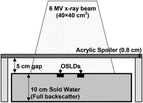
The ability of the 8 mm beam spoiler to improve surface dose to the same extent as the 5 mm bolus placed on the opposing side of the animal was also experimentally tested. To do this, OSLDs placed at the surface of a solid water phantom with a 40 × 40 cm2 field () with and without a beam spoiler were irradiated. Doses from the OSLDs were compared to measurements performed under reference conditions (1.5 cm depth, 100 cm SAD, no spoiler).
Figure 1. Experimental setup for assessing effect of beam spoiler. The OSLDs were irradiated with a 0.8 cm beam spoiler located 5 cm from the surface of a phantom comprised of 10 cm of Gammex® Solid Water designed to produce full back-scatter to the detectors. The spoiler is held on the side with foam blocks to limit scatter. Doses were compared to those measured without spoiler and 1.5 cm solid water buildup (e.g. 100% dose) to assess the bolus-ing effect of the acrylic spoiler.
In-vivo validation
We performed in-vivo dose measurements on each animal to verify the accuracy of dose delivery. Two nanoDot OSLDs (Landauer, Inc.; Greenwood IL) were placed on the skin surface of both flanks of each animal, for a total number of four detectors per animal. The OSLDs were read ∼24 h later using a Landauer microstar II optical reader which stimulates OSLDs emitting in the 400–700 nm optical range. We used screened OSLDs which possess a detector-specific correction factor individual to each detector. According to the manufacturer, individual screened nanoDots are accurate to a ± 5% range (representing two standard deviations) when irradiated with MV-energy photons. Thus, we would expect the standard error of four averaged OSLDs to be ∼2.5%.
The nanoDots and reader were calibrated by our laboratory following the manufacturer’s specifications. A non-linear calibration curve was obtained by irradiating sets of three nanoDots at eight different doses (0, 50, 100, 200, 400, 700, 100, and 1200 cGy, respectively) under calibration conditions. On the day of each irradiation, a set of two OSLDs were given a known dose to perform quality assurance of the in-vivo dosimetry system that day.
Each OSLD used for animal irradiations was averaged over four readings. We report the dose averaged for all four OSLDs as a monitor of the mean dose imparted to the animal at midline. The standard deviation was calculated using the sixteen readings (four per detector for four detectors) for each animal.
Results
Technique design
Several beam configurations were considered: an anterior-posterior (AP/PA) technique, where the beams would enter the animal ventrally and dorsally, compared to a bilateral technique where the beams enter the animal laterally from either side. Furthermore, the use of surface dose management through bolus or spoiler was also in question. Using the CT scan of the two pilot study animals and dose distributions calculated with the Varian Eclipse treatment planning software, it was determined that the bilateral technique was vastly superior to the anterior-posterior technique (See ). Indeed, the variation in the animals’ thickness varied much more in the anterior-posterior direction than in the left-right direction, due mostly to the relatively narrow thorax vs abdomen of the animals. Furthermore, the animals tend to be much wider (∼15 cm) in the anterior-posterior axis compared to the left-right axis (∼6–8 cm), which increases attenuation and further degrades dose homogeneity. As shown in , the anterior-posterior technique had both a lower minimal dose (∼85% of prescription) and a higher maximal dose (∼120% of prescription) compared to the bilateral technique (95–107%).
Figure 2. Cumulative dose volume histogram comparing dose coverage of the bilateral technique with lack of bolus/spoiler (top) or an alternative anterior/posterior irradiation geometry (bottom). In both cases, the bilateral technique achieves a much more uniform dose distribution, with minimum coverage by the 95% isodose line and a maximal dose of ∼107%, typically in the extremities (e.g. head, feet) where separation is reduced.
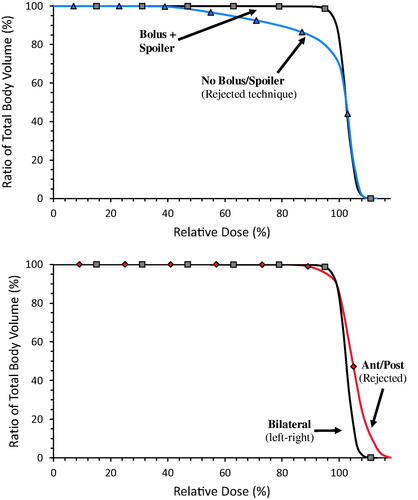
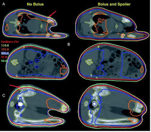
After the optimal beam arrangement was determined, the effect and importance of proper surface dose management (i.e. spoiler and bolus) was assessed and quantified. As shown by , when no surface dose management is used, only ∼2nd/3rd of the rabbit body receives the full (i.e. 100%) prescribed dose, and only ∼80% receives 95% of the prescribed dose. Using a technique with bolus + spoiler, however, delivers a dose of 95% of prescription to 98% of the rabbit body. This is shown more visually in , where the influence of the bolus/spoiler decreases the depth inside of the body at which doses reaches 95% from ∼5–6 mm to 1–2 mm.
Figure 3. Isodose distributions in the absence (left) vs in the presence (right) of bolus and spoiler for an identical prescribed dose at mid-depth at the thorax (A), abdomen (B), and pelvis (C). Without bolus, the depth in which the dose reaches 95% is of the order of ∼0.6–0.7 cm. With bolus, the entire animal is covered by at least 90%, with 95% occurring within the first 0.1–0.2 cm below the surface. Of particular note, some of the bones (contoured in yellow) are not entirely covered by the 90% and 95% isodoses in the absence of bolus (see arrows).
Organ doses in therepresentative animal
One of the pilot animals’ organs was contoured on the CT scan by an experienced dosimetrist. Analysis of the statistics of these organ-specific doses (see ) show that, when using bolus and spoiler, the mean dose for each organ ranges from 102.1 to 105.5%, with minimum doses ranging from 95.8 to 104% and maximum doses ranging from 103.7 to 110.6%. In general, organs located in the thorax (bronchus, heart) where lung inhomogeneities reduce attenuation have higher doses while organs closer to the abdomen (stomach, bowel) where attenuation is highest have slightly lower doses. Since the MU calculations were performed to deliver a dose of 100% at the animal midline, where attenuation is highest, organ doses tend to slightly exceed 100% in general.
Figure 4. DVH values for the min/95%/median/95%/max organ doses in the single representative scanned animal for the bilateral technique with or without surface dose management (i.e. bolus and spoiler). Dose to organs is generally similar between both cases except for the bowel and, more importantly, the bones which harbor the all-important bone marrow where >5% of the volumes lie in areas covered by <90% of the prescribed dose.
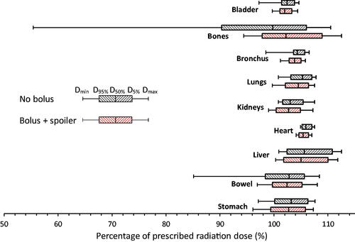
A comparison of the organ doses with and without surface dose management shows that there is little difference in organs at depth. This is expected, as surface dose management techniques such as the spoiler and bolus serve mainly to increase dose within the first few mm of the body without affecting dose at depth. The only organs which show some difference are the bowel and bones, where minimum dose increases from 84.3% of the prescribed dose to 95.8%. However, a closer look at the dose-volume histograms shows that >99% of the bowel are covered with at least 95% of the prescribed radiation dose, such that the difference is highly unlikely to lead to different biological outcomes. In the case of the bones, however, >5% of the bones receive a dose <90% of the prescribed dose, with some areas receiving as little as ∼50% of the prescribed dose. Since bones encompass the bone marrow, all important in the incidence of the hematopoietic syndrome, the use of surface dose management seems necessary to ensure reproducible biological outcomes.
Irradiation technique
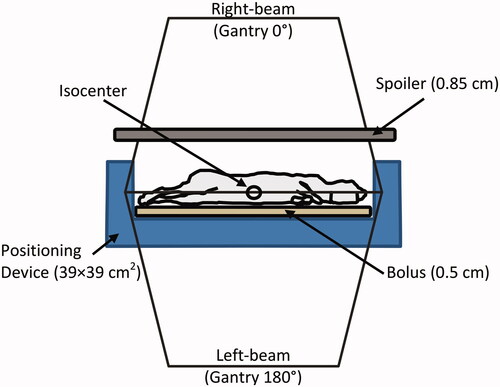
The bilateral irradiation technique settings were optimized using the CT simulation image and the Varian Eclipse TPS and are described in this section. As shown in , the animals are laid in a specially designed positioning device in the left decubitus (i.e. lying on its left side) position. The positioning device is comprised of a Vac-LokTM positioning cushion (CIVCO Radiotherapy, Coralville IA), which is malleable pillow comprising a number of microbeads (radiation attenuation is negligible) which becomes rigid when a vacuum is applied. This cushion is shaped as a 39 × 39 cm2 square opening lined with a layer of 0.5 cm of tissue equivalent bolus in the bed base and on its walls, creating a total 38 × 38 cm2 opening. The rabbit head and buttocks are positioned on the diagonal of the square opening.
There are several advantages of the decubitus position. First, the body position is symmetric laterally, and gravity serves to make the animal thickness more homogeneous along the length and width of the entire animal which improves dose homogeneity. Second, the position is very easy to achieve with short setup time, as it allows for the extension of the neck to permit unobstructed airways and ease in positioning a rectal probe to allow for the required monitoring the animal’s peripheral blood oxygen saturation in blood throughout the procedure.
Once positioning is established, the animal thickness at the abdominal level (halfway between the top of the head and the bottom of the buttocks) is measured. The animal abdominal separation measurement is subsequently used to calculate the number of monitor units (MU) required for adequate irradiation (see Supplemental data). The abdominal separation measurement is also used to position the linear accelerator isocenter (100 cm SAD) at the animal midline using the optical distance indicator (ODI). The positioning device is then moved laterally and longitudinally until the light field covers the entirety of the 38 × 38 cm2 area in the positioning device.
Two beam-modifying devices, the 0.5 cm tissue equivalent bolus placed below the animal, and a 0.85 cm acrylic spoiler placed ∼5 cm above the animal, increase the surface dose of the animal. As shown in , these beam-modifying devices dramatically improve the dose distribution in the animal, allowing a homogeneous dose delivery.
This is a true isocentric technique, in contrast to flipping the animal’s exposed side between beams or using an extended SSD technique where the treatment couch must be moved in between beams. The advantages offered by an isocentric technique is mainly that delivery is quicker, as the gantry can be rotated in between fields from outside the room. An added advantage is more robust delivery. Indeed, in non-isocentric techniques the setup in both fields are independent from one another and errors can compound, whereas they will tend to cancel each other in an isocentric technique.
Phantom validation
Before live animal irradiations began, pre-study validation of our approach was performed on a plastic phantom following two methods – one using an ionization chamber in a rabbit-sized (10 × 10 × 30 cm3) phantom, and a second with OSLDs to measure the effect of the spoiler on improving surface dose. The dose measured using the ionization chamber in the rabbit phantom was within ≤1.5% of the intended dose as calculated by hand calculations, and within 1.0% of that calculated by the treatment planning software.
The dose at the surface of a solid-water phantom using OSLDs was measured both in the absence (68% of dmax) and in the presence (92% of dmax) of a radiation beam spoiler. Since the percent depth dose for this energy is 91.8% at a depth of 5 mm, it was concluded that the spoiler provides surface coverage similar to 5 mm of water-equivalent bolus. Note that this estimate of surface dose does not predict the surface dose to animals irradiated under parallel-opposed pair beam configuration, as the opposing beam does not experience the buildup effect when exiting the animal on the opposing side. Thus, the dose at 5 mm is most probably closer to the 95% level predicted by the planning system ().
Figure 5. Irradiation geometry. The linear accelerator is calibrated to deliver dose at the Isocenter. The mid-depth of the animal is placed at isocenter, and two lateral beams (above and below the animal, at gantry 0° and 180°, respectively) are used. The animals are held within the radiation field’s maximal 40 × 40 cm2. Dose at shallow depths in the animal is enhanced using the bolus (underneath animal) and beam spoiler (∼5 cm above animal). The isocentric design of the irradiation technique allows for rotating the gantry between beam deliveries without requiring manual adjustments to the setup in mid-delivery.
In-vivo validation
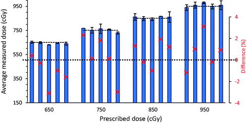
Finally, in-vivo validation was performed by measuring surface doses for each animal using 4 OSLDS and comparing these measurements to the prescribed dose (See ). The average of all OSLD surface dose measurements over all animals was within +0.13% of the intended dose. On an individual basis, animal average doses ranged from −3.06 to + 3.09%. No animal received a dose higher than 3.1% from the prescribed dose.
Figure 6. Average in-vivo OSLD dose measurements for all 20 rabbits exposed at 4 different prescribed dose levels (dashed lines). Error bars indicate the measurement standard deviation. Red crosses represent the difference between measured and prescribed dose (dotted line and scale to the right). No measurement exceeded 3.1% from prescribed dose, even though OSLD measurement statistical uncertainty is ∼5% in individual detectors (2.5% for mean of 4 OSLDs).
Discussion
To the authors’ knowledge, this study represents the first detailed description of a total body irradiation technique for rabbit models since the foundational radiation biology papers published in the 1950s. To the authors’ knowledge, the only paper to show a similar level of details regarding radiation dosimetry in total body rabbit irradiations dates from 1952, and describes a 190 kVp x-ray beam filtered with 0.8 mm Cu + 1.0 mm Al added filtration (Lennox et al. Citation1952). As this was a low-energy kilovoltage x-ray technique, field homogeneity was limited by the lack of a flattening filter and the heel effect, which is why reported dose distributions ranged from 60 to 80% when not near the abdomen, and as high as 105% at the surface near the central axis. In contrast, our technique covers the entire animal with a dose ranging from 95 to 107%, with much of the volume receiving >105% being restricted to the extremities (e.g. head, feet) where there is lack of attenuation.
As far as the authors are aware, no other study reports organ-specific doses for rabbit TBI techniques. However, our team recently published a similar study in non-human primates (NHPs) (Prado et al. Citation2017) which reports mean organ doses of 104.2–107.8% of prescribed doses when irradiated using a TBI technique with 2.5% bone marrow sparing (tibiae). Our mean doses of 102.1–105.5% compare very favorably with this TBI technique. The ability to CT scan each animal and to measure the organ doses using a given irradiation technique could provide valuable information to research involving radiation exposure and the resulting biological effects specific to the organs.
The main advantages of the proposed methodology is its robustness, convenience, and reproducibility. Owing to careful control over animal positioning, the delivered dose depends only on a single parameter (i.e. animal thickness at mid-depth) which allows the MUs to be pre-calculated. This reduces the risk of calculation or transcription error. Furthermore, use of the immobilization device minimizes the possibility of errors in positioning of the animal body, while the isocentric radiation delivery technique reduces variations due to treatment machine positioning. Finally, in-vivo measurements confirm that the technique is accurate and reproducible.
Our results also suggest that beyond the details that are absolutely necessary to replicate a study, there is value in using 3-D dose calculation treatment planning systems to prospectively design, validate, and implement an accurate and robust irradiation technique in animal models. Traditionally, hand-calculations have been used to calculate dose to midline, and dose throughout the body has been presumed equivalent to the prescribed irradiation dose. However, little attention was paid to the effect of tissue inhomogeneities or animal geometry on dose distributions. As seen from , an identical radiation dose delivered to the animal’s midline can show striking differences in the geometric distribution of the radiation dose, depending on the precise details of the irradiation technique design and geometry. The differences in dose distributions due to change in delivery technique, will alter the dose in the organs. The biological responses due to these differences have not been measured, but it is reasonable to infer that a different dose distribution will induce a difference in observed biological effects. While the need for improvement in physics and dosimetry reporting must first be addressed in the more basic experimental details necessary to reproduce a particular experiment (Pedersen et al. Citation2016; Stone et al. Citation2016; Draeger et al. Citation2020), our study showcases the potential for much more detailed and in-depth comparison between radiation studies in future research.
Conclusion
Using modern dosimetry techniques such as CT simulation and 3-D radiation therapy dose calculation algorithm, a highly reproducible, easily deliverable and robust total body irradiation technique for leporine models has been developed and validated. The advantages are speed (<10 minute set-up and irradiation delivery time per animal), dose accuracy (no animal was measured to receive an average dose >3.1% from prescription), and its convenient methodology relying on only a single thickness measurement). To the authors’ knowledge, this work represents the first comprehensive development of a total body irradiation technique for rabbit models of ARS.
Moreover, this study provides a proof of principle in the powerful role that modern techniques can play in creating and improving irradiation techniques in radiation biology. In this study, 3-D dose distributions calculated using a CT scan were used to design, optimize, and validate a highly accurate study. Furthermore, these three dimensional dose distribution showed the importance of using proper surface dose management (bolus, spoiler) which not only produced a more homogeneous dose distribution in the animal but also reduced variability in individual organ doses. It is the authors’ hope that this study will inspire other investigators to fully utilize modern tools and techniques to improve the technique and reporting of radiation dosimetry in radiation biology.
Rabbit_Paper_sup_materials_appendix_1.docx
Download MS Word (30.7 KB)Acknowledgements
The authors dedicate this article to the memory of Dr Karl Prado, who developed the preclinical physics and dosimetry program in radiobiology at the University of Maryland.
Disclosure statement
The authors declare no conflicts of interest related to the data generated within this study. Drs. Isabel L. Jackson and Zeljko Vujaskovic are scientific advisors to Humanetics Corporation.
Data availability
Raw data were generated at the University of Maryland School of Medicine. The authors confirm that all data supporting the findings of this study will be archived at the UMSOM for up to 5-years post-publication, and are available from the corresponding author (Yannick Poirier), upon reasonable request, and with approval by the Biomedical Advanced Research and Development Authority, Office of the Assistant Secretary for Preparedness and Response, Department of Health and Human Services. While the Biomedical Advanced Research and Development Authority funded the study, the study’s conclusions are those of its authors alone.
Additional information
Funding
Notes on contributors
Yannick Poirier
Yannick Poirier is an Assistant Professor and Board-certified Clinical Medical Physicist in the Department of Radiation Oncology, Division of Medical Physics and Division of Translational Radiation Sciences at the University of Maryland School of Medicine. He oversees the dosimetry and physics of all radiation biological studies of the Division of Translational Radiation Sciences.
Charlotte Prado
Charlotte Prado was a Board-certified Medical Dosimetrist at the Division of Translational Radiation Sciences at the University of Maryland School of Medicine. She has participated in a large number of rabbit and non-human primate studies over the years, contouring organs, performing treatment planning system dose calculations in whole body or whole thorax lung irradiations
Karl Prado
Karl Prado was a Full Professor and a Board-certified Clinical Medical Physicist in the Department of Radiation Oncology, Division of Medical Physics, University of Maryland School of Medicine. He was Associate Director of the Division of Medical Physics and developed the dosimetry of irradiation protocols in non-human primates and murine biological studies of the Division of Translational Radiation Sciences.
Emily Draeger
Emily Draeger was a Research Medical Physicist in the Division of Translational Radiation Sciences at the University of Maryland School of Medicine during the course of the study. She is currently an Assistant Professor at the Department of Therapeutic Radiology at Yale University School of Medicine.
Isabel L Jackson
Isabel Lauren Jackson is an Associate Professor, Radiation Biologist, and Deputy Director of the Division of Translational Radiation Sciences, Department of Radiation Oncology, at the University of Maryland School of Medicine. She is the Director of the division's Medical Counter Measures program.
Zeljko Vujaskovic
Zeljko Vujaskovic is a Full Professor in the Clinical Division and Director of the Division of Translational Radiation Sciences, Department of Radiation Oncology, University of Medicine School of Medicine.
References
- Almond PR, Biggs PJ, Coursey BM, Hanson WF, Huq MS, Nath R, Rogers DWO. 1999. AAPM’s TG-51 protocol for clinical reference dosimetry of high-energy photon and electron beams. Med Phys. 26(9):1847–1870.
- Boone J, Strauss K, Cody D, McCollough C, McNitt-Gray M, Toth T. 2011. Size-specific dose estimates (SSDE) in pediatric and adult body CT examinations: Report of the AAPM Therapy Physics Committee Task Group No. 204. City Park, MD.
- Brooke M. 1062. The effect of total body x-irradiation of the rabbit on the rejection of homologous skin grafts and on the immune response. J Immunol. 88:419–425.
- Desrosiers M, Dewerd L, Deye J, Lindsay P, Murphy MK, Mitch M, Macchiarini F, Stojadinovic S, Stone H. 2013. The Importance of Dosimetry Standardization in Radiobiology. J Res Natl Inst Stand Technol. 118:403–418.
- Dinges MM, Gregerson DS, Tripp TJ, McCormick JK, Schlievert PM. 2003. Effects of total body irradiation and cyclosporin a on the lethality of toxic shock syndrome toxin-1 in a rabbit model of toxic shock syndrome. J Infect Dis. 188(8):1142–1145.
- Draeger E, Sawant A, Johnstone C, Koger B, Becker S, Vujaskovic Z, Jacksol I-L, Poirier Y. 2020. A dose of reality: how 20 years of incomplete physics and dosimetry reporting in radiobiology studies may have contributed to the reproducibility crisis. Int J Radiat Oncol Biol Phys. 106(2):243–252.
- Gibbons JP, Antolak JA, Followill DS, Huq MS, Klein EE, Lam KL, Palta JR, Roback DM, Reid M, Khan FM. 2014. Monitor unit calculations for external photon and electron beams: Report of the AAPM Therapy Physics Committee Task Group No. 71. Med Phys. 41:1–34.
- Gratwohl A, John L, Baldomero H, Roth J, Tichelli A, Nissen C, Lyman SD, Wodnar-Filipowicz A. 1998. FLT-3 ligand provides hematopoietic protection from total body irradiation in rabbits. Blood. 92(3):765–769.
- Holmes T, Serago C, Arjomandy B, Liu C, Yin F-F, Aguirre F, Hanley J, Sandin C, Bayouth J, Ma L, et al. 2009. Task Group 142 report: Quality assurance of medical acceleratorsa). Med Phys. 36(9):4197–4212.
- Hussain A, Villarreal-Barajas E, Brown D, Dunscombe P. 2010. Validation of the eclipse AAA algorithm at extended SSD. J Appl Clin Med Phys. 11(3):3213–3100.
- Kazi AM, MacVittie TJ, Lasio G, Lu W, Prado KL. 2014. The MCART radiation physics core: the quest for radiation dosimetry standardization. Health Phys. 106(1):97–105.
- Lennox B, Dempster WJ, Boag AJ. 1952. Suppression of the Tuberculin reaction in rabbits by total body irradiation with x-rays. Br J Exp Pathol. 33(4):380–389.
- Mesbahi A, Zakariaee SS. 2013. Effect of anode angle on photon beam spectra and depth dose characteristics for X-RAD320 orthovoltage unit. Rep Pract Oncol Radiother. 18(3):148–152.
- Pedersen KH, Kunugi KA, Hammer CG, Culberson WS, DeWerd LA. 2016. Radiation biology irradiator dose verification survey. Radiat Res. 185(2):163–168.
- Prado C, MacVittie TJ, Bennett AW, Kazi A, Farese AM, Prado K. 2017. Organ doses associated with partial-body irradiation with 2.5% Bone marrow sparing of the non-human primate: a retrospective study. Radiat Res. 188(6):615–625.
- Richter M, Abdou NI, Midgley RD. 1970. Cells involved in cell-mediated and transplantation immunity in the rabbit. I. The noninvolvement of the bone marrow antigen reactive cell in the transplant rejection reaction. Proc Natl Acad Sci USA. 65(1):70–73.
- Stone HB, Bernhard EJ, Coleman CN, Deye J, Capala J, Mitchell JB, Brown JM. 2016. Preclinical data on efficacy of 10 drug-radiation combinations: evaluations, concerns, and recommendations. Transl Oncol. 9(1):46–56.
