
Abstract
Purpose
Harmful effects of ionizing radiation on the Central Nervous System (CNS) are a concerning outcome in the field of cancer radiotherapy and form a major risk for deep space exploration. Both acute and chronic CNS irradiation induce a complex network of molecular and cellular alterations including DNA damage, oxidative stress, cell death and systemic inflammation, leading to changes in neuronal structure and synaptic plasticity with behavioral and cognitive consequences in animal models. Due to this complexity, countermeasure or therapeutic approaches to reduce the harmful effects of ionizing radiation include a wide range of protective and mitigative strategies, which merit a thorough comparative analysis.
Materials and methods
We reviewed current approaches for developing countermeasures to both targeted and non-targeted effects of ionizing radiation on the CNS from the molecular and cellular to the behavioral level.
Results
We focus on countermeasures that aim to mitigate the four main detrimental actions of radiation on CNS: DNA damage, free radical formation and oxidative stress, cell death, and harmful systemic responses including tissue death and neuroinflammation. We propose a comprehensive review of CNS radiation countermeasures reported for the full range of irradiation types (photons and particles, low and high linear energy transfer) and doses (from a fraction of gray to several tens of gray, fractionated and unfractionated), with a particular interest for exposure conditions relevant to deep-space environment and radiotherapy. Our review reveals the importance of combined strategies that increase DNA protection and repair, reduce free radical formation and increase their elimination, limit inflammation and improve cell viability, limit tissue damage and increase repair and plasticity.
Conclusions
The majority of therapeutic approaches to protect the CNS from ionizing radiation have been limited to acute high dose and high dose rate gamma irradiation, and few are translatable from animal models to potential human application due to harmful side effects and lack of blood-brain barrier permeability that precludes peripheral administration. Therefore, a promising research direction would be to focus on practical applicability and effectiveness in a wider range of irradiation paradigms, from fractionated therapeutic to deep space radiation. In addition to discovering novel therapeutics, it would be worth maximizing the benefits and reducing side effects of those that already exist. Finally, we suggest that novel cellular and tissue models for developing and testing countermeasures in the context of other impairments might also be applied to the field of CNS responses to ionizing radiation.
Introduction
Development of medical countermeasures against ionizing radiation has far-ranging implications from reducing damage to the healthy tissue during radiotherapy, to limiting the hazards posed by space radiation in deep space exploration, to protecting public health in the event of a nuclear emergency. Our review focuses on countermeasures for irradiation conditions relevant to radiotherapy (low-linear energy transfer (LET) ionizing radiation, such as gamma rays and X-rays, but also high-LET particle radiation, with an increase use of proton and carbon ions (Ray et al. Citation2018), at fractionated doses ranging from 10 to 50 Gy total (Barani and Larson Citation2015; Lumniczky et al. Citation2017) and space environment: primarily galactic cosmic rays, which is the most damaging and hardest to shield component of the deep-space radiation environment, but also gamma rays in the context of low-Earth orbit missions. The radiation doses of interest to model the exposure conditions of astronauts onboard of the International Space Station (ISS) are between 0.1 to 0.4 mGy/day of gamma rays, contributing to the total dose of about 0.15 Gy for a year-long mission, corresponding to the longest time in orbit so far (Beheshti et al. Citation2018). In addition, high-LET particle radiation is becoming increasingly important for deep space exploration beyond the protective magnetic field of the Earth on lunar and Mars missions (Nelson Citation2016), where astronauts are exposed to a total dose of around 0.4 mGy/day of galactic cosmic rays (Hassler et al. Citation2014), composed of about 90% protons, 9% helium particles and 1% high mass and high charge particles (mainly from 12C to 56Fe).
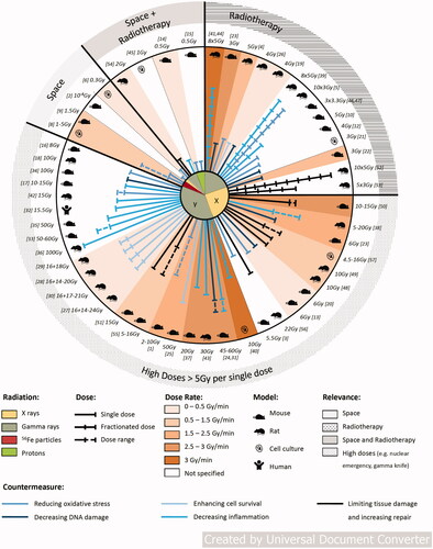
Countermeasures for extreme irradiation conditions (unfractionated doses above 2 Gy) have been extensively reported even though they present limited relevance for human CNS protection. Such doses can be emitted in the event of nuclear emergencies (Chernobyl Citation2002; Wong et al. Citation1993) and in nuclear facilities (Gillies et al. Citation2017; Azizova et al. Citation2020)), but also in novel radiotherapy treatments, such as gamma knife surgery (Colaco et al. Citation2016; Hasegawa et al. Citation2017). They are referenced in this review as potential directions for future applications targeting more relevant radiation types and doses, with the assumption that some protective pathways might be extended across ranges of irradiation conditions ().
Figure 1. Summary representation of the discussed countermeasures. Numbers refer to the following studies: 1. (Nair and Nair Citation2013b), 2. (Mohammad et al. Citation2014), 3. (Wang et al. Citation2018), 4. (Guelman et al. Citation2003), 5. (Lamproglou et al. Citation2003), 6. (Guelman et al. Citation2005), 7. (Erol et al. Citation2004), 8. (Limoli et al. Citation2007), 9. (Manda et al. Citation2008), 10. (Leu et al. Citation2017), 11. (Villasana et al. Citation2013), 12. (Allen et al. Citation2014), 13. (Lu et al. Citation2018), 14. (Parihar et al. Citation2015), 15. (Chmielewski et al. Citation2016), 16. (Weitzel et al. Citation2015), 17. (Raber et al. Citation2017), 18.(Ündeğer et al. Citation2004), 19. (El-Missiry et al. Citation2018), 20. (Manda et al. Citation2007), 21. (Facchino et al. Citation2010), 22. (Yang et al. Citation2009), 23. (Yang et al. Citation2011), 24. (Jiang, Perez-Torres, et al. Citation2014), 25. (Pena et al. Citation2000), 26. (El-Missiry et al. Citation2018), 27. (Andratschke et al. Citation2004), 28. (Nieder et al. Citation2006), 29. (Nieder, Andratschke, et al. Citation2005), 30. (Nieder, Price, et al. Citation2005), 31. (Jiang, Perez-Torres, et al. Citation2014), 32. (Gonzalez et al. Citation2007), 33. (Jiang, Engelbach, et al. Citation2014), 34. (Belarbi et al. Citation2013), 35. (Yang et al. Citation2018), 36. (Erbayraktar et al. Citation2006), 37. (Ansari et al. Citation2007), 38. (Yuan et al. Citation2003), 39. (Zhao et al. Citation2007), 40. (Schnegg et al. Citation2012), 41. (Greene-Schloesser et al. Citation2014), 42. (Desmarais et al. Citation2015), 43. (Kim et al. Citation2004), 44. (Lee et al. Citation2012), 45. (Tikka et al. Citation2001), 46. (Feng et al. Citation2016), 47. (Feng et al. Citation2018), 48. (Baulch et al. Citation2016), 49. (Smith et al. Citation2020), 50. (Liao et al. Citation2017), 51. (Wang et al. Citation2016), 52. (Piao et al. Citation2015), 53. (Zhou et al. Citation2015), 54. (Bala et al. Citation2017), 55. (Oh et al. Citation2013), 56. (Sun et al. Citation2013), 57. (Prager et al. Citation2016).
Although the central nervous system is traditionally not considered to be the most radiosensitive organ, its damage can be particularly devastating to the health and the quality of life, and is difficult to repair. Acute high-dose radiation during radiotherapy induces bystander damage to CNS, leading to reduced hippocampal neurogenesis and development of neuroinflammation throughout the cortex and hippocampus, which are associated with cognitive and memory deficits, particularly harmful to the developing brain in children and adolescents (Mizumatsu et al. Citation2003; Monje et al. Citation2003; Rooney and Laack Citation2013). Acute exposure to galactic cosmic rays or their components causes similar impairments both in vitro and in vivo: increased neuroinflammation, neuronal damage and cognitive deficits similar to accelerated aging (Cekanaviciute et al. Citation2018).
Radiation-induced damage to CNS, as to any other organ, can be classified as a combination of targeted effects of direct DNA damage and non-targeted effects that are primarily mediated by oxidative stress responses and cause cellular damage and death eventually leading to damage at tissue and organ levels (Heuskin et al. Citation2016). Thus, CNS responses to radiation, as shown in , can also be analyzed and mitigated at different levels: from molecular (DNA damage, reactive oxygen species), to cellular (cell membrane damage, cell death), to vascular leakage and disrupted electrochemical connections between neurons, to tissue and organ damage that eventually culminates in behavioral deficits (Greene-Schloesser and Robbins Citation2012).
Figure 2. Representation of radiation-induced responses of the CNS. [1–4] = (Belka et al. Citation2001; Satyamitra et al. Citation2007; Lowe et al. Citation2009; Baluchamy et al. Citation2010; Beckhauser et al. Citation2016), [5–16] = (Fournier and Taucher-Scholz Citation2004; Limoli et al. Citation2004; Kim et al. Citation2006; Al-Jahdari et al. Citation2008; Eriksson and Stigbrand Citation2010; Chakraborti et al. Citation2012; Parihar and Limoli Citation2013; Shirai et al. Citation2013; Kempf et al. Citation2014; Parihar et al. Citation2015), [17–24] = (Clatworthy et al. Citation1999; Sannita et al. Citation2007; Machida et al. Citation2010; Sanchez et al. Citation2010; Marty et al. Citation2014; Rudobeck et al. Citation2014; Sokolova et al. Citation2015), [25–46] = (Tofilon and Fike Citation2000; Vazquez and Kirk Citation2000; van Vulpen et al. Citation2002; Maier Citation2003; Mizumatsu et al. Citation2003; Lyubimova and Hopewell Citation2004; Raber et al. Citation2004; Rola et al. Citation2004; Casadesus et al. Citation2005; Rola et al. Citation2005; Hwang et al. Citation2006; Fike et al. Citation2009; Huang et al. Citation2010; Moravan et al. Citation2011; Kadir et al. Citation2012; Monje and Dietrich Citation2012; York et al. Citation2012; Rivera et al. Citation2013; Greene-Schloesser et al. Citation2014; Morganti et al. Citation2014; Hur and Yoon Citation2017), [47–60] = (Brouwers and Poplack Citation1990; Hall et al. Citation2004; Butler and Haser Citation2006; Rosi et al. Citation2008; Zeltzer et al. Citation2009; Liu et al. Citation2010; Britten et al. Citation2012; Greene-Schloesser and Robbins Citation2012; Armstrong et al. Citation2013; Greene-Schloesser, Moore, and Robbins Citation2013; Kumar et al. Citation2013; Britten et al. Citation2014; Makale et al. Citation2017; Acharya et al. Citation2019).
![Figure 2. Representation of radiation-induced responses of the CNS. [1–4] = (Belka et al. Citation2001; Satyamitra et al. Citation2007; Lowe et al. Citation2009; Baluchamy et al. Citation2010; Beckhauser et al. Citation2016), [5–16] = (Fournier and Taucher-Scholz Citation2004; Limoli et al. Citation2004; Kim et al. Citation2006; Al-Jahdari et al. Citation2008; Eriksson and Stigbrand Citation2010; Chakraborti et al. Citation2012; Parihar and Limoli Citation2013; Shirai et al. Citation2013; Kempf et al. Citation2014; Parihar et al. Citation2015), [17–24] = (Clatworthy et al. Citation1999; Sannita et al. Citation2007; Machida et al. Citation2010; Sanchez et al. Citation2010; Marty et al. Citation2014; Rudobeck et al. Citation2014; Sokolova et al. Citation2015), [25–46] = (Tofilon and Fike Citation2000; Vazquez and Kirk Citation2000; van Vulpen et al. Citation2002; Maier Citation2003; Mizumatsu et al. Citation2003; Lyubimova and Hopewell Citation2004; Raber et al. Citation2004; Rola et al. Citation2004; Casadesus et al. Citation2005; Rola et al. Citation2005; Hwang et al. Citation2006; Fike et al. Citation2009; Huang et al. Citation2010; Moravan et al. Citation2011; Kadir et al. Citation2012; Monje and Dietrich Citation2012; York et al. Citation2012; Rivera et al. Citation2013; Greene-Schloesser et al. Citation2014; Morganti et al. Citation2014; Hur and Yoon Citation2017), [47–60] = (Brouwers and Poplack Citation1990; Hall et al. Citation2004; Butler and Haser Citation2006; Rosi et al. Citation2008; Zeltzer et al. Citation2009; Liu et al. Citation2010; Britten et al. Citation2012; Greene-Schloesser and Robbins Citation2012; Armstrong et al. Citation2013; Greene-Schloesser, Moore, and Robbins Citation2013; Kumar et al. Citation2013; Britten et al. Citation2014; Makale et al. Citation2017; Acharya et al. Citation2019).](/cms/asset/190feff7-3498-41b9-bf8e-ab21474e4119/irab_a_1820598_f0002_c.jpg)
Current approaches to CNS radioprotection usually consist of eliminating radiation-induced reactive oxygen species (ROS), increasing DNA protection and repair, and targeting the downstream effects by limiting inflammation and increasing cell survival and tissue repair. In general, countermeasures can be classified as either primarily protective or mitigative (Rosenthal et al. Citation2011), depending on the timing of the administration. Radioprotectors are given prior to irradiation as preventive measures, while mitigators refer to treatments started after irradiation, prior to clinical evidence of radiation injury. Here we focus on five typical countermeasures, each of which can be either primarily protective (e.g. reducing DNA damage) or mitigative (e.g. stimulating tissue repair), or combine both effects (e.g. reducing inflammation and oxidative stress, increasing cell survival) ().
Table 1. Summary of the discussed countermeasure agents classified into five main approaches (targeting reactive oxygen species, DNA damage, cell survival, inflammation and tissue repair), according to the type of CNS impairment to be treated.
Commonly applied countermeasure approaches for limiting ionizing radiation-induced CNS damage
Reducing oxidative stress
Oxidative stress and ROS production are particularly frequent targets for currently available CNS countermeasures against both therapeutic and space radiation. ROS is a general term including superoxide (O2·-), hydrogen peroxide (H2O2) and hydroxyl radicals (·OH), which are generated by ionizing radiation-induced water radiolysis both within and outside the cell. In addition to ROS, ionizing radiation also stimulates nitric oxide synthase, generating reactive nitrogen species (RNS) (Routledge et al. Citation1994; Hall and Giaccia Citation2006), that have lower diffusion coefficients and higher short-range reactivity compared to ROS (Frongillo Citation1998; Lide Citation2004). RNS are particularly important in the CNS, because neuronal nitric oxide synthase (nNOS) is constitutively active in neurons where it participates in synaptic plasticity (Förstermann and Sessa Citation2012), and can thus be subverted for RNS production. Even comparatively low doses of ionizing radiation generate sufficient ROS and RNS to damage nucleic acids, proteins and lipids (Halliwell and Aruoma Citation1991; Wiseman and Halliwell Citation1996; Mikkelsen and Wardman Citation2003). When ROS and RNS levels exceed the cellular antioxidant defense capacities, oxidative stress can induce permanent cellular and physiological damage via apoptosis (in case of high levels of DNA damage that cannot be repaired, or failures in DNA repair processes), carcinogenesis (in case of mutations in cell cycle regulation genes) and phosphorylation/dephosphorylation imbalance (Spitz et al. Citation2004). In addition, in brain tissues, redox balance has been reported to play a major role in neurogenesis (Huang et al. Citation2012) (which is the formation and integration of new neurons that occurs in the hippocampus and subventricular zone in adult humans and rodents, but also in the olfactory bulb in rodents); neural stem cell proliferation and differentiation (Iqbal et al. Citation2017) and neuronal reprogramming (Klempin et al. Citation2017). Radiation-mediated oxidative stress can be reduced either by administering pharmaceutical antioxidants, or by activating existing cellular antioxidative mechanisms. Supplementary Table 1 compares the experimental conditions and main results of different mitigation approaches targeting the reduction of oxidative stress.
The brain is particularly vulnerable to oxidative stress due to the high abundance of polyunsaturated fatty acids in neuronal cellular and mitochondrial membranes and synaptic protein complexes (Joshi and Praticò Citation2014; Shichiri Citation2014). Membrane disruption not only leads to cell death by stimulating apoptosis and autophagy, but also further damages the DNA due to the reactions of highly reactive byproducts, such as malondialdehyde, with DNA nucleotides. Lipid peroxidation may alter membrane characteristics, leading to disrupted neuronal transport and synaptic transmission. Further disturbance to synaptic plasticity may be caused by oxidation of post-synaptic components, for example, cysteine groups of N-methyl-D-aspartate (NMDA) receptors (Lu et al. Citation2001).
In rodent models of therapeutic gamma irradiation, cellular and DNA damage caused by membrane lipid peroxidation has to some degree been decreased by compounds developed from natural phytochemicals. For example, the polyhydroxy-phenolic compound gallic acid, when administered one hour prior to irradiation, was demonstrated to reduce peroxide and increase antioxidant enzyme levels, concurrently decreasing DNA damage and increasing DNA repair (Nair and Nair Citation2013a). These molecular and cellular radioprotective effects were associated with positive behavioral outcomes: partial recovery of radiation-induced loss of body weight, and increased survival, reaching 80% at 12 days post-irradiation compared to 30% for untreated irradiated mice (Nair and Nair Citation2013a).
A more general countermeasure to reduce oxidative stress in animal models in response to simulated therapeutic or space radiation is the direct elimination of free radicals by antioxidant compounds. This approach often utilizes antioxidants that are widely available in healthy diets, such as dried plums (Schreurs et al. Citation2016), rhubarb (Lu et al. Citation2015), watermelon juice (Mohammad et al. Citation2014) or possibly, epimedium extracts (Wang et al. Citation2018). Besides nutrition-based antioxidants, the only currently FDA-approved drug to prevent oxidative stress after irradiation is amifostine (Ethylol®), usually administered before radiation exposure. It is mainly used for its free radical scavenging properties, but the protective mechanism of amifostine also involves the modulation of natural antioxidant enzymes, induction of cellular hypoxia, DNA protection and repair acceleration (Kouvaris et al. Citation2007) The radioprotective effects of amifostine on the CNS through intraperitoneal or intrathecal administration have been demonstrated with increased survival and improved behavioral outcomes in newborn and young rats (Guelman et al. Citation2003; Lamproglou et al. Citation2003), as well as protection of rat cerebellar granular cells in vitro (Guelman et al. Citation2005). However, the clinical use of amifostine is limited to the protection against xerostomia induced by radiotherapy and is frequently associated with severe side effects (Rades et al. Citation2004).
In addition to amifostine, other free radical scavengers have also been demonstrated to reduce radiation-induced neurodegeneration and behavioral impairments in irradiated rodents. For example, following therapeutic irradiation paradigm of 2 sequences of 3.6 Gy, melatonin was shown to significantly reduce edema, necrosis and neuronal degeneration in rat parietal cortex, while vitamin E only had a significant effect on necrosis (Erol et al. Citation2004). Meanwhile, when used as a countermeasure against simulated space (heavy ion) radiation lipoic acid induced a significant decrease in intracellular ROS level in vitro in rat neural precursor cells, with higher performances for post-irradiation treatment compared to pre-irradiation treatment (Limoli et al. Citation2007). Similarly in vivo lipoic acid significantly mitigated spatial memory impairments and cerebellar cell death in space radiation component 56Fe-irradiated mice (Manda et al. Citation2008). Prevention of spatial memory loss in lipoic acid-treated 56Fe-irradiated mice was also demonstrated by Villasana et al (Villasana et al. Citation2013), but this was accompanied with significant inhibition of novel object recognition and conditioned fear memory responses, suggesting that the use of lipoic acid as an antioxidant might induce other cognitive side effects.
An alternative method of reducing oxidative stress after irradiation is to increase the natural cellular expression of antioxidants by either pharmacological or genetic means. One such pharmacological tool is the chemical compound difluoromethylornithine (DMFO), which was shown to increase the levels of two antioxidant (thioredoxin 1 and peroxiredoxin 3) enzymes in the hippocampus and significantly improve spatial memory in mice subjected to combined 4 Gy gamma-irradiation and traumatic brain injury in order to better simulate the cognitive impacts in the context of a nuclear attack (Allen et al. Citation2014). These results are particularly promising given that DMFO was simply administered through enriched water, starting 24 h post-recovery from the traumatic brain injury. However, the potential therapeutic benefits of DMFO or thioredoxin 1/peroxiredoxin 3 directly in other radiation contexts, including therapy and space radiation, remain to be discovered.
Cellular responses to oxidative stress include the upregulation of various natural antioxidants, such as superoxide dismutases (SOD), glutathione peroxidase (GHS) and catalase, which reduce ROS by converting superoxide and hydroxide ions to water (Smith et al. Citation2017). In the context of CNS, the disruption of the glutathione-glutamate homeostasis by oxidative stress (Koga et al. Citation2011) can lead to synaptic dysfunction and has been associated with epilepsy (Sedlak et al. Citation2019). Augmenting the levels of cellular antioxidants by intraperitoneal administration of glutathione (GSH) in tumor-bearing mice before or after 6 Gy X-ray therapeutic radiation model improved mouse cognitive performance in the water maze (Lu et al. Citation2018). GSH is particularly promising for radiotherapy as it shows high performance when administered post-irradiation and does not interfere with the efficiency of the tumor treatment. Genetic upregulation of antioxidants has also been used in transgenic mice overexpressing human catalase localized to the mitochondria (Parihar et al. Citation2015; Chmielewski et al. Citation2016) in space radiation paradigms. Catalase overexpression reduced ROS, increased neuronal arborization and dendritic complexity, and improved performances in object recognition tests in mice following space-relevant low-dose 0.5 Gy proton irradiation. However, this option cannot be directly translated to human radioprotection and would require targeting catalase by a pharmacological agent instead.
Finally, ionizing radiation causes oxidative stress not only by the formation of ROS from water radiolysis in the cytoplasm, but also by mitochondrial dysfunction. Because of their significant spatial occupation in the cell (typically between 4 to 25% (Leach et al. Citation2001)), mitochondria are a likely target of radiation impact. ROS already exist in mitochondria as by-products of oxidative phosphorylation (Kim et al. Citation2005), and can be amplified by ionizing radiation leading to mutations in mitochondrial DNA and to disturbed expression of critical proteins for mitochondrial and cellular functions (Azzam et al. Citation2012). Moreover, the high proximity between mitochondria near the nucleus (Davis and Clayton Citation1996) allows easy nuclear propagation of the oxidative signal from the irradiated mitochondrion. Interestingly, oxidative damage in mitochondrial DNA is several-fold higher than in nuclear DNA (Richter Citation1992), probably because of the proximity of mitochondrial DNA to ROS, the lack of protective histone proteins for mitochondrial DNA and less efficient DNA repair mechanisms (Wiseman and Halliwell Citation1996).
The main natural radioprotectant in mitochondria is an enzyme called Manganese Superoxide Dismutase (MnSOD) (Guo et al. Citation2003). Thus, artificial elevation of MnSOD levels is a logical radioprotective approach (Rosenthal et al. Citation2011). Numerous SOD mimetics have been synthesized with a lower molecular weight compared to native SODs, in order to increase their cell permeability and circulating half-time (Bonetta Citation2018). MnBuOE appears as the most promising MnSOD mimetic compound for radiotherapy use and is currently in a phase 2 trial (NCT02655601). It has been demonstrated to reduce neuronal damage and demyelination and improve motor proficiency of 8 Gy gamma-irradiated mice (Weitzel et al. Citation2015), notably acting both as a neuroprotector and as a radiosensitizer on glioblastoma cells, especially when administered 1 week prior to irradiation (Leu et al. Citation2017). Another promising MnSOD mimetic, EUK-207, similarly demonstrated significant mitigation of cognitive impairments in 15 Gy gamma-irradiated mice (Raber et al. Citation2017), and was effective even post-irradiation.
Decreasing DNA damage
A critical consequence of ionizing radiation exposure is DNA damage, which can lead to cellular damage, cell death and accumulation of mutations that eventually contribute to carcinogenesis. Ionizing radiation causes DNA damage either in a targeted manner, by energy deposition along the path traversed by the radiation beam, or in a non-targeted manner, by ROS, RNS and peroxidized lipids formed during oxidative stress (Marnett Citation2002; Islam Citation2017; Sage and Shikazono Citation2017). During X-ray and gamma radiation two thirds of DNA damage are estimated to be targeted and the remaining one-third non-targeted (Sage and Shikazono Citation2017). The different strategies and experimental conditions for countermeasures to radiation-induced DNA damage in the brain are summarized in . In general, targeted DNA damage has proven difficult to prevent, unless by shielding, which is not always possible in a therapeutic setting. As a result, research in radiobiology has been more focused on preventing non-targeted DNA damage and on repairing its outcomes.
Figure 3. Flowchart of the main discussed processes of radiation-induced targeted and non-targeted DNA damage effects, and associated countermeasures. RIBE: radiation-induced bystander effects, CNPs: anti-histone antibody complexed nanoparticles, R-Cu: resveratrol-copper, ROS: reactive oxygen species, RNS: reactive nitrogen species, SSBs: single-stranded breaks, DSBs: double-stranded breaks, DDR: DNA damage response, BER: base excision repair, HR: homologous recombination, NHEJ: non-homologous end joining. Numbers refer to the following studies: 18. (Ündeğer et al. Citation2004), 19. (El-Missiry et al. Citation2018), 20. (Manda et al. Citation2007), 21. (Facchino et al. Citation2010), 22. (Yang et al. Citation2009), 23. (Yang et al. Citation2011), 24. (Jiang et al. Citation2014) Supplementary Table 2 provides additional information regarding the model, irradiation, administration conditions and main results for each of the discussed countermeasures.
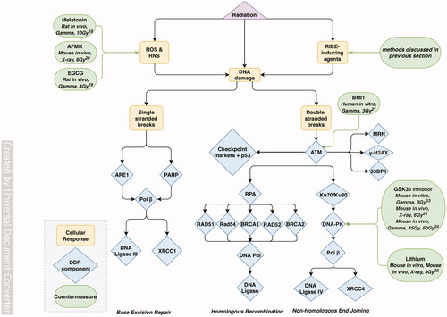
The collective ensemble of pathways and proteins that participate in DNA repair after radiation-induced DNA damage is called the DNA damage response (DDR). DDR is responsible for dealing with radiation induced single strand breaks, which occur more frequently, as well as double strand breaks, which are less frequent, but more dangerous to the cell (Santivasi and Xia Citation2014; O'Connor Citation2015; Delia and Mizutani Citation2017). Single strand breaks undergo repair through base excision repair, (Wallace Citation2014; O'Connor Citation2015), while double strand breaks are mainly repaired through either homologous recombination or non-homologous end joining (Santivasi and Xia Citation2014; O'Connor Citation2015). Homologous recombination is a highly accurate DDR process in which a homologous sister chromatid is used as a template for repairing the DSB site, but it is slow, depends on an undamaged sister chromatid, and can only occur in S phase of the cell cycle (Kobayashi et al. Citation2008; Shrivastav et al. Citation2008; Lord and Ashworth Citation2012; Santivasi and Xia Citation2014; O'Connor Citation2015; Delia and Mizutani Citation2017). Non-homologous end joining is faster, because it occurs in all phases of the cell cycle, but is more error-prone because it repairs double stranded breaks by simply ligating the ends of the lesion together (Kobayashi et al. Citation2008; Shrivastav et al. Citation2008; Lord and Ashworth Citation2012). Regardless of the type of DDR, failure to repair DNA damage, or mistakes during repair, can lead to genomic instability, tumorigenesis or cell death via signaling by transcription factor p53 (Santivasi and Xia Citation2014; Delia and Mizutani Citation2017).
Most of the success in preventing radiation-induced DNA damage in brain tissues has been achieved by free radical scavenging and mitigation of ROS production, described in detail in the previous section. These approaches focus on clearing cells of detrimental free radicals and ROS before they have the chance to damage nucleic acids. For example, administration of melatonin or epigallocatechin-3-gallate prior to simulated therapeutic irradiation has been shown to reduce DNA damage in brains of rats irradiated with respectively 10 Gy and 4 Gy gamma rays (Ündeğer et al. Citation2004; El-Missiry et al. Citation2018); pretreatment with melatonin also reduced DNA damage in mice exposed to 6 Gy X-rays (Manda et al. Citation2007).
Instead of DNA damage prevention, other countermeasures focus on enhancing DNA repair in damaged cells to rescue them from cell death or genomic instability. Tumor cells such as glioblastoma have been found to be particularly efficient in repairing DNA damage and avoiding cell death (Lord and Ashworth Citation2012). Consequently, studying what makes these cancer cells radioresistant and emulating their DDR mechanisms has been an advantageous strategy for the discovery of countermeasures aimed at DNA damage repair. For example, BMI1, a gene typically known for its importance in stem cell maintenance, has been found to be significantly upregulated in highly radioresistant glioblastoma cells as compared to normal brain cells (Bruggeman et al. Citation2007). This finding inspired the study of human neural stem cells (NSCs) infected with lentivirus to overexpress BMI1, which led to faster DNA repair in vitro (Facchino et al. Citation2010); although viral gene expression and the facts that it would have to be limited to the bystander cells avoiding tumor cells, not to mention a potential side effect of tumorigenesis, limit its therapeutic applicability.
Another approach of targeting DNA repair mechanisms is repurposing the agents that have been neuroprotective in other CNS injuries, such as lithium, which protects the brain during stroke and oxidative stress (Dell'Osso et al. Citation2016). Indeed, lithium-based pharmaceuticals have been demonstrated to enhance the repair of double stranded DNA breaks in mouse hippocampal neurons in vitro and in vivo (Yang et al. Citation2009). Since lithium is an inhibitor of glycogen synthase kinase 3 beta isoform (GSK3b), direct inhibition of GSK3b using a small molecule SB216763 has similarly accelerated DNA repair in mouse hippocampal neurons and in vivo (Yang et al. Citation2011) in response to 3-6Gy simulated radiotherapy irradiation. However, GSK3b inhibition might have improved the cellular health in a less specific manner as well, due to it being involved in multiple Wnt/β-catenin signaling pathways that regulate trophic support to cells and the cell cycle. A notable advantage of radioprotection through lithium treatment and through GSK3b inhibition is that the protective effects did not extend to tumor cells: mouse glioma cells (GL261) and human glioma cells (D54) showed no significantly different repair kinetics between treated and untreated groups following 3 Gy of gamma irradiation. This difference in protective potential may be explained by impairments of GSK3b signaling in tumor cells. The ability to selectively protect non-tumor cells during radiotherapy treatments is highly necessary to prevent necrosis of healthy tissue as well as to avoid increasing malignancy and metastases of existing tumors (Lord and Ashworth Citation2012; Santivasi and Xia Citation2014). Moreover, the inhibition of GSK3 by small molecule SB415286 was also shown to downregulate inflammatory responses for reduction of mouse brain necrosis (Jiang, Perez-Torres, et al. Citation2014).
In summary, although DNA damage is widely studied as a biomarker and as a metric, the DDR has not been extensively or effectively targeted as a countermeasure itself, especially with regard to the CNS. The majority of published studies instead aim to reduce non-targeted DNA damage through antioxidant and anti-inflammatory mechanisms. On the other hand, it is important to consider that increasing DNA repair just-enough to prevent cell death may be harmful for the tissue and the whole organism by retaining DNA mutations and increasing carcinogenesis. Therefore, an alternative approach to radioprotection would be enhancing overall tissue health by diminishing DNA repair so that injured cells die quickly (Zhou et al. Citation2015). It conveys a potential benefit to the whole tissue if injured cells are silenced quickly, before they can harm surrounding cells through bystander effects or initiate carcinogenesis through accrued genomic instability (Jeggo Citation2009; Zhou et al. Citation2015).
Enhancing cell survival
As has been observed in radiotherapy, cellular exposure to high doses of ionizing radiation or prolonged irradiation can lead to increasing damage until cell death. Four main modes of cell death have been reported so far in the context of irradiation responses and are represented in (Eriksson and Stigbrand Citation2010): apoptosis (programmed and rapid death), necrosis (membrane disruption and cell swelling), autophagy (self-consumption of the cell) and senescence (essentially defined by a permanent growth arrest and alteration of neighboring cell functions). Indeed, the senescence phenotype can spread into the microenvironment of the senescent cell through the release of signaling molecules (Campisi Citation2013), which (same as apoptosis) can be beneficial when limited to eliminating the potentially cancerous surrounding cells. The specific mode of cell death gets determined by the type and dose of ionizing radiation, and also by the type and functions of the cell (Abend Citation2003). Apoptosis is a controlled cell death process that causes the least possible damage to the organism. When its control cannot be implemented, cell death occurs through necrosis or autophagy, inducing damage to neighbor cells; while senescence is experienced by irradiated cells following high levels of DNA damage (Abend Citation2003). The different approaches targeting these cell death mechanisms to enhance the survival rate of irradiated cells are summarized in Supplementary Table 3.
Figure 4. Flowchart of the main discussed processes of radiation-induced cell cycle modifications, and associated countermeasures. AMPK: AMP-activated protein kinase, JNK: Jun N-terminal kinase, BAX: Bcl-2-associated X protein, BAD: Bcl-2-associated death promoter protein, TSC2: tuberous sclerosis complex-2, mTOR: mammalian target of rapamycin, ASM: acid sphingomyelinase, PI3K: phosphatidylinositol-3 kinase, Akt: serine/threonine-specific protein kinase, PKC: Protein kinase C, IGF-1: insulin-like growth factor-1, MAPK: mitogen-activated protein kinase, ERK: extracellular signal-regulated kinase, EGCG: epigallocathechin-3-gallate, FGF: fibroblast growth factor, PDGF: platelet-derived growth factor. Numbers refer to the following studies: 25. (Pena et al. Citation2000), 26. (El-Missiry et al. Citation2018), 27. (Andratschke et al. Citation2004), 28. (Nieder et al. Citation2006), 29. (Nieder, Andratschke, et al. Citation2005), 30. (Nieder, Price, et al. Citation2005) Supplementary Table 3 provides additional information regarding the model, irradiation, administration conditions and main results for each of the discussed countermeasures.
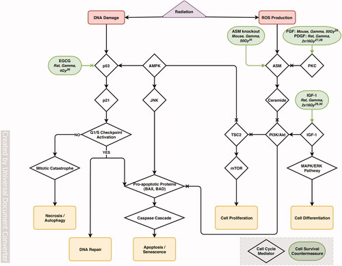
Transcription factor p53 has an essential role in the induction of cell death and is often referred to as the guardian of the genome (Efeyan and Serrano Citation2007). According to the extent of damaged DNA and cell type, p53 activates either DNA damage repair genes or apoptosis and senescence genes (Eriksson and Stigbrand Citation2010). Recently, therapeutic inhibition of p53-induced apoptosis was demonstrated through oral administration of epigallocathechin-3-gallate (EGCG), the main polyphenol found in green tea, in 4 Gy gamma-irradiated rats (El-Missiry et al. Citation2018). EGCG is a promising neuroprotective compound due to its ability to pass through the blood-brain barrier (Pogačnik et al. Citation2016). However, this approach needs to be further investigated in order to evaluate the associated detrimental effects of p53 inhibition, because it has been reported that even though p53 mutation or deletion allows cells to evade apoptosis, they instead undergo several cycles of cell division with severe DNA damage eventually inducing necrosis or autophagy (Eriksson and Stigbrand Citation2010), which would be even worse for the organism.
Complementary to inhibiting cell death, a number of studies have investigated the use of growth factors to prevent cellular damage during irradiation. Intrathecal administration of PDGF (platelet-derived growth factor) in the first 4 days after therapeutic gamma irradiation was shown to significantly reduce myelopathy in rats for 12 months afterwards (Andratschke et al. Citation2004). Nonetheless, treatment efficacy was highly dependent on the administered PDGF dose; and a different administration method (than intrathecal injection) and time (after irradiation) might be more relevant for human applications. However, late administration of PDGF not only did not improve outcomes, but worsened them by accelerating tissue damage (Nieder et al. Citation2006).
Meanwhile, another growth factor, IGF-1 (Insulin-like Growth Factor 1), which has the advantage of crossing the blood-brain barrier (Pan and Kastin Citation2000), has similarly reduced myelopathy in 12-month long studies on therapeutic gamma irradiated rats (Nieder, Price, et al. Citation2005; Nieder, Zimmermann, et al. Citation2005). However, since IGF is a major activator of cell growth and survival (Valenciano et al. Citation2012), its administration carries the risk of inducing unwanted stimulation of tumor growth and reducing the efficacy of radiotherapeutic treatments. Thus, overall, the modulation of cell cycle processes is a complex approach for radioprotective measures since it has the risk of negatively influence the balance between cell repair, apoptosis and proliferation. Specifically, the side effects associated with growth factors suggest that cell and tissue survival might better be targeted by alternative approaches, such as localized stem cell therapy, instead of changing the growth factor levels in the irradiated organism.
Reducing inflammation
Inflammation is one of the most important responses to ionizing radiation exposure that can contribute to tissue impairments years after the irradiation. Inflammatory responses to radiation are complex and include vascular damage, immune cell migration and release of inflammatory regulators, as represented in . As described in previous sections, DNA damage together with the generation of ROS and RNS in irradiated cells induces cell death through different mechanisms such as apoptosis, necrosis, autophagy and senescence. The inflammatory response is in part determined by the mechanism of cell death: while necrosis, autophagy and senescence are associated with rapid loss of cell membrane integrity that induces inflammation-stimulating danger signals (Lasry and Ben-Neriah Citation2015; Qian et al. Citation2017), apoptosis is a programmed cellular suicide that stimulates phagocytes to produce anti-inflammatory cytokines (Rock and Kono Citation2008), unless the apoptotic cell is not cleared by the phagocytes sufficiently fast and undergoes secondary necrosis that again induces proinflammatory responses (Multhoff and Radons Citation2012).
Figure 5. Flowchart of the main discussed processes of radiation-induced inflammatory responses, and associated countermeasures. EPO: erythropoietin, HIF-1α: hypoxia-inducible factor α, VEGF: vascular endothelial growth factor, CXCR4: CXC motif chemokine receptor 4, CCR2: CC motif chemokine receptor 2, ACE: angiotensin-converting enzyme, Ang II: angiotensin II, TNF-α: tumor necrosis factor α, VCAM-1: vascular cell adhesion protein 1, ICAM-1: intercellular adhesion molecule 1, GSK-3: glycogen synthase kinase 3, DAMPs: danger-associated molecular patterns, TLRs: toll-like receptors, NF-κB: nuclear factor NF-κB, AP-1: activator protein 1, PPAR: proliferator-activated receptor, COX-2: cyclooxygenase-2, iNOS: inducible nitric oxide synthase, NLRP3: NLR Family Pyrin Domain Containing 3, IL-1: interleukin-1, IFN-γ: interferon-γ, TGF-β: transforming growth factor β. Numbers refer to the following studies: 31. (Jiang, Perez-Torres, et al. Citation2014), 32. (Gonzalez et al. Citation2007), 33. (Jiang, Engelbach, et al. Citation2014), 34. (Belarbi et al. Citation2013), 35. (Yang et al. Citation2018), 36. (Erbayraktar et al. Citation2006), 37. (Ansari et al. Citation2007), 38. (Yuan et al. Citation2003), 39. (Zhao et al. Citation2007), 40. (Schnegg et al. Citation2012), 41. (Greene-Schloesser et al. Citation2014), 42. (Desmarais et al. Citation2015), 43. (Kim et al. Citation2004), 44. (Lee et al. Citation2012) Supplementary Table 4 provides additional information regarding the model, irradiation, administration conditions and main results for each of the discussed countermeasures.
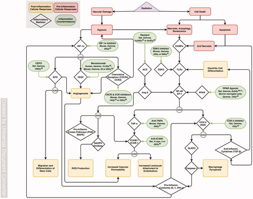
A severe consequence of acute high dose ionizing radiation in the CNS that might be used for brain tumor treatment is inflammatory damage to the vasculature. The damage of vascular tissues following irradiation dysregulates oxygen diffusion between the tissue and blood vessels, in part via vascular endothelial growth factor (VEGF) expression, leading to tissue hypoxia and ultimately, necrosis of bystander non-cancerous brain tissue. Thus, VEGF expression inhibitor bevacizumab (Avastin®, Genentech) has been used to treat radiation-induced brain necrosis in 15 patients (Gonzalez et al. Citation2007), and has efficiently decreased it in 50–60 Gy gamma ray (‘gamma knife’ model) irradiated mice (Jiang, Engelbach, et al. Citation2014). However, bevacizumab treatment has been reported to cause side effects during prolonged administration, including vessel overpruning, deep vein thrombosis and focal mineralization (Jeyaretna et al. Citation2011; Levin et al. Citation2011; Duan et al. Citation2017). Moreover, other studies have demonstrated the recurrence of radiation necrosis after stopping bevacizumab treatment and drug-resistance for re-treatment after discontinuation (Furuse et al. Citation2011; Zhuang et al. Citation2016).
These reasons have initiated a search for alternative approaches to reduce brain necrosis after therapeutic irradiation, including pharmaceutical blocking of cytokine and chemokine signaling. Genetic knockout of chemokine receptor CCR2 was shown to be partially efficient, since it has prevented 10 Gy gamma ray-induced cognitive impairments and rescued synaptic plasticity, but was not sufficient to prevent the overall loss of newborn neurons (Belarbi et al. Citation2013). Pharmaceutic targeting of cytokine/receptor HIF-1a-CXCR4 signaling pathway using topotecan and AMD3100 (Yang et al. Citation2018) showed similar outcomes. This pathway increases cell growth, invasiveness and endothelial cell recruitment, leading to angiogenesis (Kircher et al. Citation2018), and also enhances hypoxia (Yang et al. Citation2018), thus, blocking it could theoretically prevent both radiation-induced brain damage and tumor growth. Both topotecan and AMD3100 indeed significantly reduced brain necrosis and lesion volumes after 50 Gy irradiation (Yang et al. Citation2018). However, the mechanisms behind this effect remained unknown, because HIF-1α expression was unchanged.
Another HIF-1α activated inflammatory mediator is the cytokine erythropoietin (EPO), which acts synergistically with VEGF to enhance injury-induced angiogenesis (Wang et al. Citation2004). Significant therapeutic advantages of EPO are its ability to cross the brain-blood barrier in neuroprotective amounts (Brines et al. Citation2000) and to increase the tightness of the barrier and provide protection against VEGF-induced leakiness both in vitro and in vivo (Martínez-Estrada et al. Citation2003; Üzüm et al. Citation2006). Carbamylated EPO (CEPO) was developed by Erbayraktar et al. to limit its side effect of thrombosis (Erbayraktar et al. Citation2006). CEPO-treated rats have indeed shown a reduction in brain necrosis, and improved forelimb reflex movements following 100 Gy gamma-irradiation modeling gamma knife-based tumor excision. This study further contributes to the perspectives of stimulating angiogenesis to reduce damage in irradiated non-cancerous tissue.
Complementary approaches have also been investigated to preserve irradiated tissues by decreasing microvascular permeability to pro-inflammatory immune cells that are activated elsewhere in the body and could further damage the brain tissue. In particular, radiation is responsible for the adhesion of leukocytes and alteration of endothelial cell tight junctions that form the protective blood-brain barrier (Frank and Lisanti Citation2008). This radiation-induced permeability in a simulated radiotherapy setup was efficiently limited by targeting the TNF-α signaling pathway mediated by NF-κB that stimulates the expression of leukocyte adhesion molecules such as VCAM-1 and ICAM-1, using anti-TNF-α (Ansari et al. Citation2007) or anti-ICAM-1 antibodies (Yuan et al. Citation2003). In addition, the beneficial effects of NF-κB inhibition have been demonstrated by using agonists of proliferator-activated receptors (PPARs)-alpha and –gamma (Zhao et al. Citation2007; Schnegg et al. Citation2012; Greene-Schloesser et al. Citation2014), which inhibit NF-κB activity (Daynes and Jones Citation2002) and improve cognitive performance in irradiated rats. However, these cognitive benefits were obtained together with a side effect of decreased locomotor behavior, while the PPAR-alpha treatment did not provide protection against the radiation-induced reduction in neurogenesis and increase in microglial activation. Nevertheless, PPAR-agonists remain promising radioprotective agents especially as they have demonstrated antitumor properties in addition to their neuroprotection (Tachibana et al. Citation2008).
Alternative approaches to limit radiation-induced neuroinflammation by reducing cell proliferation and migration are to utilize antagonists to cyclooxygenases (COX) or glycogen synthase kinases (Jope et al. Citation2007; Nuvoli and Galati Citation2013). COX-2 inhibition indeed reduced inflammation and increased survival in 15 Gy-irradiated rat glioma model (Desmarais et al. Citation2015).
In radiotherapy, the effectiveness of anti-inflammatory neuroprotective countermeasures is in general limited, because brain tumors are often treated by promoting systemic inflammation for example, by inhibiting Transforming Growth Factor-β (TGF-β) signaling (Hardee et al. Citation2012) (Tran et al. Citation2007). Targeting multiple detrimental mechanisms that are induced by ionizing radiation may provide a more successful therapeutic solution. Such a target that modulates both radiation-induced inflammatory responses and oxidative stress is the renin-angiotensin system (RAS), where the angiotensin-converting enzyme (ACE) produces multiple angiotensin peptides with oxidative and pro-inflammatory characteristics (Hanna et al. Citation2002; Suzuki et al. Citation2003; Robbins et al. Citation2010; Satou et al. Citation2018). The first ACE-inhibitor for targeting radiotherapy-induced brain damage (Ramipril) was proposed in 2004 (Kim et al. Citation2004), when it prevented optic nerve damage induced by 30 Gy gamma-irradiation in rats. In 2012, Ramipril was also shown to improve cognitive performances in irradiated rats (novel object recognition task), together with significant decrease in radiation-induced microglial activation and increased neurogenesis, when continuously administrated for the time of the experiment starting 3 days prior to irradiation (Lee et al. Citation2012).
Finally, neuroinflammation caused by therapeutic gamma radiation or simulated space high-LET radiation can be limited by targeting microglia using a small molecule inhibitor. Temporary microglial depletion with reconstitution has been shown to limit neuronal and synaptic damage as well as cognitive outcomes in adult mice irradiated with 9 Gy gamma rays (Acharya et al. Citation2016) as well as with 0.15 − 1 Gy 4He ions (Krukowski et al. Citation2018).
Both neuronal DNA damage (Tikka et al. Citation2001) and neuroinflammation, specifically, microglial activation, can be effectively suppressed using antibiotics: minocycline (Hanlon, Raghupathi, and Huh 2017), doxycycline (Santa-Cecília et al. Citation2016) and ceftriaxone (Lujia et al. Citation2014). These antibiotics present documented safety for human applications and ability to penetrate the blood-brain barrier. However, they also have multiple off-target effects on the rest of the body and its microbiome, which limit their usefulness as countermeasures, especially for chronic administration. Thus, selective targeting of microglia is a more promising approach to reduce CNS damage after irradiation.
Healthy microglia and monocyte populations are stimulated by colony-stimulating factor 1 (CSF-1) (De et al. Citation2014) and therefore can be inhibited using PLX3397, a small molecule inhibitor of the CSF1 receptor. PLX3397 has recently been demonstrated to prevent radiation-induced memory deficits by reducing microglia activation and monocyte accumulation in mouse models following therapeutic-relevant fractionated whole-brain irradiation (Feng et al. Citation2016, Citation2018). Importantly, PLX3397 administration started as late as one week after radiation exposure showed effective results, further increasing the therapeutic value of PLX3397, which is currently in clinical trials. Another inhibitor of the same CSF-1 pathway of microglial activation, a small molecule PLX5622, has also been demonstrated to protect against CNS irradiation by reducing synapse loss and preserving dendritic spines, as well as reducing memory impairments (Krukowski, Feng, Paladini, Chou, Sacramento, Grue, Riparip, Jones, Campbell-Beachler, and Nelson Citation2018; Feng et al. Citation2016; Acharya et al. Citation2016).
Limiting tissue damage and increasing repair
Ionizing radiation causes damage at the tissue-to-organism level through the accumulation of all the outcomes discussed in the previous sections: DNA damage, oxidative stress, vascular damage, cell death, hypoxia and inflammation. Tissue damage in the CNS can manifest as impaired neurogenesis, depleted populations or inhibited functions of specific cell types, chronic inflammation, and progressive white matter degeneration. Physiological impairments, such as chronic CNS inflammation and neuronal loss, lead to cognitive deficits that can last for years after irradiation in animal models and human patients (Prasanna et al. Citation2014; Parihar et al. Citation2015; Burns et al. Citation2016). Tissue damage is particularly important in CNS responses to space radiation, including the components of galactic cosmic rays: it combines neuronal damage, neuroinflammation, and cognitive and behavioral changes primarily associated with loss of social, recognition and spatial memory (Cekanaviciute et al. Citation2018) and remains to be solved before embarking upon long duration spaceflight and space habitation in future lunar and Mars missions. However, the majority of countermeasures have been investigated in radiotherapy models and remain to be applied to simulated space radiation.
All reviewed countermeasures targeting radiation-induced tissue damage are summarized in Supplementary Table 5. One increasingly investigated approach irradiation is cell transplants into mouse or rat brain after simulated radiotherapy. Mesenchymal stem cell (MSC) transplants into mouse brains 2 days after 15 Gy X-ray exposure have been shown to reduce inflammation, cell death and cognitive deficits one month later (Liao et al. Citation2017). A similar study combined MSC transplants with antihypertensive drug nimodipine in mice after 15 Gy of gamma radiation, with more success than MSC transplants alone (Wang et al. Citation2016). In comparison, Baulch et al. (Baulch et al. Citation2016) conducted transplant experiments with microvesicles (MVs) derived from human neural stem cells (hNSC), rather than with the stem cells themselves. After irradiation with 10 Gy X-ray followed by MV transplants 2 days later into the hippocampus, MV-treated rats scored significantly higher than untreated rats on cognitive tasks at one month post-irradiation. Additionally, MV-treated rats were found to have increased dendritic complexity and less activated microglia in the hippocampus, cortex, and amygdala (Baulch et al. Citation2016). More recently, the same group demonstrated that unilateral transplantation of extracellular vesicles from human neuronal stem cells (hNSCs) into rat hippocampi protected the dendritic morphology in both hemispheres of the brain, suggesting the potential of EVs for distal paracrine signaling (Smith et al. Citation2020).
Another strategy for CNS tissue repair after radiotherapy in rodents is the transplantation of oligodendrocyte precursor cells (OPCs), which are important for myelination of axons. Rats exposed to 22 Gy X-ray radiation and transplanted with OPCs 4 months later exhibited decreased demyelination of axons and increased forelimb function 2 months after transplantation compared to rats that did not receive OPC transplants (Sun et al. Citation2013). Similarly, OPCs and O4+ oligodendrocyte precursor transplantation into rats 4 weeks after exposure to fractionated X-ray irradiation with a total dose of 50 Gy spread out over two weeks improved learning and memory when transplanted into corpus callosum, motor functions when transplanted into the cerebellum, and remyelination regardless of location (Piao et al. Citation2015).
Taken together, these transplant studies demonstrate a potential for repopulating the brain, reducing cellular damage, and mitigating cognitive decline. However, it is essential to combine these studies with assessing the potential side effects, such as tumorigenesis by stem cells and glial precursors, and epileptogenesis due to abnormal incorporation of new neurons. Therefore, cell-derived factors or microvesicles might be a better solution that avoids the side effects, although the delivery method may have to be improved to allow them to pass through the blood-brain barrier and remove the requirement for multiple times of administration.
In addition, in rodent models of radiotherapy, neuronal tissue damage is attenuated and neurogenesis is increased by some of the previously discussed countermeasures that reduce oxidative stress and inflammation. For example, pretreatments of 2 Gy gamma ray irradiated rats with antioxidant SBL-1 also reduced neurodegeneration in the cortex, amygdala and hippocampus (Bala et al. Citation2017). Another antioxidant, baicalein, has been shown to exert similar protective effects in mice when administered prior to irradiation with 5 Gy of gamma radiation, increasing cognitive performance, neuronal differentiation and neurogenesis (Oh et al. Citation2013). Finally, a common food antioxidant and anti-inflammatory compound resveratrol has been shown to protect mouse hippocampal slice cultures both before and after exposure to doses up to 16 Gy X-ray radiation by increasing neurogenesis (Prager et al. Citation2016), though it has not been used in vivo. Neurogenesis was also increased by oral administration of the neuroprotective compound NSI-189 through pro-neurogenic and anti-inflammatory actions (Allen et al. Citation2018).
Potential future directions for developing and testing CNS countermeasures against ionizing radiation
Analysis of the advantages and drawbacks of currently available countermeasures to protect CNS against ionizing radiation in the context of radiotherapy and spaceflight suggests directions for future countermeasure development. Optimal countermeasures would combine multiple approaches with a focus on reducing oxidative stress, limiting neuroinflammation and restoring tissue health. In addition, to be therapeutically efficient the countermeasures have to be administered peripherally, ideally without the need for repeated or ongoing administration, and with low probability of detrimental side effects (such as carcinogenesis or dysregulated immune responses in the rest of the body), potentially achieved by high tissue and cell type specificity. While post-irradiation delivery of treatment is essential to combat acute high irradiation due to a nuclear event, and would be desirable to combat space radiation outcomes, in medical radiotherapy preventative measures could be easily employed as well.
Drug repurposing has recently been successfully employed in immune context and may provide novel radiation countermeasures as well. In addition to agnostic repurposing purely based on analysis of medical records (Himmelstein et al. Citation2017), specific targets could be selected from CNS disorders with overlapping effects, including neurodegeneration due to aging, Alzheimer’s and Parkinson’s disease, acute injury responses such as stroke and traumatic brain injury, and neuroinflammation involving systemic changes in the rest of the body as well as the brain such as multiple sclerosis. The key functions affected by these disorders are likely to overlap with the ones most in need of protection to improve the quality of life in both patients and astronauts: memory and cognitive skills as well as sensorimotor abilities. Similarly, exposure to ionizing radiation, especially to high LET particles that are the elements of simulated galactic cosmic rays, could be conceived as a model of accelerated aging, neuroinflammation and neurodegeneration (Cekanaviciute et al. Citation2018), thus novel radiation countermeasures may be repurposed for neurological disorders.
Developing more effective radiation countermeasures may be facilitated by new research techniques and model systems. For example, personalized medicine approaches that would take into account individual epidemiological and genomic susceptibility to ionizing radiation would be more likely to increase efficiency and reduce side effects, and would be suitable for applications in radiotherapy and space travel. They would also reveal more information about fundamental biological mechanisms regulating radiation responses, which could be utilized for a more general countermeasure development. On a more limited scale, incorporating demographic factors such as gender, age and comorbidities into research had been reported in comparatively few papers discussed here, but would significantly advance the applicability of the results.
Furthermore, recent technological advancements have expanded the model systems to include personalized tissues/organs-on-a-chip that can be utilized to test radiation countermeasures in human tissues instead of animal models, and individualized using induced pluripotent stem cell-derived cells to evaluate the outcomes and infer potential side effects for a particular subject. Such human CNS models include multicellular brain organoids (Sloan et al. Citation2018) and high-throughput neuron/astrocyte co-cultures (Wevers et al. Citation2016) as well as models of the blood-brain barrier (Wevers et al. Citation2018).
Finally, the CNS is not an isolated system, but responds to ionizing radiation exposure together with the rest of the body, therefore, systemic countermeasures may have beneficial CNS effects as well, especially by reducing inflammation and oxidative damage. One of the most unusual approaches to limit brain inflammation has been by using metabolites produced by the gut microbiome, such as tryptophan derivatives that have the advantage of passing through the blood-brain barrier and are associated with few side effects.
SupplementaryTable5.docx
Download MS Word (25.6 KB)SupplementaryTable4.docx
Download MS Word (30.3 KB)SupplementaryTable3.docx
Download MS Word (19.9 KB)SupplementaryTable2.docx
Download MS Word (21.7 KB)SupplementaryTable1.docx
Download MS Word (31.8 KB)Acknowledgements
The authors thank the two reviewers for providing helpful comments and suggestions to improve this manuscript.
Disclosure statement
No potential conflict of interest is reported by the author(s).
Additional information
Funding
Notes on contributors
Eloise Pariset
Eloise Pariset, Ph.D., is a Postdoctoral Research Fellow in the Space Biosciences Research Branch at NASA Ames Research Center, Mountain View, CA.
Sherina Malkani
Sherina Malkani is a former Research Associate in the Space Biosciences Research Branch at NASA Ames Research Center, Mountain View, CA, and is currently a PhD student at Rice University, Houston, TX.
Egle Cekanaviciute
Egle Cekanaviciute, Ph.D., is a Research Scientist in the Space Biosciences Research Branch at NASA Ames Research Center, Mountain View, CA.
Sylvain V. Costes
Sylvain V. Costes, Ph.D., is a Senior Research Scientist in the Space Biosciences Research Branch at NASA Ames Research Center, Mountain View, CA, and the Project Manager for NASA GeneLab space omics database.
References
- Abend M. 2003. Reasons to reconsider the significance of apoptosis for cancer therapy. Int J Radiat Biol. 79(12):927–941.
- Acharya MM, Baulch Peter JE, Klein Al Anoud M, Baddour D, Apodaca Eniko LA, Kramár A, Alikhani L, Garcia Jr C, Angulo MC, Batra RS. 2019. New concerns for neurocognitive function during deep space exposures to chronic, low dose-rate, neutron radiation. eNeuro. 6(2):ENEURO.0094–19.2019.
- Acharya MM, Green KN, Allen BD, Najafi AR, Syage A, Minasyan H, Le MT, Kawashita T, Giedzinski E, Parihar VK, et al. 2016. Elimination of microglia improves cognitive function following cranial irradiation. Sci Rep. 6:31545.
- Al-Jahdari WS, Suzuki Y, Yoshida Y, Noda S-e, Shirai K, Saito S, Goto F, Nakano T. 2008. Growth cone collapse and neurite retractions: an approach to examine X-irradiation affects on neuron cells. J Radiat Res. 49(5):481–489.
- Allen AR, Eilertson K, Sharma S, Baure J, Allen B, Leu D, Rosi S, Raber J, Huang TT, Fike JR. 2014. Delayed administration of alpha-difluoromethylornithine prevents hippocampus-dependent cognitive impairment after single and combined injury in mice. Radiat Res. 182(5):489–498.
- Allen BD, Acharya MM, Lu C, Giedzinski E, Chmielewski NN, Quach D, Hefferan M, Johe KK, Limoli CL. 2018. Remediation of radiation-induced cognitive dysfunction through oral administration of the neuroprotective compound NSI-189. Radiat Res. 189(4):345–353.
- Andratschke NH, Nieder C, Price RE, Rivera B, Tucker SL, Ang KK. 2004. Modulation of rodent spinal cord radiation tolerance by administration of platelet-derived growth factor. Int J Radiat Oncol Biol Phys. 60(4):1257–1263.
- Ansari R, Gaber MW, Wang B, Pattillo CB, Miyamoto C, Kiani MF. 2007. Anti-TNFA (TNF-α) treatment abrogates radiation-induced changes in vascular density and tissue oxygenation. Radiat Res. 167(1):80–86.
- Armstrong GT, Reddick WE, Petersen RC, Santucci A, Zhang N, Srivastava D, Ogg RJ, Hillenbrand CM, Sabin N, Krasin MJ, et al. 2013. Evaluation of memory impairment in aging adult survivors of childhood acute lymphoblastic leukemia treated with cranial radiotherapy. J Natl Cancer Inst. 105(12):899–907.
- Azizova TV, Bannikova MV, Grigoryeva ES, Rybkina VL, Hamada N. 2020. Occupational exposure to chronic ionizing radiation increases risk of Parkinson's disease incidence in Russian Mayak workers. Int J Epidemiol. 49(2):435–447.
- Azzam EI, Jay-Gerin J-P, Pain D. 2012. Ionizing radiation-induced metabolic oxidative stress and prolonged cell injury. Cancer Lett. 327(1–2):48–60.
- Bala M, Gupta V, Prasad J. 2017. A standardized Hippophae extract (SBL-1) counters neuronal tissue injuries and changes in neurotransmitters: implications in radiation protection. Pharm Biol. 55(1):1833–1842.
- Baluchamy S, Zhang Y, Ravichandran P, Ramesh V, Sodipe A, Hall JC, Jejelowo O, Gridley DS, Wu H, Ramesh GT. 2010. Differential oxidative stress gene expression profile in mouse brain after proton exposure. In Vitro Cell Dev Biol Anim. 46(8):718–725.
- Barani IJ, Larson DA. 2015. Radiation therapy of glioblastoma. Cancer Treat Res. 163:49–73.
- Baulch JE, Acharya MM, Allen BD, Ru N, Chmielewski NN, Martirosian V, Giedzinski E, Syage A, Park AL, Benke SN, et al. 2016. Cranial grafting of stem cell-derived microvesicles improves cognition and reduces neuropathology in the irradiated brain. Proc Natl Acad Sci USA. 113(17):4836–4841.
- Beckhauser TF, Francis-Oliveira J, De Pasquale R. 2016. Reactive oxygen species: physiological and physiopathological effects on synaptic plasticity. J Exp Neurosci. 10(Suppl 1):23–48.
- Beheshti A, Miller J, Kidane Y, Berrios D, Gebre SG, Costes SV. 2018. NASA GeneLab project: bridging space radiation omics with ground studies. Radiat Res. 189(6):553–559.
- Belarbi K, Jopson T, Arellano C, Fike JR, Rosi S. 2013. CCR2 deficiency prevents neuronal dysfunction and cognitive impairments induced by cranial irradiation. Cancer Res. 73(3):1201–1210.
- Belka C, Budach W, Kortmann RD, Bamberg M. 2001. Radiation induced CNS toxicity-molecular and cellular mechanisms. Br J Cancer. 85(9):1233–1239.
- Bonetta R. 2018. Potential therapeutic applications of MnSODs and SOD-mimetics. Chemistry. 24(20):5032–5041.
- Brines ML, Ghezzi P, Keenan S, Agnello D, de Lanerolle NC, Cerami C, Itri LM, Cerami A. 2000. Erythropoietin crosses the blood-brain barrier to protect against experimental brain injury. Proc Natl Acad Sci USA, 97: 10526–10531.
- Britten RA, Davis LK, Jewell JS, Miller VD, Hadley MM, Sanford LD, Machida M, Lonart G. 2014. Exposure to mission relevant doses of 1 GeV/Nucleon (56)Fe particles leads to impairment of attentional set-shifting performance in socially mature rats. Radiat Res. 182(3):292–298.
- Britten RA, Davis LK, Johnson AM, Keeney S, Siegel A, Sanford LD, Singletary SJ, Lonart G. 2012. Low (20 cGy) doses of 1 GeV/u (56)Fe-particle radiation lead to a persistent reduction in the spatial learning ability of rats. Radiat Res. 177(2):146–151.
- Brouwers P, Poplack D. 1990. Memory and learning sequelae in long-term survivors of acute lymphoblastic leukemia: association with attention deficits. Am J Pediatr Hematol Oncol. 12(1):174–181.
- Bruggeman SW, Hulsman D, Tanger E, Buckle T, Blom M, Zevenhoven J, van Tellingen O, van Lohuizen M. 2007. Bmi1 controls tumor development in an Ink4a/Arf-independent manner in a mouse model for glioma. Cancer Cell. 12(4):328–341.
- Burns TC, Awad AJ, Li MD, Grant GA. 2016. Radiation-induced brain injury: low-hanging fruit for neuroregeneration. Neurosurg Focus. 40(5):E3
- Butler RW, Haser JK. 2006. Neurocognitive effects of treatment for childhood cancer. Ment Retard Dev Disabil Res Rev. 12(3):184–191.
- Campisi J. 2013. Aging, cellular senescence, and cancer. Annu Rev Physiol. 75:685–705.
- Casadesus G, Shukitt-Hale B, Stellwagen HM, Smith MA, Rabin BM, Joseph JA. 2005. Hippocampal neurogenesis and PSA-NCAM expression following exposure to 56Fe particles mimics that seen during aging in rats. Exp Gerontol. 40(3):249–254.
- Cekanaviciute E, Rosi S, Costes SV. 2018. Central nervous system responses to simulated galactic cosmic rays. Int J Mol Sci. 19(11):3669.
- Chakraborti A, Allen A, Allen B, Rosi S, Fike JR. 2012. Cranial irradiation alters dendritic spine density and morphology in the hippocampus. PloS One. 7(7):e40844.
- Chernobyl NEA. 2002. Assessment of radiological and health impact. 2002 Update of Chernobyl: ten years on. Paris: Nuclear Energy Agency.
- Chmielewski NN, Caressi C, Giedzinski E, Parihar VK, Limoli CL. 2016. Contrasting the effects of proton irradiation on dendritic complexity of subiculum neurons in wild type and MCAT mice. Environ Mol Mutagen. 57(5):364–371.
- Clatworthy AL, Noel F, Grose E, Cui M, Tofilon PJ. 1999. Ionizing radiation-induced alterations in the electrophysiological properties of Aplysia sensory neurons. Neurosci Lett. 268(1):45–48.
- Colaco RJ, Martin P, Harriet MK, James BY, Chiang VL. 2016. Does immunotherapy increase the rate of radiation necrosis after radiosurgical treatment of brain metastases? J Neurosurg. 125(1):17–23.
- Davis AF, Clayton DA. 1996. In situ localization of mitochondrial DNA replication in intact mammalian cells. J Cell Biol. 135(4):883–893.
- Daynes RA, Jones DC. 2002. Emerging roles of PPARS in inflammation and immunity. Nat Rev Immunol. 2(10):748–759.
- De I, Nikodemova M, Steffen MD, Sokn E, Maklakova VI, Watters JJ, Collier LS. 2014. CSF1 overexpression has pleiotropic effects on microglia in vivo. Glia. 62(12):1955–1967.
- Delia D, Mizutani S. 2017. The DNA damage response pathway in normal hematopoiesis and malignancies. Int J Hematol. 106(3):328–334.
- Dell'Osso L, Del Grande C, Gesi C, Carmassi C, Musetti L. 2016. A new look at an old drug: neuroprotective effects and therapeutic potentials of lithium salts. Neuropsychiatr Dis Treat. 12:1687–1703.
- Desmarais G, Charest G, Fortin D, Bujold R, Mathieu D, Paquette B. 2015. Cyclooxygenase-2 inhibitor prevents radiation-enhanced infiltration of F98 glioma cells in brain of Fischer rat. Int J Radiat Biol. 91(8):624–633.
- Duan C, Perez-Torres CJ, Yuan L, Engelbach JA, Beeman SC, Tsien CI, Rich KM, Schmidt RE, Ackerman JJH, Garbow JR. 2017. Can anti-vascular endothelial growth factor antibody reverse radiation necrosis? A preclinical investigation. J Neurooncol. 133(1):9–16.
- Efeyan A, Serrano M. 2007. p53: guardian of the genome and policeman of the oncogenes. Cell Cycle. 6(9):1006–1010.
- El-Missiry MA, Othman AI, El-Sawy MR, Lebede MF. 2018. Neuroprotective effect of epigallocatechin-3-gallate (EGCG) on radiation-induced damage and apoptosis in the rat hippocampus. Int J Radiat Biol. 94(9):798–808.
- Erbayraktar S, de Lanerolle N, de Lotbinière A, Knisely PS, Erbayraktar Z, Yilmaz O, Cerami A, Coleman TR and Brines M. 2006. Carbamylated erythropoietin reduces radiosurgically-induced brain injury. Mol Med. 12: 74–80.
- Eriksson D, Stigbrand T. 2010. Radiation-induced cell death mechanisms. Tumour Biol. 31(4):363–372.
- Erol FS, Topsakal C, Ozveren MF, Kaplan M, Ilhan N, Ozercan IH, Yildiz OG. 2004. Protective effects of melatonin and vitamin E in brain damage due to gamma radiation: an experimental study. Neurosurg Rev. 27(1):65–69.
- Facchino S, Abdouh M, Chatoo W, Bernier G. 2010. BMI1 confers radioresistance to normal and cancerous neural stem cells through recruitment of the DNA damage response machinery. J Neurosci. 30(30):10096–10111.
- Feng X, Jopson TD, Paladini MS, Liu S, West BL, Gupta N, Rosi S. 2016. Colony-stimulating factor 1 receptor blockade prevents fractionated whole-brain irradiation-induced memory deficits. J Neuroinflammation. 13(1):215
- Feng X, Liu S, Chen D, Rosi S, Gupta N. 2018. Rescue of cognitive function following fractionated brain irradiation in a novel preclinical glioma model. ELife. 7:e38865.
- Fike JR, Rosi S, Limoli CL. 2009. Neural precursor cells and central nervous system radiation sensitivity. Semin Radiat Oncol. 19(2):122–132.
- Förstermann U, Sessa WC. 2012. Nitric oxide synthases: regulation and function. Eur Heart J. 33(7):829–837.
- Fournier C, Taucher-Scholz G. 2004. Radiation induced cell cycle arrest: an overview of specific effects following high-LET exposure. Radiother Oncol. 73 (Suppl 2):S119–S22.
- Frank PG, Lisanti MP. 2008. ICAM-1: role in inflammation and in the regulation of vascular permeability. Am J Physiol Heart Circ Physiol. 295(3):H926–H27.
- Frongillo Y. 1998. Carlo simulation of fast electron and proton tracks in liquid water – II. Nonhomogeneous chemistry. Radiat Phys Chem. 51:245–254.
- Furuse M, Kawabata S, Kuroiwa T, Miyatake S-I. 2011. Repeated treatments with bevacizumab for recurrent radiation necrosis in patients with malignant brain tumors: a report of 2 cases. J Neurooncol. 102(3):471–475.
- Gillies M, Richardson DB, Cardis E, Daniels RD, O'Hagan JA, Haylock R, Laurier D, Leuraud K, Moissonnier M, Schubauer-Berigan MK, et al. 2017. Mortality from circulatory diseases and other non-cancer outcomes among nuclear workers in France, the United Kingdom and the United States (INWORKS). Radiat Res. 188(3):276–290.
- Gonzalez J, Kumar AJ, Conrad CA, Levin VA. 2007. Effect of bevacizumab on radiation necrosis of the brain. Int J Radiat Oncol Biol Phys. 67(2):323–326.
- Greene-Schloesser D, Moore E, Robbins ME. 2013. Molecular pathways: radiation-induced cognitive impairment. Clin Cancer Res. 19(9):2294–2300.
- Greene-Schloesser D, Payne V, Peiffer AM, Hsu F-C, Riddle DR, Zhao W, Chan MD, Metheny-Barlow L, Robbins ME. 2014. The peroxisomal proliferator-activated receptor (PPAR) α agonist, fenofibrate, prevents fractionated whole-brain irradiation-induced cognitive impairment. Radiat Res. 181(1):33–44.
- Greene-Schloesser D, Robbins ME. 2012. Radiation-induced cognitive impairment-from bench to bedside. Neuro-oncology. 14(suppl 4):iv37–iv44.
- Guelman LR, Cabana JI, M. del Lujan Pagotto R, Zieher LM. 2005. Ionizing radiation-induced damage on developing cerebellar granule cells cultures can be prevented by an early amifostine post-treatment. Int J Dev Neurosci. 23(1):1–7.
- Guelman LR, Zorrilla Zubilete MA, Rios H, Zieher LM. 2003. WR-2721 (amifostine, ethyol) prevents motor and morphological changes induced by neonatal X-irradiation. Neurochem Int. 42(5):385–391.
- Guo G, Yan-Sanders Y, Lyn-Cook BD, Wang T, Tamae D, Ogi J, Khaletskiy A, Li Z, Weydert C, Longmate JA, et al. 2003. Manganese superoxide dismutase-mediated gene expression in radiation-induced adaptive responses. Mol Cell Biol. 23(7):2362–2378.
- Hall EJ, Giaccia AJ. 2006. Radiobiology for the Radiologist. 6th ed. Philadelphia (PA): Lippincott Williams & Wilkins.
- Hall P, Adami H-O, Trichopoulos D, Pedersen NL, Lagiou P, Ekbom A, Ingvar M, Lundell M, Granath F. 2004. Effect of low doses of ionising radiation in infancy on cognitive function in adulthood: Swedish population based cohort study. BMJ. 328(7430):19.
- Halliwell B, Aruoma OI. 1991. DNA damage by oxygen-derived species. Its mechanism and measurement in mammalian systems. FEBS Lett. 281(1–2):9–19.
- Hanna IR, Taniyama Y, Szöcs K, Rocic P, Griendling KK. 2002. NAD(P)H oxidase-derived reactive oxygen species as mediators of angiotensin II signaling. Antioxid Redox Signal. 4(6):899–914.
- Hardee ME, Marciscano AE, Medina-Ramirez CM, Zagzag D, Narayana A, Lonning SM, Barcellos-Hoff MH. 2012. Resistance of glioblastoma-initiating cells to radiation mediated by the tumor microenvironment can be abolished by inhibiting transforming growth factor-β. Cancer Res. 72(16):4119–4129.
- Hasegawa T, Kato T, Yamamoto T, Iizuka H, Nishikawa T, Ito H, Kato N. 2017. Multisession gamma knife surgery for large brain metastases. J Neurooncol. 131(3):517–524.
- Hassler DM, Zeitlin C, Wimmer-Schweingruber RF, Ehresmann B, Rafkin S, Eigenbrode JL, Brinza DE, Weigle G, Böttcher S, Böhm E, et al. 2014. Mars' surface radiation environment measured with the Mars Science Laboratory's Curiosity rover. Science. 343(6169):1244797.
- Heuskin AC, Osseiran AI, Tang J, Costes SV. 2016. Simulating space radiation-induced breast tumor incidence using automata. Radiat Res. 186(1):27–38.
- Himmelstein DS, Lizee A, Hessler C, Brueggeman L, Chen SL, Hadley D, Green A, Khankhanian P, Baranzini SE. 2017. Systematic integration of biomedical knowledge prioritizes drugs for repurposing. ELife. 6:e26726.
- Huang L, Smith A, Badaut J, Obenaus A. 2010. Dynamic characteristics of 56Fe-particle radiation-induced alterations in the rat brain: magnetic resonance imaging and histological assessments. Radiat Res. 173(6):729–737.
- Huang T-T, Zou Y, Corniola R. 2012. Oxidative stress and adult neurogenesis—effects of radiation and superoxide dismutase deficiency. Semin Cell Dev Biol. 23:738–744.
- Hur W, Yoon SK. 2017. Molecular pathogenesis of radiation-induced cell toxicity in stem cells. IJMS. 18(12):2749.
- Hwang S-Y, Jung J-S, Kim T-H, Lim S-J, Oh E-S, Kim J-Y, Ji K-A, Joe E-H, Cho K-H, Han I-O. 2006. Ionizing radiation induces astrocyte gliosis through microglia activation. Neurobiol Dis. 21(3):457–467.
- Iqbal M, Ariff E. Eftekharpour 2017. Regulatory role of redox balance in determination of neural precursor cell fate. Stem Cells Int. 2017:1–13. 2017.
- Islam MT. 2017. Radiation interactions with biological systems. Int J Radiat Biol. 93(5):487–493.
- Jeggo PA. 2009. Risks from low dose/dose rate radiation: what an understanding of DNA damage response mechanisms can tell us. Health Phys. 97(5):416–425.
- Jeyaretna D, Sanjeeva WT, Curry TT, Batchelor A, Stemmer-Rachamimov SR., Plotkin 2011. Exacerbation of cerebral radiation necrosis by bevacizumab. J Clin Oncol. 29(7):e159–e162.
- Jiang X, Engelbach JA, Yuan L, Cates J, Gao F, Drzymala RE, Hallahan DE, Rich KM, Schmidt RE, Ackerman JJH, et al. 2014. Anti-VEGF antibodies mitigate the development of radiation necrosis in mouse brain. Clin Cancer Res. 20(10):2695–2702.
- Jiang X, Perez-Torres CJ, Thotala D, Engelbach JA, Yuan L, Cates J, Gao Robert F, Drzymala E, Rich Robert KM, Schmidt E, et al. 2014. A GSK-3β inhibitor protects against radiation necrosis in mouse brain. Int J Radiat Oncol Biol Phys. 89(4):714–721.
- Jope RS, Yuskaitis CJ, Beurel E. 2007. Glycogen Synthase Kinase-3 (GSK3): inflammation, diseases, and therapeutics. Neurochem Res. 32(4–5):577–595.
- Joshi YB, Praticò D. 2014. Lipid peroxidation in psychiatric illness: overview of clinical evidence. Oxid Med Cell Longevity. 2014:1–5.
- Kadir T, Birol Sarica F, Ozgur K, Cekinmez M, Nur AM. 2012. Delayed radiation myelopathy: Differential diagnosis with positron emission tomography/computed tomography examination. Asian J Neurosurg. 7(4):206–209.
- Kempf SJ, Casciati A, Buratovic S, Janik D, von Toerne C, Ueffing M, Neff F, Moertl S, Stenerlöw B, Saran A, et al. 2014. The cognitive defects of neonatally irradiated mice are accompanied by changed synaptic plasticity, adult neurogenesis and neuroinflammation. Mol Neurodegener. 9:57.
- Kim A, Murphy MP, Oberley TD. 2005. Mitochondrial redox state regulates transcription of the nuclear-encoded mitochondrial protein manganese superoxide dismutase: a proposed adaptive response to mitochondrial redox imbalance. Free Radic Biol Med. 38(5):644–654.
- Kim GJ, Chandrasekaran K, Morgan WF. 2006. Mitochondrial dysfunction, persistently elevated levels of reactive oxygen species and radiation-induced genomic instability: a review. Mutagenesis. 21(6):361–367.
- Kim JH, Brown SL, Kolozsvary A, Jenrow KA, Ryu S, Rosenblum ML, Carretero OA. 2004. Modification of radiation injury by ramipril, inhibitor of angiotensin-converting enzyme, on optic neuropathy in the rat. Radiat Res. 161(2):137–142.
- Kircher M, Herhaus P, Schottelius M, Buck AK, Werner RA, Wester H-J, Keller U, Lapa C. 2018. CXCR4-directed theranostics in oncology and inflammation. Ann Nucl Med. 32(8):503–511.
- Klempin F, Gertz K, Kronenberg G. 2017. Redox homeostasis: unlocking the bottleneck in glia-to-neuron conversion. Stem Cell Investig. 4:7.
- Kobayashi J, Iwabuchi K, Miyagawa K, Sonoda E, Suzuki K, Takata M, Tauchi H. 2008. Current topics in DNA double-strand break repair. J Radiat Res. 49(2):93–103.
- Koga M, Serritella AV, Messmer MM, Hayashi-Takagi A, Hester LD, Snyder SH, Sawa A, Sedlak TW. 2011. Glutathione is a physiologic reservoir of neuronal glutamate. Biochem Biophys Res Commun. 409(4):596–602.
- Kouvaris JR, Kouloulias VE, Vlahos LJ. 2007. Amifostine: the first selective-target and broad-spectrum radioprotector. Oncologist. 12(6):738–747.
- Krukowski K, Feng X, Paladini MS, Chou A, Sacramento K, Grue K, Riparip LK, Jones T, Campbell-Beachler M, Nelson G, et al. 2018. Temporary microglia-depletion after cosmic radiation modifies phagocytic activity and prevents cognitive deficits. Sci Rep. 8(1):7857.
- Kumar M, Haridas S, Trivedi R, Khushu S, Manda K. 2013. Early cognitive changes due to whole body γ-irradiation: a behavioral and diffusion tensor imaging study in mice. Exp Neurol. 248:360–368.
- Lamproglou I, Djazouli K, Boisserie G, Patin PH, Mazeron JJ, Baillet F. 2003. Radiation-induced cognitive dysfunction: the protective effect of ethyol in young rats. Int J Radiat Oncol Biol Phys. 57(4):1109–1115.
- Lasry A, Ben-Neriah Y. 2015. Senescence-associated inflammatory responses: aging and cancer perspectives. Trends Immunol. 36(4):217–228.
- Leach JK, Van Tuyle G, Lin PS, Schmidt-Ullrich R, Mikkelsen RB. 2001. Ionizing Radiation-induced, Mitochondria-dependent Generation of Reactive Oxygen/Nitrogen. Cancer Res. 61(10):3894–3901.
- Lee TC, Greene-Schloesser D, Payne V, Diz DI, Hsu F-C, Kooshki M, Mustafa R, Riddle DR, Zhao W, Chan MD, et al. 2012. Chronic administration of the angiotensin-converting enzyme inhibitor, ramipril, prevents fractionated whole-brain irradiation-induced perirhinal cortex-dependent cognitive impairment. Radiat Res. 178(1):46–56.
- Leu D, Spasojevic I, Nguyen H, Deng B, Tovmasyan A, Weitner T, Sampaio RS, Batinic-Haberle I, Huang TT. 2017. CNS bioavailability and radiation protection of normal hippocampal neurogenesis by a lipophilic Mn porphyrin-based superoxide dismutase mimic, MnTnBuOE-2-PyP5. Redox Biol. 12:864–871.
- Levin VA, Bidaut L, Hou P, Kumar AJ, Wefel JS, Bekele BN, Grewal J, Prabhu S, Loghin M, Gilbert MR, et al. 2011. Randomized double-blind placebo-controlled trial of bevacizumab therapy for radiation necrosis of the central nervous system. Int J Radiat Oncol Biol Phys. 79(5):1487–1495.
- Liao H, Wang H, Rong X, Li E, Xu RH, Peng Y. 2017. Mesenchymal stem cells attenuate radiation-induced brain injury by inhibiting microglia pyroptosis. Biomed Res Int. 2017:1–11.
- Lide DR. 2004. CRC handbook of chemistry and physics. Boca Raton (FL): CRC press.
- Limoli CL, Giedzinski E, Baure J, Rola R, Fike JR. 2007. Redox changes induced in hippocampal precursor cells by heavy ion irradiation. Radiat Environ Biophys. 46(2):167–172.
- Limoli CL, Giedzinski E, Rola R, Otsuka S, Palmer TD, Fike JR. 2004. Radiation response of neural precursor cells: linking cellular sensitivity to cell cycle checkpoints, apoptosis and oxidative stress'. Radiat Res. 161(1):17–27.
- Liu Y, Xiao S, Liu J, Zhou H, Liu Z, Xin Y, Suo WZ. 2010. An experimental study of acute radiation-induced cognitive dysfunction in a young rat model. AJNR Am J Neuroradiol. 31(2):383–387.
- Lord CJ, Ashworth A. 2012. The DNA damage response and cancer therapy. Nature. 481(7381):287–294.
- Lowe XR, Bhattacharya S, Marchetti F, Wyrobek AJ. 2009. Early brain response to low-dose radiation exposure involves molecular networks and pathways associated with cognitive functions, advanced aging and Alzheimer's disease. Radiat Res. 171(1):53–65.
- Lu C, Chan SL, Haughey N, Lee WT, Mattson MP. 2001. Selective and biphasic effect of the membrane lipid peroxidation product 4-hydroxy-2,3-nonenal on N-methyl-D-aspartate channels. J Neurochem. 78(3):577–589.
- Lu K, Zhang C, Wu W, Zhou M, Tang Y, Peng Y. 2015. Rhubarb extract has a protective role against radiation-induced brain injury and neuronal cell apoptosis. Mol Med Rep. 12(2):2689–2694.
- Lu L, Li Z, Zuo Y, Zhao L, Liu B. 2018. Radioprotective activity of glutathione on cognitive ability in X-ray radiated tumor-bearing mice. Neurol Res. 40(9):758–766.
- Lujia Y, Xin L, Shiquan W, Yu C, Shuzhuo Z, Hong Z. 2014. Ceftriaxone pretreatment protects rats against cerebral ischemic injury by attenuating microglial activation-induced IL-1β expression. Int J Neurosci. 124(9):657–665.
- Lumniczky K, Szatmári T, Sáfrány G. 2017. Ionizing radiation-induced immune and inflammatory reactions in the brain. Front Immunol. 8:517.
- Lyubimova N, Hopewell JW. 2004. Experimental evidence to support the hypothesis that damage to vascular endothelium plays the primary role in the development of late radiation-induced CNS injury. BJR. 77(918):488–492.
- Machida M, Lonart G, Britten RA. 2010. Low (60 cGy) doses of (56)Fe HZE-particle radiation lead to a persistent reduction in the glutamatergic readily releasable pool in rat hippocampal synaptosomes. Radiat Res. 174(5):618–623.
- Maier SF. 2003. Bi-directional immune-brain communication: Implications for understanding stress, pain, and cognition. Brain Behav Immun. 17(2):69–85.
- Makale MT, McDonald CR, Hattangadi-Gluth J, Kesari S. 2017. Brain irradiation and long-term cognitive disability: current concepts. Nat Rev Neurol. 13(1):52–64.
- Manda K, Ueno M, Anzai K. 2008. Memory impairment, oxidative damage and apoptosis induced by space radiation: ameliorative potential of alpha-lipoic acid. Behav Brain Res. 187(2):387–395.
- Manda K, Ueno M, Anzai K. 2007. AFMK, a melatonin metabolite, attenuates X-ray-induced oxidative damage to DNA, proteins and lipids in mice. J Pineal Res. 42(4):386–393.
- Marnett LJ. 2002. Oxy radicals, lipid peroxidation and DNA damage. Toxicology. 181-182:219–222.
- Martínez-Estrada OM, Rodríguez-Millán E, González-De Vicente E, Reina M, Vilaró S, Fabre M. 2003. Erythropoietin protects the in vitro blood-brain barrier against VEGF-induced permeability. Eur J Neurosci. 18(9):2538–2544.
- Marty VN, Vlkolinsky R, Minassian N, Cohen T, Nelson GA, Spigelman I. 2014. Radiation-induced alterations in synaptic neurotransmission of dentate granule cells depend on the dose and species of charged particles. Radiat Res. 182(6):653–665.
- Mikkelsen RB, Wardman P. 2003. Biological chemistry of reactive oxygen and nitrogen and radiation-induced signal transduction mechanisms. Oncogene. 22(37):5734–5754.
- Mizumatsu S, Monje ML, Morhardt DR, Rola R, Palmer TD, Fike JR. 2003. Extreme sensitivity of adult neurogenesis to low doses of X-irradiation. Cancer Res. 63(14):4021–4027.
- Mohammad MK, Mohamed MI, Zakaria AM, Abdul Razak HR, Saad WM. 2014. Watermelon (Citrullus lanatus (Thunb.) Matsum. and Nakai) juice modulates oxidative damage induced by low dose X-ray in mice. Biomed Res Int. 2014:1–6.
- Monje M, Dietrich J. 2012. Cognitive side effects of cancer therapy demonstrate a functional role for adult neurogenesis. Behav Brain Res. 227(2):376–379.
- Monje ML, Toda H, Palmer TD. 2003. Inflammatory blockade restores adult hippocampal neurogenesis. Science. 302(5651):1760–1765.
- Moravan MJ, Olschowka JA, Williams JP, O'Banion MK. 2011. Cranial irradiation leads to acute and persistent neuroinflammation with delayed increases in T-cell infiltration and CD11c expression in C57BL/6 mouse brain. Radiat Res. 176(4):459–473.
- Morganti JM, Jopson TD, Liu S, Gupta N, Rosi S. 2014. Cranial irradiation alters the brain’s microenvironment and permits CCR2+ macrophage infiltration. PloS One. 9(4):e93650.
- Multhoff G, Radons J. 2012. Radiation, inflammation, and immune responses in cancer. Front Oncol. 2:58
- Nair GG, Nair CKK. 2013b. Radioprotective effects of gallic acid in mice. BioMed Res Int. 2013:1–13.
- Nair GG, Nair CK. 2013a. Radioprotective effects of gallic acid in mice. Biomed Res Int. 2013:953079.
- Nelson GA. 2016. Space radiation and human exposures, a primer. Radiat Res. 185(4):349–358.
- Nieder C, Andratschke N, Price RE, Rivera B, Kian Ang K. 2005. Evaluation of insulin-like growth factor-1 for prevention of radiation-induced spinal cord damage. Growth Factors. 23(1):15–18.
- Nieder C, Price RE, Rivera B, Andratschke N, Ang KK. 2005. Effects of insulin-like growth factor-1 (IGF-1) and amifostine in spinal cord reirradiation. Strahlenther Onkol. 181(11):691–695.
- Nieder C, Wiedenmann N, Andratschke N, Molls M. 2006. Current status of angiogenesis inhibitors combined with radiation therapy. Cancer Treat Rev. 32(5):348–364.
- Nieder C, Zimmermann FB, Adam M, Molls M. 2005. The role of pentoxifylline as a modifier of radiation therapy. Cancer Treat Rev. 31(6):448–455.
- Nuvoli B, Galati R. 2013. Cyclooxygenase-2, epidermal growth factor receptor, and aromatase signaling in inflammation and mesothelioma. Mol Cancer Ther. 12(6):844–852.
- O'Connor MJ. 2015. Targeting the DNA damage response in cancer. Mol Cell. 60:547–560.
- Oh SB, Park HR, Jang YJ, Choi SY, Son TG, Lee J. 2013. Baicalein attenuates impaired hippocampal neurogenesis and the neurocognitive deficits induced by γ-ray radiation. Br J Pharmacol. 168(2):421–431.
- Pan W, Kastin AJ. 2000. Interactions of IGF-1 with the blood-brain barrier in vivo and in situ. Neuroendocrinology. 72(3):171–178.
- Parihar VK, Limoli CL. 2013. Cranial irradiation compromises neuronal architecture in the hippocampus. Proc Natl Acad Sci USA. 110(31):12822–12827.
- Parihar VK, Pasha J, Tran KK, Craver BM, Acharya MM, Limoli CL. 2015. Persistent changes in neuronal structure and synaptic plasticity caused by proton irradiation. Brain Struct Funct. 220(2):1161–1171.
- Pena LA, Fuks Z, Kolesnick RN. 2000. Radiation-induced apoptosis of endothelial cells in the murine central nervous system: protection by fibroblast growth factor and sphingomyelinase deficiency. Can Res. 60:321–327.
- Piao J, Major T, Auyeung G, Policarpio E, Menon J, Droms L, Gutin P, Uryu K, Tchieu J, Soulet D, et al. 2015. Human embryonic stem cell-derived oligodendrocyte progenitors remyelinate the brain and rescue behavioral deficits following radiation. Cell Stem Cell. 16(2):198–210.
- Pogačnik L, Pirc K, Palmela I, Skrt M, Kim KS, Brites D, Brito MA, Ulrih NP, Silva RFM. 2016. Potential for brain accessibility and analysis of stability of selected flavonoids in relation to neuroprotection in vitro. Brain Res. 1651:17–26.
- Prager I, Patties I, Himmelbach K, Kendzia E, Merz F, Muller K, Kortmann RD, Glasow A. 2016. Dose-dependent short- and long-term effects of ionizing irradiation on neural stem cells in murine hippocampal tissue cultures: neuroprotective potential of resveratrol. Brain Behav. 6(10):e00548.
- Prasanna PG, Ahmed MM, Stone HB, Vikram B, Mehta MP, Coleman CN. 2014. Radiation-induced brain damage, impact of Michael Robbins' work and the need for predictive biomarkers. Int J Radiat Biol. 90(9):742–752.
- Qian M, Fang X, Wang X. 2017. Autophagy and inflammation. Clin Transl Med. 6(1):24.
- Raber J, Davis MJ, Pfankuch T, Rosenthal R, Doctrow SR, Moulder JE. 2017. Mitigating effect of EUK-207 on radiation-induced cognitive impairments. Behav Brain Res. 320:457–463.
- Raber J, Rola R, LeFevour A, Morhardt D, Curley J, Mizumatsu S, VandenBerg SR, Fike JR. 2004. Radiation-induced cognitive impairments are associated with changes in indicators of hippocampal neurogenesis. Radiat Res. 162(1):39–47.
- Rades D, Fehlauer F, Bajrovic A, Mahlmann B, Richter E, Alberti W. 2004. Serious adverse effects of amifostine during radiotherapy in head and neck cancer patients. Radiother Oncol. 70(3):261–264.
- Ray S, Cekanaviciute E, Lima IP, Sørensen BS, Costes SV. 2018. Comparing photon and charged particle therapy using DNA damage biomarkers. Int J Part Ther. 5(1):15–24.
- Richter C. 1992. Reactive oxygen and DNA damage in mitochondria. Mutat Res. 275(3–6):249–255.
- Rivera PD, Shih H-Y, LeBlanc JA, Cole MG, Amaral WZ, Mukherjee S, Zhang S, Lucero MJ, DeCarolis NA, Chen BPC, et al. 2013. Acute and fractionated exposure to high-LET 56Fe HZE-particle radiation both result in similar long-term deficits in adult hippocampal neurogenesis. Radiat Res. 180(6):658–667.
- Robbins ME, Zhao W, Garcia-Espinosa MA, Diz DI. 2010. Renin-angiotensin system blockers and modulation of radiation-induced brain injury. Curr Drug Targets. 11(11):1413–1422.
- Rock KL, Kono H. 2008. The inflammatory response to cell death. Annu Rev Pathol. 3:99–126.
- Rola R, Raber J, Rizk A, Otsuka S, VandenBerg SR, Morhardt DR, Fike JR. 2004. Radiation-induced impairment of hippocampal neurogenesis is associated with cognitive deficits in young mice. Exp Neurol. 188(2):316–330.
- Rola R, Sarkissian V, Obenaus A, Nelson GA, Otsuka S, Limoli CL, Fike JR. 2005. High-LET radiation induces inflammation and persistent changes in markers of hippocampal neurogenesis. Radiat Res. 164(4 Pt 2):556–560.
- Rooney JW, Laack NN. 2013. Pharmacological interventions to treat or prevent neurocognitive decline after brain radiation. CNS Oncol. 2(6):531–541.
- Rosenthal RA, Doctrow SR, Callaway WB. 2011. Superoxide dismutase mimics. Antioxid Redox Signaling. 14:1173.
- Rosenthal RA, Fish B, Hill RP, Huffman KD, Lazarova Z, Mahmood J, Medhora M, Molthen R, Moulder JE, Sonis ST, et al. 2011. Salen Mn complexes mitigate radiation injury in normal tissues. Anticancer Agents Med Chem. 11(4):359–372.
- Rosi S, Andres-Mach M, Fishman KM, Levy W, Ferguson RA, Fike JR. 2008. Cranial irradiation alters the behaviorally induced immediate-early gene arc (activity-regulated cytoskeleton-associated protein). Cancer Res. 68(23):9763–9770.
- Routledge MN, Wink DA, Keefer LK, Dipple A. 1994. DNA sequence changes induced by two nitric oxide donor drugs in the supF assay. Chem Res Toxicol. 7(5):628–632.
- Rudobeck E, Nelson GA, Sokolova IV, Vlkolinský R. 2014. (28)silicon radiation impairs neuronal output in CA1 neurons of mouse ventral hippocampus without altering dendritic excitability. Radiat Res. 181(4):407–415.
- Sage E, Shikazono N. 2017. Radiation-induced clustered DNA lesions: repair and mutagenesis. Free Radic Biol Med. 107:125–135.
- Sanchez MC, Nelson GA, Green LM. 2010. Effects of protons and HZE particles on glutamate transport in astrocytes, neurons and mixed cultures. Radiat Res. 174(6a):669–678.
- Sannita WG, Peachey NS, Strettoi E, Ball SL, Belli F, Bidoli V, Carozzo S, Casolino M, Di Fino L, Picozza P, et al. 2007. Electrophysiological responses of the mouse retina to 12C ions. Neurosci Lett. 416(3):231–235.
- Santa-Cecilia FV, Socias B, Ouidja MO, Sepulveda-Diaz JE, Acuna L, Silva RL, Michel PP, Del-Bel E, Cunha TM, Raisman-Vozari R. 2016. Doxycycline suppresses microglial activation by inhibiting the p38 MAPK and NF-kB signaling pathways. Neurotox Res. 29(4):447–459.
- Santivasi WL, Xia F. 2014. Ionizing radiation-induced DNA damage, response, and repair. Antioxid Redox Signal. 21(2):251–259.
- Satou R, Penrose H, Gabriel Navar L. 2018. Inflammation as a regulator of the renin-angiotensin system and blood pressure. Curr Hypertens Rep. 20(12):100.
- Satyamitra M, Tofilon P, Camphausen K. 2007. Persistent neuronal DNA damage after brain irradiation as detected by γ-H2AX expression in histological sections. Can Res. 67:1066.
- Schnegg CI, Kooshki M, Hsu F-C, Sui G, Robbins ME. 2012. PPARδ prevents radiation-induced proinflammatory responses in microglia via transrepression of NF-κB and inhibition of the PKCα/MEK1/2/ERK1/2/AP-1 pathway. Free Radic Biol Med. 52(9):1734–1743.
- Schreurs AS, Shirazi-Fard Y, Shahnazari M, Alwood JS, Truong TA, Tahimic CG, Limoli CL, Turner ND, Halloran B, and RK. Globus.2016. Dried plum diet protects from bone loss caused by ionizing radiation. Sci Rep. 6:21343.
- Sedlak TW, Paul BD, Parker GM, Hester LD, Snowman AM, Taniguchi Y, Kamiya A, Snyder SH, and Sawa A. 2019. The glutathione cycle shapes synaptic glutamate activity. Proc Natl Acad Sci. 116:2701–2706.
- Shichiri M. 2014. The role of lipid peroxidation in neurological disorders. J Clin Biochem Nutr. 54(3):151–160.
- Shirai K, Mizui T, Suzuki Y, Okamoto M, Hanamura K, Yoshida Y, Hino M, Noda S-e, Al-Jahdari WS, Chakravarti A, et al. 2013. X irradiation changes dendritic spine morphology and density through reduction of cytoskeletal proteins in mature neurons. Radiat Res. 179(6):630–636.
- Shrivastav M, De Haro LP, Nickoloff JA. 2008. Regulation of DNA double-strand break repair pathway choice. Cell Res. 18(1):134–147.
- Sloan SA, Andersen J, Pașca AM, Birey F, Pașca SP. 2018. Generation and assembly of human brain region-specific three-dimensional cultures. Nat Protoc. 13(9):2062–2085.
- Smith SM, Giedzinski E, Angulo MC, Lui T, Lu C, Park AL, Tang S, Martirosian V, Ru N, Chmielewski NN, et al. 2020. Functional equivalence of stem cell and stem cell‐derived extracellular vesicle transplantation to repair the irradiated brain. Stem Cells Transl Med. 9(1):93–105.
- Smith TA, Kirkpatrick DR, Smith S, Smith TK, Pearson T, Kailasam A, Herrmann KZ, Schubert J, Agrawal DK. 2017. Radioprotective agents to prevent cellular damage due to ionizing radiation. J Transl Med. 15(1):232.
- Sokolova IV, Schneider CJ, Bezaire M, Soltesz I, Vlkolinsky R, Nelson GA. 2015. Proton radiation alters intrinsic and synaptic properties of CA1 pyramidal neurons of the mouse hippocampus. Radiat Res. 183(2):208–218.
- Spitz DR, Azzam EI, Li JJ, Gius D. 2004. Metabolic oxidation/reduction reactions and cellular responses to ionizing radiation: a unifying concept in stress response biology. Cancer Metastasis Rev. 23(3–4):311–322.
- Sun Y, Xu CC, Li J, Guan XY, Gao L, Ma LX, Li RX, Peng YW, Zhu GP. 2013. Transplantation of oligodendrocyte precursor cells improves locomotion deficits in rats with spinal cord irradiation injury. PloS One. 8(2):e57534.
- Suzuki Y, Ruiz-Ortega M, Lorenzo O, Ruperez M, Esteban V, Egido J. 2003. Inflammation and angiotensin II. Int J Biochem Cell Biol. 35(6):881–900.
- Tachibana K, Yamasaki D, Ishimoto K, Doi T. 2008. The role of PPARs in cancer. PPAR Res. 2008:1–15.
- Tikka T, Usenius T, Tenhunen M, Keinänen R, Koistinaho J. 2001. Tetracycline derivatives and ceftriaxone, a cephalosporin antibiotic, protect neurons against apoptosis induced by ionizing radiation. J Neurochem. 78(6):1409–1414.
- Tofilon PJ, Fike JR. 2000. The radioresponse of the central nervous system: a dynamic process. Radiat Res. 153(4):357–370.
- Tran T-T, Uhl M, Ma JY, Janssen L, Sriram V, Aulwurm S, Kerr I, Lam A, Webb HK, Kapoun AM, et al. 2007. Inhibiting TGF-beta signaling restores immune surveillance in the SMA-560 glioma model . Neuro-oncology. 9(3):259–270.
- Ündeğer Ü, Giray B, Zorlu AF, Öge K, Baçaran N. 2004. Protective effects of melatonin on the ionizing radiation induced DNA damage in the rat brain. Exp Toxicol Pathol. 55(5):379–384.
- Üzüm G, Sarper Diler A, Bahçekapılı N, Ziylan YZ. 2006. Erythropoietin prevents the increase in blood–brain barrier permeability during pentylentetrazol induced seizures. Life Sci. 78(22):2571–2576.
- Valenciano A, Henríquez-Hernández LA, Moreno M, Lloret M, Lara PC. 2012. Role of IGF-1 receptor in radiation response. Transl Oncol. 5(1):1–9.
- van Vulpen M, Kal HB, Taphoorn MJB, El-Sharouni SY. 2002. Changes in blood-brain barrier permeability induced by radiotherapy: implications for timing of chemotherapy? Oncol Rep. 9(4):683–688.
- Vazquez ME, Kirk E. 2000. In vitro neurotoxic effects of 1 GeV/n iron particles assessed in retinal explants. Adv Space Res. 25(10):2041–2049.
- Villasana LE, Rosenthal RA, Doctrow SR, Pfankuch T, Zuloaga DG, Garfinkel AM, Raber J. 2013. Effects of alpha-lipoic acid on associative and spatial memory of sham-irradiated and 56Fe-irradiated C57BL/6J male mice. Pharmacol Biochem Behav. 103(3):487–493.
- Wallace SS. 2014. Base excision repair: a critical player in many games. DNA Repair. 19:14–26.
- Wang GH, Liu Y, Wu XB, Lu Y, Liu J, Qin YR, Li T, Duan HF. 2016. Neuroprotective effects of human umbilical cord-derived mesenchymal stromal cells combined with nimodipine against radiation-induced brain injury through inhibition of apoptosis. Cytotherapy. 18(1):53–64.
- Wang L, Zhang Z, Wang Y, Zhang R, Chopp M. 2004. Treatment of stroke with erythropoietin enhances neurogenesis and angiogenesis and improves neurological function in rats. Stroke. 35(7):1732–1737.
- Wang SW, Ren BX, Qian F, Luo XZ, Tang X, Peng XC, Huang JR, Tang FR. 2018. Radioprotective effect of epimedium on neurogenesis and cognition after acute radiation exposure. Neurosci Res. 145:46–53.
- Weitzel DH, Tovmasyan A, Ashcraft KA, Rajic Z, Weitner T, Liu C, Li W, Buckley AF, Prasad MR, Young KH, et al. 2015. Radioprotection of the brain white matter by Mn(III) n-Butoxyethylpyridylporphyrin-based superoxide dismutase mimic MnTnBuOE-2-PyP5+. Mol Cancer Ther. 14(1):70–79.
- Wevers NR, Kasi DG, Gray T, Wilschut KJ, Smith B, van Vught R, Shimizu F, Sano Y, Kanda T, Marsh G, et al. 2018. A perfused human blood-brain barrier on-a-chip for high-throughput assessment of barrier function and antibody transport. Fluids Barriers CNS. 15(1):23.
- Wevers NR, van Vught R, Wilschut KJ, Nicolas A, Chiang C, Lanz HL, Trietsch SJ, Joore J, 2016. and P. Vulto. High-throughput compound evaluation on 3D networks of neurons and glia in a microfluidic platform. Sci Rep. 6:38856.
- Wiseman H, Halliwell B. 1996. Damage to DNA by reactive oxygen and nitrogen species: role in inflammatory disease and progression to cancer. Biochem J. 313(1):17–29.
- Wong FL, Yamada M, Sasaki H, Kodama K, Akiba S, Shimaoka K, Hosoda Y. 1993. Noncancer disease incidence in the atomic bomb survivors: 1958–1986. Radiat Res. 135(3):418–430.
- Yang ES, Nowsheen S, Wang T, Thotala DK, Xia F. 2011. Glycogen synthase kinase 3beta inhibition enhances repair of DNA double-strand breaks in irradiated hippocampal neurons. Neuro-oncology. 13(5):459–470.
- Yang ES, Wang H, Jiang G, Nowsheen S, Fu A, Hallahan DE, Xia F. 2009. Lithium-mediated protection of hippocampal cells involves enhancement of DNA-PK-dependent repair in mice. J Clin Invest. 119(5):1124–1135.
- Yang R, Duan C, Yuan L, Engelbach JA, Tsien CI, Beeman SC, Perez-Torres CJ, Ge X, Rich KM, Ackerman JJH, et al. 2018. Inhibitors of HIF-1α and CXCR4 mitigate the development of radiation necrosis in mouse brain. Int J Radiat Oncol Biol Phys. 100(4):1016–1025.
- York JM, Blevins NA, Meling DD, Peterlin MB, Gridley DS, Cengel KA, Freund GG. 2012. The biobehavioral and neuroimmune impact of low-dose ionizing radiation. Brain Behav Immun. 26(2):218–227.
- Yuan H, Gaber MW, McColgan T, Naimark MD, Kiani MF, Merchant TE. 2003. Radiation-induced permeability and leukocyte adhesion in the rat blood-brain barrier: modulation with anti-ICAM-1 antibodies. Brain Res. 969(1–2):59–69.
- Zeltzer LK, Recklitis C, Buchbinder D, Zebrack B, Casillas J, Tsao JCI, Lu Q, Krull K. 2009. Psychological status in childhood cancer survivors: a report from the Childhood Cancer Survivor Study. J Clin Oncol. 27(14):2396–2404.
- Zhao W, Payne V, Tommasi E, Diz DI, Hsu F-C, Robbins ME. 2007. Administration of the peroxisomal proliferator-activated receptor gamma agonist pioglitazone during fractionated brain irradiation prevents radiation-induced cognitive impairment. Int J Radiat Oncol Biol Phys. 67(1):6–9.
- Zhou Y, Niu J, Li S, Hou H, Xu Y, Zhang W, Jiang Y. 2015. Radioprotective effects of valproic acid, a histone deacetylase inhibitor, in the rat brain. Biomed Rep. 3(1):63–69.
- Zhuang H, Yuan X, Sun D, Bian J, Chang JY, Yuan Z, Wang P. 2016. Acquired-resistance of bevacizumab treatment for radiation brain necrosis: a case report. Oncotarget. 7(11):13265–13268.