
Abstract
Purpose: Intentional or accidental exposure of relatively large as well as localized areas of the skin to ionizing radiation can lead to severe damage of many of its cellular components and cutaneous radiation syndrome. Patients can be treated with an invasive surgical procedure coupled with autologous cell therapy. However, this approach remains perfectible, especially for muscle repair. Indeed, a severe underlying muscle defect persists, in particular because of the damage to the satellite cells which ensure muscle regeneration. To overcome these shortcomings, a solution could be to develop new therapeutic strategies based on pharmacological treatments to improve post-irradiation muscle regeneration. In this study, we focus on the Hedgehog signaling pathway as a target, due to its involvement in myogenesis.
Materials and methods: To evaluate the benefit of the pro-myogenic Hedgehog signaling pathway modulation, recombinant Sonic Hedgehog (rShh; agonist) or Cyclopamine (antagonist) were used in a stable cell line of mouse C2C12 myoblasts exposed to radiation (X-rays; 5 Gy). Our in vitro studies were carried out under either proliferation or differentiation conditions. Proliferation, migration, survival (apoptosis) and expression of myogenic genes/proteins were evaluated.
Results: A high dose of radiation was shown to exert a serious negative impact in our in vitro model of mouse muscle progenitors after irradiation in proliferation or differentiation conditions. Interestingly, Hh pathway stimulation by rShh promotes the proliferation of myoblasts and their survival while its blockade by Cyclopamine significantly increases cell differentiation toward mature myotubes.
Conclusion: These data suggest that, after irradiation, the sequence of activation and inhibition of the Hh pathway could allow rescue and proliferation of satellite cells, followed by their differentiation to regenerate new fibers. On the basis of these encouraging in vitro results, the second phase of our study will involve the in vivo validation of this treatment in a new murine model of ultra-localized muscle irradiation.
Introduction
Therapeutic treatment, especially radiotherapy, or radiological accidents, can cause local high-dose radiation exposure that first affects physiological barriers such as skin and subcutaneous musculature (Lefaix and Delanian Citation2005). Depending on the dose, the type of radiation and the affected tissue volume, a cutaneous radiation syndrome (CRS) may result, causing severe, highly inflammatory, degenerative lesions as it progresses (Hopewell Citation1990; Lefaix and Delanian Citation2005).
The current gold-standard treatment for CRS consists in evaluating the irradiated area, excision, grafting a skin flap over the lesion and simultaneously injecting autologous bone marrow mesenchymal stromal cells (BM-MSC). This treatment stops the progression of CRS and allows partial tissue repair (Lataillade et al. Citation2007; Bey et al. Citation2010; Reyes et al. Citation2016). Nevertheless, the surgery and BM-MSC collection are highly invasive and perfectible procedures. New therapeutic strategies are needed to treat CRS in its early stages and promote post-irradiation tissue regeneration, especially muscle.
Muscle repair is a complex process that involves the regeneration of damaged fibers by new ones formed from particular stem cells identified in 1961 by Mauro and known as satellite cells (SC) (Mauro Citation1961; Zammit et al. Citation2006). These progenitors, interspersed between the plasma membrane and the basal layer of fibers, can be activated from their quiescent state following a traumatic event to proliferate and differentiate into mature myoblasts, which fuze to reconstitute myotubes. These newly-formed structures merge into myofibers and regenerate a functional muscle. The different stages of differentiation, fusion and maturation are orchestrated by a cascade of myogenic regulatory factors (MRF). SC markers Pax3 and Pax7 disappear after the early stages of activation. Then, in the intermediate stages, Myf5 and MyoD are necessary for myoblast commitment toward muscle cell differentiation. Myogenin (MyoG gene) plays a role in the late phases of fusion and in the synthesis of Myosin, essential for muscle functionality (Hawke and Garry Citation2001). Other mature proteins are also synthesized at the end of the process, such as beta-enolase (ENO3 gene) which is involved especially in the storage of glycogen.
Different treatments are being evaluated to improve post-irradiation muscle repair, such as the use of BM-MSC (Linard et al. Citation2018) or mesenchymal stromal cells derived from adipose tissue (Riccobono et al. Citation2016). Among the targets with high therapeutic potential, the Hedgehog (Hh) signaling pathway seems to be particularly promising and is extensively dealt with in the literature (Bürglin Citation2008; Teperino et al. Citation2014; Giarretta et al. Citation2019). Three physiological activators are known to date: Sonic (Shh), Desert (Dhh) and Indian Hh (Ihh). Various biological roles, including neurogenesis, angiogenesis and myogenesis, have been assigned to Hh proteins in normal physiological conditions, mainly during development, from Drosophila to mammals and therefore humans. Indeed, Shh is a morphogen that regulates major stages in the formation of skeletal muscle during embryogenesis (Anderson et al. Citation2012; Hu et al. Citation2012). It has also been shown that the Hedgehog pathway is activated after muscle trauma, such as ischemia, and has a major role in tissue regeneration by promoting myogenesis, neoangiogenesis and limiting inflammation (Renault, Vandierdonck, et al. Citation2013; Yao et al. Citation2014; Caradu et al. Citation2018). Activation of this pathway also increases repair after muscle injury, even in the context of myodystrophy (Piccioni et al. Citation2014). However, it seems that this benefit of Hh pathway activation decreases with age (Renault, Robbesyn, et al. Citation2013).
Here, we investigated the effects of the modulation of the Hh metabolic pathway on the ability of C2C12, a murine skeletal myoblast cell line, to survive, proliferate and differentiate after high-dose radiation exposure (Blau et al. Citation1983; McMahon et al. Citation1994; Bajaj et al. Citation2011). Following a single 5 Gy dose of X-ray radiation, changes were evaluated in cells where the Hh pathway was either stimulated by murine recombinant Shh or blocked by the selective inhibitor Cyclopamine, under basal or differentiation conditions. The aim of our study is to acquire data on the therapeutic potential of the activation or inhibition of the Hh pathway for muscle regeneration after irradiation (Heretsch et al. Citation2010; Hadden Citation2014).
Materials and methods
Cell culture
The mouse myoblast cell line C2C12 was purchased from the American Type Culture Collection (ATCC, Manassas, VA). Cell cultures were maintained in basal (proliferation) medium consisting of Dulbecco’s modified Eagle’s medium with high glucose concentration (4.5 g/L), GlutaMAXTM supplement and pyruvate (DMEM, ThermoFischer Scientific, Waltham, MA) supplemented with 10% Fetal Bovine Serum (FBS, ThermoFischer Scientific) and 1% Penicillin/Streptomycin (Pen/Strep, ThermoFischer Scientific) in a humidified atmosphere with 5% CO2, at 37 °C. Medium was changed every two or three days to keep cells in a proliferating stage. At 50–60% confluence, cells were resuspended in freezing medium and frozen in liquid nitrogen or reseeded for experiments.
For induction of myogenic differentiation, cells were cultured in differentiation medium consisting of DMEM supplemented with 2% Horse Serum (HS, ThermoFischer Scientific) and 1% Pen/Strep.
Before experiments, cells were grown in proliferation medium for four days (up to 50–60% confluence), detached by trypsin action and plated at high density (12,000 cells/cm2) for RT-qPCR, Western Blot and apoptosis (TUNEL) analysis and at low density (800 cells/cm2) for proliferation assays. They were kept in proliferation medium 3 days prior to irradiation. Just after radiation exposure, cells were placed either in basal or differentiation medium alone or in medium containing mouse recombinant amino-terminal Shh peptide from R&D Systems (Minneapolis, Minnesota, USA) at a final concentration of 4 µg/mL (stock at 160 µg/mL in 0.1% BSA in PBS) to stimulate the Hh pathway or 3-keto-N-aminoethyl aminocaproyl dihydrocinnamoyl cyclopamine (Cyclopamine, Santa Cruz Biotechnology, Dallas, TX) at a final concentration of 3 µM (stock at 1 mM in ethanol) for specific inhibition of Hh signaling through interaction with Smo.
Radiation exposure
To investigate the effect of ionizing radiation, C2C12 cells were irradiated with a SARRP X-ray generator (Small Animal Radiation Research Platform, XStrahl, Brownhills, UK) calibrated to deliver a homogeneous 5 Gy dose. Briefly, after the SARRP had been preheated, it was calibrated with a PTW sensor (PTW, Freiburg, Germany) at the atmospheric pressure and temperature of the day. Then, cells were placed at the bottom of the platform, which was operated at 220 kV, 13 mA, with 0.55 mm Cu filtration at a dose rate of about 0.7 Gy/min.
Indeed, after exposure of the cells to doses of 2, 5 and 10 Gy, the dose of 5 Gy was chosen because it had the maximum deleterious effect on the expression of myogenesis late genes (Myogenin, Myosin) while impacting moderately cell survival (data not shown).
Immediately after irradiation, cells were placed in basal or differentiation medium and treated.
Proliferation assay
Cell counting
Proliferation of cells was evaluated by counting cells at different times. Cells were plated in 24-well plates at low density in triplicates for 3 days before being irradiated. They were detached at days 0, 2, 4 and 7 by trypsin action and counted on Malassez type C-chip disposable hemocytometer (NanoEntek, Seoul, Korea), under microscope.
PrestoBlueTM analysis
The C2C12 proliferation/viability assay was performed with PrestoBlueTM Cell Viability Reagent (PB, Invitrogen by ThermoFischer scientific), according to the manufacturer’s protocol. Cells were seeded at low density in 48-well plates (4–5 wells/condition). Three days later, cells were irradiated and treated as described previously. On days 0, 2, 4 and 7, they were washed with Dulbecco’s phosphate-buffered saline without calcium and magnesium (DPBS, ThermoFischer Scientific) and incubated for 90 min in 300 µL of PB 1X reagent, prepared in basal or differentiation medium among culture condition. After incubation, 100 µL of the PB solution from each well of the assay plates was transferred to a new well in a 96-well reading plate, in duplicates, and the absorbance was measured using a Mithras LB 940 spectrophotometer (Berthold Technologies, Bad Wildbad, Germany) at a wavelength of 550 nm. After measurement, remaining PB was removed from the 48-well assay plates and replaced with suitable culture medium until the next measurement day.
Quantitative RT-PCR
C2C12 total RNA was isolated using RNeasy® Plus Micro Kit (Qiagen, Melbourne, Australia) according to the manufacturer’s protocol, from cells cultured at high density, in triplicates, in 48-wells plates. RNA concentrations were evaluated with a NanodropTM One spectrophotometer (Thermo Fischer Scientific).
Five hundred nanograms of total RNA were reverse-transcribed using a High-Capacity cDNA Reverse Transcription Kit (Applied Biosystems by ThermoFischer Scientific). Assuming 100% efficient reverse transcription, 20 ng of cDNA were used for each well and each PCR. Complementary DNAs were amplified in the 7900HT Fast Real-Time PCR System using specific TaqmanTM Gene Expression Assays and TaqmanTM Fast Advanced Master Mix (Applied Biosystems).
Amplified genes and references were: Pax3 (Paired-box 3, Id: Mm00435491_m1), MyoG (Myogenin; Id: Mm00446194_m1), MyHC (Myosin Heavy Chain; Id: Mm01249941_m1), ENO3 (Beta-enolase; Id: Mm00468267_m1) and HPRT (Hypoxanthine-guanine phosphoribosyl transferase; Id: Mm00446968_m1). All the assays were designed to cross large introns to avoid genomic DNA amplification.
Results are expressed in relative gene expression (2−ΔΔCt) with HPRT as housekeeping gene and Day 0 or Basal/0 Gy/Day 4 as the control condition (as specified in the figure legend).
Western blot analysis
For Western Blot (WB), C2C12 cells were plated in 6-well plates at high density. Before WB, cells were washed with DPBS, harvested in Tissue Protein Extraction Reagent (T-PER, ThermoFischer Scientific) and protein concentrations were determined with a PierceTM BCA Protein Assay Kit (ThermoFischer Scientific) according to the manufacturer’s protocol. Then, 10 µg of protein were separated by electrophoresis on a 4–15% Mini-PROTEAN TGX Stain-Free Gel in Tris-Glycine-SDS Buffer 1X (Bio-Rad, Hercules, CA, USA). Proteins were blotted onto a 0.2 µm PVDF membrane with Trans-Blot® TurboTM Transfer System (Bio-Rad). After saturation for 30 min at room temperature with a solution of 5% BSA, 0.05% Tween 20 in PBS, the membrane was incubated with the primary antibody overnight at 4 °C. Mouse anti-Myogenin antibody (sc-12732, Santa Cruz) was used at a dilution of 1:100 and mouse anti-Myosin antibody (M4276, Sigma-Aldrich by MERK KGaA, Darmstadt, Germany) at a dilution of 1:250 in 1% BSA/0.05% Tween 20/PBS. Mouse anti beta-Actin (sc-47778, Santa Cruz Biotechnology) was used at a dilution of 1:200 in 1% BSA/0.05% Tween 20/PBS. After washing 3 times for 5 min in 0.05% Tween 20/PBS, blots were incubated for 1 h at room temperature with a peroxidase-conjugated secondary antibody (Goat anti-mouse IgG H&L (HRP), ab205719, Abcam, Cambridge, UK) at a dilution of 1:2000. Membranes were washed 3 times for 5 min in 0.05% Tween 20/PBS and blots were developed using the chemiluminescent Clarity Max ECL Western Blotting Substrate and the Chemidoc XRS + detection system (Bio-Rad).
TUNEL assay and immunofluorescent cell staining
Apoptosis level was determined in each condition by TUNEL technique, using Click-ItTM Plus TUNEL Assay (Invitrogen by Thermo Fischer Scientific). In this assay, detection is based on a copper catalyzed covalent reaction between an Alexa FluorTM 488 picolyl azide dye and a modified dUTP which is incorporated at the 3′-OH ends of fragmented DNA. C2C12 cells were plated in 48-well plates in duplicates, as described above. Cells were fixed in 4% paraformaldehyde for 15 min at room temperature, permeabilized in 0.25% Triton X-100 and incubated for 1 h at 37 °C with TdT Reaction Buffer according to the manufacturer’s protocol. They were then incubated for 30 min at 37 °C with TUNEL label and enzyme, washed 2 times for 5 min with 3% BSA and finally samples were covered with mounting medium containing DAPI (Abcam) directly in the culture plates.
Fluorescent signals were measured using a Leica DMi6000B inverted microscope (Leica mycrosystems, Wetzlar, Germany) and the analysis software MetaMorph® (Molecular Devices, San Jose, CA, USA). Analyses were performed with Fiji software, to measure the percentage of labeled apoptotic cells (Alexa FluorTM 488) relative to the total number of cells (DAPI).
For each experiment, ten fields were analyzed for each condition (2 wells/condition, 5 fields/well, ×100 magnification).
For immunofluorescent staining, after fixation and permeabilization as described above, cells were blocked in 1% BSA and incubated for 1 h with primary antibodies. Mouse anti-MyoD1 antibody (MA5-12902, Thermo Fischer Scientific) was used at a dilution of 1:100, Mouse anti-Myogenin antibody (sc-12732, Santa Cruz) at a dilution of 1:100 and mouse anti-Myosin antibody (M4276, Sigma-Aldrich) at a dilution of 1:250 in 1% BSA/0.05% Tween 20/PBS. Cells were then washed 3 times for 5 min in 1% BSA/0.05% Tween 20/PBS and incubated for 1 h at room temperature with the secondary antibody Goat anti-mouse AlexaFluor 488 (Abcam, ab150113) at a dilution of 1:250 in 1% BSA/0.05% Tween 20/PBS. After being washed 3 times for 5 min in 1% BSA/0.05% Tween 20/PBS, cells were then covered with mounting medium containing DAPI (ab104139, abcam) and fluorescent signals were measured as for the TUNEL experiment.
Statistical analysis
Graphical analysis is presented as mean ± SEM. Each study was repeated at least three times independently, with the intra-experiment replicates indicated above.
As C2C12 cells could not be synchronized at the start of each experiment by a serum depletion of the basal medium, which would have led to cell differentiation, measured amplitudes vary between the independent experiments for each type of analysis.
Nevertheless, the comparison profiles between the different conditions are similar. To overcome these amplitude differences while maintaining a high statistical power, the differences between the experimental conditions were evaluated by a calculation of Z-scores followed by One Way ANOVA or Mann-Whitney test according to the number of values and their distribution (normal or not). *p < 0.05, **p < 0.01, ***p < 0.001 or #p < 0.05, ##p < 0.01, ###p < 0.001.
Results
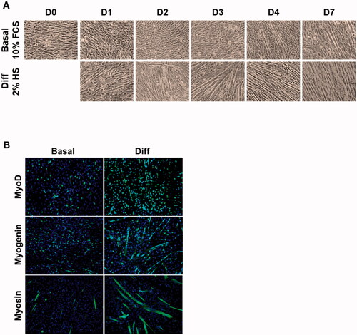
In our model, pre-myoblasts were found to differentiate and fuze in vitro when the 10% fetal calf serum present in the basal growth medium (, top)) was replaced by 2% horse serum (, bottom)). Under these conditions, centronucleated myotubes appeared from day 2 and were well-developed by day 7 (). In the basal medium, only a few myotubes were present (). Indeed, C2C12 cells, like SCs, can differentiate in vitro in basal medium, albeit more slowly than in differentiation medium, and when strong confluence is reached.
Figure 1. Observation of C2C12 proliferation, differentiation, and fusion. (A) Cells were cultured in basal or differentiation condition for 7 days and observed under light microscope (× 100). (B) Cells were cultured in proliferation or differentiation medium for 4 days and revealed by immunostaining with MyoD1, myogenin or myosin antibody (green). Nuclei are labeled using DAPI (blue).
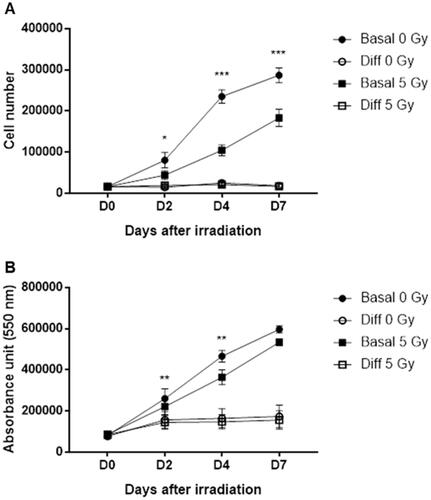
First, we investigated the effects of radiation exposure on cell proliferation for seven days. Direct observation of C2C12 proliferation by cell counting () showed a significant decrease after radiation exposure in the basal medium. Cell number was respectively 56% and 37% lower 4 and 7 days after irradiation. The decrease was less prominent when measured using PrestoBlueTM (), a reagent which indicates mitochondrial metabolic activity and therefore mainly proliferation. Nevertheless, it clearly reflects the deleterious effects of the 5 Gy radiation exposure on cells. With this second technique, absorbance decreased by 22% and 10% respectively 4 and 7 days after irradiation in the medium containing 10% FBS. Under the differentiation conditions, proliferation was stopped with or without irradiation.
Figure 2. Effects of 5 Gy irradiation on C2C12 proliferation in basal or differentiation conditions. C2C12 cells were grown for 3 days, irradiated and counted 2, 4 and 7 days after irradiation. (A) Cells were counted under microscope or (B) proliferation/viability assay was performed with PrestoBlueTM. *p < 0.05, ** p < 0.01, *** p < 0.001 (ANOVA) denotes (A) the number of cells or (B) the level of absorbance in irradiated C1C12 vs. the same measure in control non-irradiated C2C12.
Analysis was also performed for myogenic marker expression over time (). It can be observed that without radiation in differentiation conditions early markers such as Pax3 decreased almost immediately compared to day 0 and reached a low point with a level of expression 4.4 times lower on day 4 (). Conversely, late markers such as Myogenin, Myosin or Enolase increased noticeably until day 7. Indeed, in our model, MyoG expression was maximal on day 4 (× 116 vs day 0) and remained at the same level on day 7 (), while ENO3 continued to increase on day 7 (× 4.3 vs day0, ), as did MyHC (× 382, data not shown).
Figure 3. Effects of 5 Gy irradiation over time in basal or differentiation conditions on relative expression of (A) Pax3, (B) MyoG, (C) ENO3. Cells were grown for 3 days prior to irradiation and then cultured in basal or differentiation conditions. RT-qPCR was performed 2, 4 and 7 days after irradiation and the relative expression (2−ΔΔCt) of (A) Pax3, (B) MyoG and (C) ENO3 was measured. Expression is normalized to the HPRT reference gene and to the day 0 condition. (D) Cells were grown 3 days prior to irradiation then cultured in proliferation or differentiation medium for 4 days and revealed by immunostaining with myogenin antibody (green). (E) Western Blot of myogenin was performed with cells cultured in the same way. *p < 0.05, **p < 0.01, ***p < 0.001 (ANOVA) denotes level of absorbance in irradiated C1C12 vs. level in the same condition but non-irradiated C2C12, #p < 0.05, ##p < 0.01, ###p < 0.001 (ANOVA) indicate significant difference between groups below the vertical dotted lines.
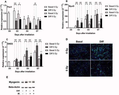
In basal conditions, still without irradiation, cells can also differentiate and fuze under strong confluence (, top). This can be observed also with a slow decrease in early gene expression and increase in late gene expression in DMEM supplemented with 10% FBS, compared to differentiation conditions. It is worth noting that, even though the increase in MyoG expression was slower, it reached the same level obtained in differentiation conditions at day 7 (). This is why, in addition to the data obtained by analyzing cell proliferation (see above), day 4 was subsequently used to study the effects of modulation of the Hedgehog pathway on C2C12 cell proliferation and differentiation.
We then tested the effects of irradiation on myogenic differentiation. As has been shown previously (Ikeda et al. Citation2000; Hoerth and Kodym Citation2004; Sakurai et al. Citation2009), exposure to ionizing radiation is deleterious for the differentiation capacity of cells. Indeed, in DMEM supplemented with 2% HS, Pax3 expression increased 1.8 or 2.2 fold respectively at day 4 or day 7 (), while MyoG decreased by 32 or 33% () and ENO3 by 37 or 42% respectively (), at the same time points in comparison to non-irradiated cells. Even larger decreases were observed for late genes in the basal environment (62% or 73% for MyoG and 39% or 49% for ENO3, at day 4 or day 7; ). Comparable analysis was performed with MyHC (data not shown).
In order to better understand interactions between the Hh signaling pathway and the post-irradiation myogenic regeneration process, we evaluated the effects of a modulation of this metabolic pathway on the capacities of C2C12 cells to proliferate, survive and differentiate after exposure to high dose ionizing radiation. All proliferation and differentiation studies were carried out on day 4 while the survival analysis using the TUNEL method was performed at day 2 so that apoptosis had time to take effect, but apoptotic cells did not detach. For this purpose, under the same conditions of differentiation and irradiation as those used above, the cells were treated either with an agonist of the Hh pathway, the recombinant N-terminal portion of murine Shh, or with a specific antagonist, Cyclopamine.
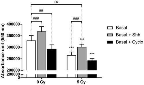
As shown above, C2C12 cell proliferation was affected by irradiation in basal medium (). In our model, the addition of exogenous Shh stimulated the proliferation by 12-13% with or without irradiation (). Blocking the Hh pathway by Cyclopamine had the opposite effect with a decrease of 11% without irradiation and 9% after a 5 Gy X-ray exposure (). In the differentiation medium, no effect was observed, as this medium almost completely blocks proliferation (data not shown).
Figure 4. Effects of Hh pathway modulation on cell proliferation in basal condition after irradiation. C2C12 cells were grown 3 days prior to irradiation, then treated with Shh 4 ug/mL or Cyclopamine 3 mM, and 4 days after irradiation proliferation/viability assay was performed with PrestoBlueTM. ***p < 0.001 (ANOVA) denotes level of absorbance in irradiated C1C12 vs. level in control non-irradiated C2C12, #p < 0.05, ##p < 0.01, ###p < 0.001 (ANOVA) indicate significant difference between groups below the vertical dotted lines.
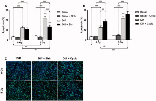
Regarding apoptosis, shows that the decrease of serum in the differentiation medium has a strong pro-apoptotic effect, with a 3.7 and 5-fold increase of apoptotic cells respectively in 0 and 5 Gy conditions, compared to basal medium condition. Irradiation led to a 26% increase (non significant, n.s.) in programmed cell death in basal medium compared to no irradiation, and 69% (p < 0.001) in differentiation medium (). When added to the basal medium, Shh led to an increase in survival with a 19% (n.s.) or 28% (n.s.) decrease in apoptosis at 0 or 5 Gy. This decrease was respectively 13% (n.s.) or 37% (p < 0.001) at the same doses when differentiation was stimulated (). On the other hand, blocking the Hh pathway had a slight deleterious effect when cells proliferate (n.s.) and a more noticeable effect with + 48% (p < 0.01) or 21% (n.s.) apoptosis at 0 or 5 Gy during differentiation ().
Figure 5. Effects of Hh pathway modulation on cell apoptosis after irradiation. Cells were grown for 3 days, irradiated, and treated with Shh 4 μg/mL or Cyclopamine 3 mM. At day 2, survival analyses were performed using TUNEL technique in basal and differentiation media for cells treated (A) with Shh 4 μg/mL or (B) with Cyclopamine 3 mM. (C) Representative fields obtained in differentiation condition are presented. Apoptotic cells are labeled with Alexa Fluor 488 (green spots) and nuclei with DAPI (blue). Fluorescence microscopy ×100, scale bar = 200 µm. ***p < 0.001 (ANOVA) denotes level of absorbance in irradiated C1C12 vs. level in the same conditions but non-irradiated C2C12, ##p < 0.01, ###p < 0.001 (ANOVA) indicate significant difference between groups below the vertical dotted lines.
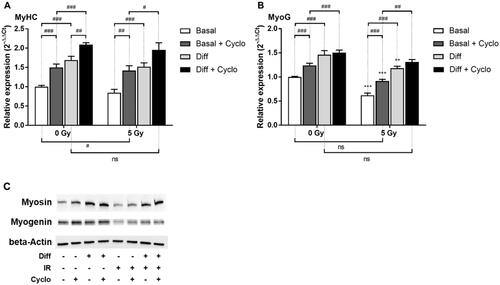
While activation of the Hh pathway seems to promote proliferation and survival of myogenic progenitors, the opposite effect was observed on differentiation (). Indeed, recombinant Shh decreased the expression of the late MyHC gene by 30 or 12% in the basal medium and by 35 or 21% under differentiation conditions, respectively at 0 or 5 Gy (). The same observation can be made with the MyoG gene (). Western blot analysis of the corresponding proteins Myosin and Myogenin confirmed these results (). Conversely, blocking the Hh pathway led to an increased differentiation of C2C12 cells (). Thus, the addition of Cyclopamine increased the expression of MyHC (; +50 or 68% in proliferative conditions and +24 or 28% in differentiation conditions, at 0 or 5 Gy) and MyoG (; +24 or 48% in proliferative condition and +3 or 11% in differentiation condition, at 0 or 5 Gy). This was also confirmed by protein expression (). Similar results were obtained with ENO3 (data not shown).
Figure 6. Effect of Hh pathway activation by Shh on C2C12 after irradiation. Cells were grown for 3 days, irradiated, treated with Shh 4 μg/mL and 4 days after irradiation RT-qPCR was performed and relative expression (2−ΔΔCt) of (A) MyoG and (B) MyHC was measured. Expression is normalized to the HPRT reference gene and to the basal 0 Gy condition. (C) WesternBlot of the corresponding proteins was performed with cells cultured in the same way. **p < 0.01, ***p < 0.001 (t-test) denotes level of absorbance in irradiated C1C12 vs. level in the same conditions but non-irradiated C2C12, #p < 0.05, ##p < 0.01, ###p < 0.001 (t-test) indicate significant difference between groups below the vertical dotted lines.
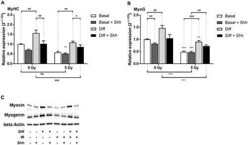
Figure 7. Effect of Hh pathway blockade by Cyclopamine on C2C12 after irradiation. Cells were grown for 3 days, irradiated, treated with Cyclopamine 3 mM and 4 days after irradiation RT-qPCR was performed and relative expression (2−ΔΔCt) of (A) MyoG and (B) MyHC was measured. Expression is normalized to the HPRT reference gene and to the basal 0 Gy condition. (C) Western Blot of the corresponding proteins was performed with cells cultured in the same way. **p < 0.01, ***p < 0.001 (t-test) denotes level of absorbance in irradiated C1C12 vs. level in the same conditions but non-irradiated C2C12, #p < 0.05, ##p < 0.01, ###p < 0.001 (t-test) indicate significant difference between groups below the vertical dotted lines.
Discussion and conclusions
Depending on cell type and stage of differentiation, mammalian cells do not all have the same level of radiosensitivity. The cells that form the fibers of mature muscle tissue, for example, are among the more radio-resistant, as are those that make up nerve fibers (Harris Citation1963; Jurdana Citation2008). With respect to other tissue types such as bone marrow, intestine, skin or many types of cancer cells which are more radiosensitive, few preclinical studies have been carried out to date with a therapeutic aim of promoting muscle repair after localized high-dose radiation exposure. And yet, it has been widely noted that when CRS develops, skin but also subcutaneous skeletal muscle are severely affected (Riccobono et al. Citation2016, Citation2018; Linard et al. Citation2018).
In humans, muscle damage and repair following irradiation are largely neglected. The benchmark clinical treatment approved in 2009 by the International Atomic Energy Agency mainly promotes skin repair in radiation-induced lesions. It consists of dosimetry-guided surgical excision of the irradiated area, completed by an autologous skin flap transplantation and repeated injections of autologous BM-MSCs (Reyes et al. Citation2016). The effectiveness of BM-MSCs, with or without the addition of cytokines, in improving skin healing has been demonstrated using irradiation models in animals (François et al. Citation2007, Citation2017; Wu et al. Citation2010). Clinically, in the case of compassionate treatment for CRS following various acute irradiation accidents, BM-MSC treatment has also proven its efficiency in skin repair (Lataillade et al. Citation2007; Bey et al. Citation2010; Lataillade et al. Citation2010). Mesenchymal stromal cells derived from the adipose tissue (Ad-MSCs) have shown similar therapeutic potential in pig (Forcheron et al. Citation2012). Two recent studies in pig models of CRS show that deep injection of BM-MSCs or Ad-MSCs could also improve post-irradiation muscle repair (Riccobono et al. Citation2016; Linard et al. Citation2018). Nevertheless, the use of these stromal cells requires harvesting autologous cells, which is invasive for the irradiated patient, and a relatively long and irreducible period of time to grow them in vitro before obtaining the cell graft.
In order to improve muscle repair, we propose to evaluate therapeutic strategies based not only on cell therapy but on radiation protection and stimulation of satellite cells (SCs) present in the muscle, near the irradiated area. SCs are undifferentiated cells specific to muscle tissue (Mauro Citation1961; Zammit et al. Citation2006). Under normal physiological conditions, they represent about 2% of the total number of nuclei present in adult skeletal muscle and ensure myogenesis during muscle growth, after intense physical effort and physiopathological or accidental trauma (Appell et al. Citation1988; Collins et al. Citation2005; Jurdana Citation2008). However, SCs are highly radiosensitive (Jurdana Citation2008; Caiozzo et al. Citation2010). In case of localized high-dose radiation exposure, their number decreases sharply, probably because of radiation-induced damage to DNA strands leading to reduced mitotic activity or even cell death. When too many SCs are lost, fusion, which is necessary for the formation of new muscle fibers, becomes impossible. In our study, we used a mouse cell model comparable to SCs, C2C12 cells (), which have the ability to proliferate, differentiate rapidly and form myotubes in vitro (McMahon et al. Citation1994; Bajaj et al. Citation2011). In the proliferation medium, a 5 Gy dose of radiation caused a significant drop in the number of cells compared to the control condition (). In the differentiation medium, cell proliferation is blocked with or without exposure to X-rays, and irradiation therefore has no effect on it in this medium. Moreover, following irradiation, the number of apoptotic cells increased, both in differentiation conditions and otherwise (). This tends to show that C2C12 cells, like SCs, exhibit a high level of sensitivity to ionizing radiation. These deleterious effects have also been demonstrated in similar cellular models, at comparable doses, after gamma or X-ray irradiation (Ikeda et al. Citation2000; Sakurai et al. Citation2009; Caiozzo et al. Citation2010). In our model, when a 5 Gy dose was used, a sufficient number of cells managed to survive and proliferate, leading to an increase in the number of cells up to day 7. Conversely, in vivo, the much lower density SCs in muscle tissue, compared to culture, does not seem sufficient to allow rapid regeneration of the muscle tissue.
An interesting strategy would therefore consist in identifying molecular targets which could be activated or inhibited so as to conserve a sufficient number of SCs and/or increase the proliferation of the surviving cells after irradiation and thereby repair radiation-induced lesions by regenerating the muscle tissue. Modulation of the Hh pathway could thus show great potential. Indeed, this pathway is involved in the regulation of many physiological functions, including myogenesis and the maintenance of numerous stem cell types, as well as carcinogenesis, and is essential in the development of many different species throughout the animal kingdom (Bürglin Citation2008). Its role in post-traumatic muscle regeneration will be of particular interest to us here because it has been shown that the Hh signaling pathway is reactivated after muscle injury. Renault et al. have shown, in particular, that myogenesis and neoangiogenesis are markedly altered in ischemia/reperfusion models in mice with a lack in Dhh, Gli1 or Gli3 expression (Renault, Robbesyn, et al. Citation2013; Renault, Vandierdonck, et al. Citation2013; Yao et al. Citation2014). Moreover, different types of cancer originate from mutations in the Patched1 (Ptch1) receptor, resulting in persistent activation of the Hh signaling pathway (Bonifas et al. Citation2001; Berman et al. Citation2003; Sanchez et al. Citation2004). In embryonal rhabdomyosarcoma, as observed frequently in mice, the lack of one allele of Ptch1 leads to hyperactivation of the signal due in particular to overexpression of Gli1 (Hahn et al. Citation1998). In models of ischemia or muscle damage caused by injection of cardiotoxin (CTX) in dystrophic Mdx mice, gene therapy with Shh strongly stimulates muscle regeneration (Piccioni et al. Citation2014). Conversely, blocking the Hh signal with cyclopamine severely limits muscle regeneration after mechanical injury or injection of CTX into the tibialis anterior muscle of C57Bl/6J mice (Straface et al. Citation2009).
Like Koleva et al. using in vitro models of C2C12 or satellite cells under basal conditions (Koleva et al. Citation2005), we have shown here that the activation of the Hh pathway by recombinant Shh stimulates proliferation () and limits apoptosis (). Quite interestingly, this increase in proliferation and cell survival was also observed after irradiation, bringing the 2 parameters to a level comparable to that of the control condition without irradiation, in basal condition. In our model, without irradiation, the decrease in the amount of serum in the differentiation medium led to increased apoptosis compared to the control condition in basal medium. This serum-dependent increase is even greater after exposure of C2C12 cells to a 5 Gy dose of radiation. Furthermore, it is important to point out that the addition of Shh under differentiation conditions increased the survival of the cells and brought it to the same level as in the absence of irradiation, as observed in proliferation medium. This prevention of programmed cell death could be due to the blocking effect of Shh on Caspase-3 activation (Koleva et al. Citation2005). After irradiation, the activation of the Hh signaling pathway thus seems to provide a major benefit for the conservation of the reserve of myogenic progenitors by promoting both the proliferation of SCs in basal medium as well as their survival in proliferation or differentiation conditions. Inhibition of the Hh pathway by Cyclopamine has the opposite effect, particularly on cell survival, which it decreases after induction of differentiation (). Given that no exogenous activator was added, this effect could be due to the inhibition of the anti-aptoptotic effect of Ihh synthesized endogenously by the cells and released into the culture medium. Indeed, no Shh expression in the C2C12 cells was detected by RT-qPCR, while Dhh expression did not vary regardless of conditions, irradiation, differentiation or treatment. On the other hand, the Ihh gene was overexpressed from day 2 to day 7 under differentiation conditions and irradiation did not seem to have any effect on the expression of Ihh (data not shown). The inhibition of the anti-apoptotic effect of Ihh by Cyclopamine, in differentiation conditions, could thus explain the decrease in cell survival compared to that observed in the medium alone.
We have thus observed an important aspect of the treatment of radiation-induced muscle damage, which entails the need both to protect myogenic progenitors against the effects of radiation and to increase their number after irradiation so as to ensure the formation of new fibers. A second aspect is crucial for muscle repair: the ability of SCs to differentiate and fuze. Indeed, it has been shown that exposure to a 4 Gy dose of X-ray radiation (on Linac, 4 MV or MBR-1520R, 150 kV) significantly reduces the formation of myotubes and the synthesis of Myosin (late differentiation markers) in C2C12 cells and primary murine SCs, under differentiation conditions (Ikeda et al. Citation2000; Hoerth and Kodym Citation2004; Sakurai et al. Citation2009).
Concerning the modulation of the Hh pathway, some studies suggest that Shh may have a pro-differentiation effect (Elia et al. Citation2007; Voronova et al. Citation2013) but the experimental procedures as well as the cell models (chick primary myoblast cultures) are quite different from those used in our study. The use of murine recombinant Shh on embryonic cells derived from chickens and different differentiation conditions could very well explain the variations in the results obtained. Under conditions similar to ours, activation of the Hh pathway has been found to be highly deleterious for the differentiation of SCs in the absence of irradiation. The formation of myotubes is greatly reduced in the presence of 4 μg/mL of recombinant Shh (Koleva et al. Citation2005). Likewise, in our cell model and under the irradiation and culture conditions described above, we have shown that the activation of the Hh pathway tends to decrease the gene and protein expression of Myogenin, Myosin and Enolase, whereas inhibition by Cyclopamine seems to increase the expression of these late markers of differentiation.
On the whole, this study thus provides new data that support modulation of the Hh signaling pathway as a means to improve post-irradiation muscle regeneration. This could lead to an original and innovative therapeutic strategy, initially using local stimulation of the Hh pathway to rescue SCs after irradiation and ensure their development near the irradiated area, followed by inhibition of the pathway to promote differentiation and muscle regeneration. Such an approach seems more appropriate than the one used by Straface et al. which consisted in injecting Cyclopamine as a first-line treatment into a muscle damaged by CTX (Straface et al. Citation2009). Indeed, our results suggest that direct inhibition of Hh signaling would not allow the pool of progenitors to regenerate and might therefore contribute to the increase in post-traumatic cell death, leading over time to increased fibrosis with a complete loss of muscle tissue functionality (irreversible muscle loss), with no possibility of regeneration.
Our laboratory is currently evaluating the therapeutic potential of alternately activating and blocking the Hh pathway in a model of ultra-localized high-dose irradiation of the gatrocnemius/soleus muscles in the paw of C57Bl/6 mice. Stimulation of the Hh signal will be carried out with recombinant Shh or microvesicles derived from cells which overexpress Shh. Vismodegib or Sonidegib, which are both powerful and specific inhibitors of the Hh pathway, will be used to replace Cyclopamine. These molecules, which are used in the clinical treatment of basal cell carcinomas, are compatible with in vivo use and exhibit superior efficacy to Cyclopamine.
Acknowledgments
We wish to express our deep thanks to the French Direction Générale de l’Armement (Paris, France) for granting our work, to IRBA Molecular Biology Unit headed by Dr Laure BARBIER for helping us in the development of RT-qPCR experiments, to IRBA Imaging Unit headed by Dr Anne-Laure FAVIER for making the cell imaging platform available, to Mrs Véronique CHASTRES for her precious advice in statistics and to Mr Daniel HENKEL for his excellent and very quick work in correcting the translation of the manuscript.
Disclosure statement
The authors report no conflict of interest. They are entirely responsible for the content of the paper.
Additional information
Funding
Notes on contributors
E. Rota Graziosi
Emmanuelle Rota Graziosi is a French engineer specialized in chemistry, biology and health. She has also obtained a double master’s degree in Medical and Translational Chemistry, University of Montpellier, France. For one year now, she has been completing a PhD in the Radiobiology Unit of the French Armed Forces Biomedical Research Institute (IRBA), Brétigny-sur-Orge, France.
S. François
Sabine François, PhD, is a specialist of cell therapy optimization for regenerative medicine, especially in cases of lesions induced by ionizing radiation. She is a project leader and develop new therapeutic approaches to treat acute radiation syndrome in the Radiobiology Unit of IRBA, Brétigny-sur-Orge, France. She is also part of the Joint Research Unit UMR1296 of French National Institute for Health and Medical Research (INSERM, Lyon, France) and French Armed Forces Biomedical Research Institute (IRBA, Brétigny-sur-Orge, France).
J. Pateux
Jérôme Pateux is a paramedical technician in IRBA, Brétigny-sur-Orge, France. He works both in the Radiobiology Unit where he is a specialist in the analysis of localized high-dose radiation effects and in the Radiation Biological Dosimetry Laboratory where he is in charge of the identification of new biomarkers for post-irradiation biological dosimetry.
M. Gauthier
Michel Gauthier is a technician specialized in gene identification and therapy in genetic diseases. He has also worked to develop therapeutic strategies for the treatment of chronic inflammatory bowel diseases. From 2020, he is a paramedical technician in the Radiobiology Unit of IRBA, Brétigny-sur-Orge, France.
X. Butigieg
Xavier Butigieg is a paramedical technician in the Imaging Unit of IRBA, Brétigny-sur-Orge, France. He is a specialist in various specific techniques like histology, bright field and fluorescence microscopy, scanning and transmission electron microscopy on the state-of-art microscopy platform of the Institute.
M. Oger
Myriam Oger, PhD, is a biomedical researcher specialized in Sciences and Technology of Information in IRBA, Brétigny-sur-Orge, France. She is a specialist in image acquisition, processing and analysis. Her fields of expertise are in microscopical and 3D microtomography images, using FIJI software and Python libraries.
M. Drouet
Michel Drouet, MD, PhD, is currently Head of Radiation Biological Effects Department and Head of Chemical, Biological, Radiobiological and Nuclear Risks Division in IRBA, Brétigny-sur-Orge, France. He is also part of the Joint Research Unit UMR1296 of French National Institute for Health and Medical Research (INSERM, Lyon, France) and French Armed Forces Biomedical Research Institute (IRBA, Brétigny-sur-Orge, France).
D. Riccobono
Diane Riccobono, MD, PhD, worked as a medical officer for two years. She performed a PhD specialized in radiobiology, in the development of advanced therapy medicinal products based on mesenchymal stromal cells and in inflammation pathways analysis after irradiation. She is also specialized in radioprotection and CBRN threat. Since 2018, Dr Diane RICCOBONO is in charge of the Radiobiology unit of IRBA Brétigny-sur-Orge, France, and is part of the Joint Research Unit UMR1296 of French National Institute for Health and Medical Research (INSERM, Lyon, France) and French Armed Forces Biomedical Research Institute (IRBA, Brétigny-sur-Orge, France).
N. Jullien
Nicolas Jullien, PhD, is a specialist in pharmacology, radiobiology and in the development of advanced therapy medicinal products based on the use of mesenchymal stromal cells obtained from bone marrow or adipose tissue. He is a biomedical researcher in the Radiobiology unit of IRBA Brétigny-sur-Orge, France.
References
- Anderson C, Williams VC, Moyon B, Daubas P, Tajbakhsh S, Buckingham ME, Shiroishi T, Hughes S, Borycki A-G. 2012. Sonic hedgehog acts cell-autonomously on muscle precursor cells to generate limb muscle diversity. Genes Dev. 26(18):2103–2117.
- Appell HJ, Forsberg S, Hollmann W. 1988. Satellite cell activation in human skeletal muscle after training: evidence for muscle fiber neoformation. Int J Sports Med. 9(4):297–299.
- Bajaj P, Reddy B, Millet L, Wei C, Zorlutuna P, Bao G, Bashir R. 2011. Patterning the differentiation of C2C12 skeletal myoblasts. Integr Biol (Camb). 3(9):897–909.
- Berman DM, Karhadkar SS, Maitra A, Montes De Oca R, Gerstenblith MR, Briggs K, Parker AR, Shimada Y, Eshleman JR, Watkins DN, et al. 2003. Widespread requirement for Hedgehog ligand stimulation in growth of digestive tract tumours. Nature. 425(6960):846–851.
- Bey E, Prat M, Duhamel P, Benderitter M, Brachet M, Trompier F, Battaglini P, Ernou I, Boutin L, Gourven M, et al. 2010. Emerging therapy for improving wound repair of severe radiation burns using local bone marrow-derived stem cell administrations. Wound Repair Regen. 18(1):50–58.
- Blau HM, Chiu CP, Webster C. 1983. Cytoplasmic activation of human nuclear genes in stable heterocaryons. Integr Biol (Camb). 32(4):1171–1180.
- Bonifas JM, Pennypacker S, Chuang PT, McMahon AP, Williams M, Rosenthal A, De Sauvage FJ, Epstein EH. 2001. Activation of expression of hedgehog target genes in basal cell carcinomas. J Invest Dermatol. 116(5):739–742.
- Bürglin TR. 2008. The Hedgehog protein family. Genome Biol. 9(11):241.
- Caiozzo VJ, Giedzinski E, Baker M, Suarez T, Izadi A, Lan M, Cho-Lim J, Tseng BP, Limoli CL. 2010. The radiosensitivity of satellite cells: cell cycle regulation, apoptosis and oxidative stress. Radiat Res. 174(5):582–589.
- Caradu C, Guy A, James C, Reynaud A, Gadeau A-P, Renault M-A. 2018. Endogenous Sonic Hedgehog limits inflammation and angiogenesis in the ischaemic skeletal muscle of mice. Cardiovasc Res. 114(5):759–770.
- Collins CA, Olsen I, Zammit PS, Heslop L, Petrie A, Partridge TA, Morgan JE. 2005. Stem cell function, self-renewal, and behavioral heterogeneity of cells from the adult muscle satellite cell niche. Cell. 122(2):289–301.
- Elia D, Madhala D, Ardon E, Reshef R, Halevy O. 2007. Sonic hedgehog promotes proliferation and differentiation of adult muscle cells: involvement of MAPK/ERK and PI3K/Akt pathways. Biochim Biophys Acta. 1773(9):1438–1446.
- Forcheron F, Agay D, Scherthan H, Riccobono D, Herodin F, Meineke V, Drouet M. 2012. Autologous adipocyte derived stem cells favour healing in a minipig model of cutaneous radiation syndrome. PLoS One. 7(2):e31694.
- François S, Eder V, Belmokhtar K, Machet M-C, Douay L, Gorin N-C, Benderitter M, Chapel A. 2017. Synergistic effect of human bone morphogenic protein-2 and mesenchymal stromal cells on chronic wounds through hypoxia-inducible factor-1 α induction. Sci Rep. 7(1):4272.
- François S, Mouiseddine M, Mathieu N, Semont A, Monti P, Dudoignon N, Saché A, Boutarfa A, Thierry D, Gourmelon P, et al. 2007. Human mesenchymal stem cells favour healing of the cutaneous radiation syndrome in a xenogenic transplant model. Ann Hematol. 86(1):1–8.
- Giarretta I, Gaetani E, Bigossi M, Tondi P, Asahara T, Pola R. 2019. The Hedgehog signaling pathway in ischemic tissues. IJMS. 20(21):5270. https://doi.org/https://doi.org/10.3390/ijms20215270.
- Hadden MK. 2014. Hedgehog pathway agonism: therapeutic potential and small-molecule development. ChemMedChem. 9(1):27–37.
- Hahn H, Wojnowski L, Zimmer AM, Hall J, Miller G, Zimmer A. 1998. Rhabdomyosarcomas and radiation hypersensitivity in a mouse model of Gorlin syndrome. Nat Med. 4(5):619–622.
- Harris RJC. 1963. Cellular basis and aetiology of late somatic effects of ionizing radiation. Cellular basis and aetiology of late somatic effects of ionizing radiation [Internet]. [accessed 2021 Oct 2]. https://www.cabdirect.org/cabdirect/abstract/19631605348.
- Hawke TJ, Garry DJ. 2001. Myogenic satellite cells: physiology to molecular biology. J Appl Physiol (1985). 91(2):534–551.
- Heretsch P, Tzagkaroulaki L, Giannis A. 2010. Cyclopamine and hedgehog signaling: chemistry, biology, medical perspectives. Angew Chem Int Ed Engl. 49(20):3418–3427.
- Hoerth E, Kodym R. 2004. Involvment of c-Abl in the radiation-induced inhibition of myoblast differentiation. Int J Radiat Biol. 80(10):729–736.
- Hopewell JW. 1990. The skin: its structure and response to ionizing radiation. Int J Radiat Biol. 57(4):751–773.
- Hu JK-H, McGlinn E, Harfe BD, Kardon G, Tabin CJ. 2012. Autonomous and nonautonomous roles of Hedgehog signaling in regulating limb muscle formation. Genes Dev. 26(18):2088–2102.
- Ikeda S, Hachisu R, Yamaguchi A, Gao YH, Okano T. 2000. Radiation retards muscle differentiation but does not affect osteoblastic differentiation induced by bone morphogenetic protein-2 in C2C12 myoblasts. Int J Radiat Biol. 76(3):403–411.
- Jurdana M. 2008. Radiation effects on skeletal muscle. Radiol Oncol. 42(1):15–22. doi:https://doi.org/10.2478/v10019-007-0034-5.
- Koleva M, Kappler R, Vogler M, Herwig A, Fulda S, Hahn H. 2005. Pleiotropic effects of sonic hedgehog on muscle satellite cells. Cell Mol Life Sci. 62(16):1863–1870.
- Lataillade J-J, Bey E, Thepenier C, Prat M, Leclerc T, Bargues L. 2010. Skin engineering for burns treatment. Bull Acad Natl Med. 194(7):1339–1351.
- Lataillade JJ, Doucet C, Bey E, Carsin H, Huet C, Clairand I, Bottollier-Depois JF, Chapel A, Ernou I, Gourven M, et al. 2007. New approach to radiation burn treatment by dosimetry-guided surgery combined with autologous mesenchymal stem cell therapy. Regen Med. 2(5):785–794.
- Lefaix JL, Delanian S. 2005. Menace terroriste nucléaire: approche médicale. Arcueil, France: John Libbey Eurotext Plc; p. 87–95.
- Linard C, Brachet M, L’homme B, Strup-Perrot C, Busson E, Bonneau M, Lataillade J-J, Bey E, Benderitter M. 2018. Long-term effectiveness of local BM-MSCs for skeletal muscle regeneration: a proof of concept obtained on a pig model of severe radiation burn. Stem Cell Res Ther. 9(1):299.
- Mauro A. 1961. SATELLITE CELL OF SKELETAL MUSCLE FIBERS. J Biophys Biochem Cytol. 9(2):493–495.
- McMahon DK, Anderson PA, Nassar R, Bunting JB, Saba Z, Oakeley AE, Malouf NN. 1994. C2C12 cells: biophysical, biochemical, and immunocytochemical properties. Am J Physiol. 266(6):C1795–C1802.
- Piccioni A, Gaetani E, Palladino M, Gatto I, Smith RC, Neri V, Marcantoni M, Giarretta I, Silver M, Straino S, et al. 2014. Sonic hedgehog gene therapy increases the ability of the dystrophic skeletal muscle to regenerate after injury. Gene Ther. 21(4):413–421.
- Renault M-A, Robbesyn F, Chapouly C, Yao Q, Vandierdonck S, Reynaud A, Belloc I, Traiffort E, Ruat M, Desgranges C, et al. 2013. Hedgehog-dependent regulation of angiogenesis and myogenesis is impaired in aged mice. Arterioscler Thromb Vasc Biol. 33(12):2858–2866.
- Renault M-A, Vandierdonck S, Chapouly C, Yu Y, Qin G, Metras A, Couffinhal T, Losordo DW, Yao Q, Reynaud A, et al. 2013. Gli3 regulation of myogenesis is necessary for ischemia-induced angiogenesis. Circ Res. 113(10):1148–1158.
- Reyes EH, Baciu F, Benderitter M, Lataillade JJ, Bey E, Trompier F, Tamarat R. 2016. Medical Response to Radiological Accidents in Latin America and International Assistance. Radiat Res. 185(4):359–365.
- Riccobono D, Agay D, François S, Scherthan H, Drouet M, Forcheron F. 2016. Contribution of INTRAMUSCULAR autologous adipose tissue-derived stem cell injections to treat cutaneous radiation syndrome: preliminary results. Health Phys. 111(2):117–126.
- Riccobono D, Nikovics K, François S, Favier A-L, Jullien N, Schrock G, Scherthan H, Drouet M. 2018. First insights into the M2 inflammatory response after adipose-tissue-derived stem cell injections in radiation-injured muscles. Health Phys. 115(1):37–48.
- Sakurai T, Ueda T, Kawai M, Tobita H, Miyakoshi J. 2009. Protective effects of insulin-like growth factor-I on the decrease in myogenic differentiation by ionizing radiation. Int J Radiat Biol. 85(2):153–158.
- Sanchez P, Hernández AM, Stecca B, Kahler AJ, DeGueme AM, Barrett A, Beyna M, Datta MW, Datta S, Ruiz I Altaba A. 2004. Inhibition of prostate cancer proliferation by interference with SONIC HEDGEHOG-GLI1 signaling. Proc Natl Acad Sci U S A. 101(34):12561–12566.
- Straface G, Aprahamian T, Flex A, Gaetani E, Biscetti F, Smith RC, Pecorini G, Pola E, Angelini F, Stigliano E, et al. 2009. Sonic hedgehog regulates angiogenesis and myogenesis during post-natal skeletal muscle regeneration. J Cell Mol Med. 13(8B):2424–2435.
- Teperino R, Aberger F, Esterbauer H, Riobo N, Pospisilik JA. 2014. Canonical and non-canonical Hedgehog signalling and the control of metabolism. Semin Cell Dev Biol. 33:81–92.
- Voronova A, Coyne E, Al Madhoun A, Fair JV, Bosiljcic N, St-Louis C, Li G, Thurig S, Wallace VA, Wiper-Bergeron N, et al. 2013. Hedgehog signaling regulates MyoD expression and activity. J Biol Chem. 288(6):4389–4404.
- Wu Y, Zhao RCH, Tredget EE. 2010. Concise review: bone marrow-derived stem/progenitor cells in cutaneous repair and regeneration. Stem Cells. 28(5):905–915.
- Yao Q, Renault M-A, Chapouly C, Vandierdonck S, Belloc I, Jaspard-Vinassa B, Daniel-Lamazière J-M, Laffargue M, Merched A, Desgranges C, et al. 2014. Sonic hedgehog mediates a novel pathway of PDGF-BB-dependent vessel maturation. Blood. 123(15):2429–2437.
- Zammit PS, Partridge TA, Yablonka-Reuveni Z. 2006. The skeletal muscle satellite cell: the stem cell that came in from the cold. J Histochem Cytochem. 54(11):1177–1191.