
Abstract
Purpose
Ionizing radiation can induce mutations in germ cells in various organisms, including fruit flies and mice. However, currently, there is no clear evidence for the transgenerational effects of radiation in humans. This review is an effort to identify possible reasons for the lack of such observations.
Methods
Literature search and narrative review.
Results
1) In both mice and humans, resting oocytes locate primarily in the cortical region of the ovary where the number of blood vessels is very low especially when young and extra-cellular material is rich, and this region is consequently hypoxic, which probably leads to immature oocytes being resistant to the cell killing and mutagenic effects of radiation. 2) In studies of spermatogonia, the mouse genes used for specific locus test (SLT) studies, which include coat color genes, were hypermutable when compared to many other genes. Recent studies which examined over 1000 segments of genomic DNA indicate that the induction rate of deletion mutation per segment was on the order of 10−6 per Gy, which is one order of magnitude lower than that obtained from the SLT data. Therefore, it appears possible that detecting any transgenerational effects of radiation following human male exposures will be difficult due to a lack of mutable marker genes. 3) Fetal malformations were examined in studies in humans, but the genetic component in such malformations is low, and abnormal fetuses are prone to undergo miscarriage which does not occur in mice, and which leads to difficulties in detecting transgenerational effects.
Conclusion
The lack of clear evidence for radiation effects in humans probably does not result from any problem in the methodologies used but may be due largely to biological properties. Currently, whole genome sequencing studies of exposed parents and offspring are planned, but ethical guidelines need to be followed to avoid discrimination, which had once happened to the atomic bomb survivors.
Introduction
It was nearly a century ago when ionizing radiation was found to induce transgenerational mutations in fruit flies (Muller Citation1927) and barley and maize (Stadler Citation1928a, Citation1928b) which were observed in the following generation. Starting from the 1950s, mutations were also found to be induced in mice. Past studies in mice clearly showed that mutations are induced in mature oocytes and at all stages of development in male germ cells, but quite importantly, no mutations were induced in immature oocytes in a resting stage (Searle Citation1974; Russell Citation1977). In human epidemiology, there is no clear unambiguous evidence for any transgenerational effects of radiation following parental exposures to radiation (Nakamura Citation2006, Citation2018, Citation2019, Nakamura et al. Citation2013, Green et al. Citation2009, Citation2010). In this context, although one might raise a recent paper on the reanalysis of untoward pregnancy outcomes in the offspring of atomic-bomb survivors (Yamada et al. Citation2021) as possible evidence, the authors concluded that ‘the estimates were imprecise for direct radiation effects, and most were not statistically significant’ and hence the evidence is still moot. In addition, whole genome sequencing studies thus far did not detect an increased frequency of deletion mutations, which is characteristic to radiation-induced event, in the offspring of irradiated mice (Adewoye et al. Citation2015, Satoh et al. Citation2020), in three Nagasaki survivor families (Horai et al. Citation2018), and in Chernobyl survivor families which include 130 offspring (Yeager et al. Citation2021). This lack of evidence in humans prompted a search for possible reasons underlying these observations.
The present review deals primarily with radiation effects on spermatogonia and resting oocytes in adults. Observations of radiation effects on fetal primordial spermatogonia or pre-dictyate oocytes in mice have indicated that mutation induction rates are similar in both sexes but tended to be somewhat lower than those seen in adult spermatogonia or maturing oocytes, and suggest that cell killing may be part of the reason for these lower observed rates. For more details, please refer to the review article by Searle (Citation1974).
Resting oocytes are radioresistant
It is not common for women to become pregnant within one year after exposure to radiation, either from accidental exposure or due to medical reasons. Since it takes nearly one year for an immature oocyte to start maturation processes and undergo ovulation (Gougeon Citation1986), the target cells at risk for radiation-induced transgenerational effects of radiation are primarily immature oocytes in a resting stage. In this regard, immature oocytes in mice (born ≥7 weeks after irradiation) are known to be sensitive to radiation-induced apoptotic death, but completely resistant to radiation-induced mutagenesis ().
Table 1. Summary of radiation mutagenesis at 7 specific loci in mouse oocytes.
Notably, only three mutants were observed among 259,683 offspring derived from irradiated immature oocytes to 400 R gamma rays (1 R = 2.6 × 10⁻⁴ C/kg), 0.6 Gy of fast neutrons (which can induce mutations equivalent to 3 Gy of X rays when delivered to spermatogonia or 2 Gy to mature oocytes) and some other exposures. The mean mutation frequency per locus is 0.17 per 105, which does not differ from the frequency of 0.16 per 105 in the control group. In contrast, the mutation frequency after irradiation of mature and maturing oocytes with 200 R of gamma rays was 10.4 per 105, about 60 times higher than the control frequency.
The structure of the ovary
Primordial follicles that are produced from oogonia become primary follicles following meiotic cell division. Follicles have a structure of oocytes surrounded by somatic cells such as granulosa cells and are initially a single cell layer (primary follicles) followed by two cell layers (secondary follicles), and then by multiple-layered follicles which include follicular fluid (antral follicles) before ovulation. The primordial follicles are the simplest form of follicles and remain quiescent ().
Figure 1. A) A schematic view of mouse ovary at day 21 after birth (Hayashi et al. Citation2020). B) A close view of mouse ovary at day 14 after birth. Note that one primary follicle (PrF) started to grow at a place very close to the ovarian surface (Choi et al. Citation2008). PF stands for primordial follicle, SF secondary follicle, and AF antral follicle.
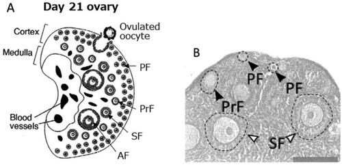
In both mice and humans, adult ovaries contain both resting immature oocytes and mature or maturing oocytes, and immature oocytes are primarily observed near the surface of the ovary, in the cortical region () (Kerr et al. Citation2006, Choi et al. Citation2008). The cortex region is rich in extracellular matrix (connective tissue) and tough, which corresponds to the tunica albuginea in the testis (one of the toughest tissues in the body), and blood vessels are rarely found. These conditions satisfy the requirements for a hypoxic environment. Nevertheless, direct measurements of partial oxygen pressure (pO2) are technically difficult and have not yet been successfully performed. On the other hand, follicles that have committed to the maturation process move toward the inner area of the ovary (medulla) which is softer and rich in blood vessels.
Resting oocytes are protected by hypoxic conditions
In recent years, it became possible to artificially induce somatically derived mouse iPS cells to become oocytes which were subsequently fertilized in vitro and transplanted into foster mothers to produce healthy offspring. However, such oocytes were at the stage of secondary follicles and capable of becoming mature oocytes, whereas conditions for creating resting oocytes were not realized. Recently, this problem was overcome by culturing these cells under hypoxic conditions (5% O2) (Shimamoto et al. Citation2019). This transformation into resting oocytes seems to also require physical force from the ovarian cortex to press the cells and the presence of FOXO3 in an intra-nuclear location. Among these requirements, the low oxygen condition seems likely to be the reason for the observed radio resistance toward mutation induction.
It is well known that the effects of radiation are affected by the presence or absence of oxygen. Under low oxygen conditions, observations of radiation effects decrease. When cells in glass ampoules were gassed for 2 min with a mixture of 95% N2 and 5% CO2 immediately prior to a radiation exposure (termed acute hypoxic conditions), and irradiation is then followed by culture under normoxic conditions, cell killing effect of radiation decrease to nearly 1/3 of that seen when irradiated in the air (21% O2). For example, after exposure to 2 Gy of gamma rays in Chinese hamster V79 cells, 30% of the cells are killed if they were irradiated in air, but the effect decreased to 10% cell death when cells were irradiated under acute hypoxic conditions. Further, irradiation under acute hypoxic conditions in the presence of the radioprotector WR1065 (an aminothiol compound) resulted in a further decrease to only 5% cell death (Grdina et al. Citation1989). Such studies led to the conclusion that low oxygen conditions are highly protective from radiation damage (e.g. Ling et al. Citation1981). In vivo, other molecules like SPINK1, which is induced under severely hypoxic conditions, may allow adjacent cells to become resistant to radiation, indicating that further protection may occur in vivo than was anticipated from in vitro studies (Suwa et al. Citation2021).
Resting oocytes do not have the characteristics of tissue stem cells, e.g. continuous cell division, but share the same requirement that they stay intact for a long period of time in an organism. Since a low oxygen microenvironment has been reported to be a common requirement for tissue stem cells (e.g. Huang et al. Citation2018), it could also be suited for resting oocytes.
Interrelationship between apoptotic cell death and mutagenesis
Resting mouse oocytes are known to be highly vulnerable to death from apoptosis following exposure to radiation. For example, as many as 90% of immature oocytes undergo apoptotic cell death with an exposure of only 0.18 Gy in 5-day-old mice (Dobson and Kwan Citation1977). Also, in irradiated adult females, resting oocytes undergo apoptosis leading to a decreased litter size of births derived from fertilization which occurred 6 to 7 weeks after the irradiation, but the surviving offspring did not show an increased mutation frequency as shown in (Russell Citation1977). The results prompted us to logically speculate that in the mouse ovarian cortex, there exists a gradient in both stromal density (highest near the surface) and oxygen levels (lowest near the surface), which caused better protection of the resting oocytes which locate closer to the ovarian surface from radiation injury due to a hypoxic microenvironment. In contrast, those resting oocytes that locate closer to the medulla are likely to be less protected and remain sensitive to radiation-induced apoptosis.
The hypersensitive nature of immature oocytes to radiation-induced apoptotic death was once a topic of arguments in the 1970s. Specifically, the possibility was raised that such a hypersensitivity might function as a protective mechanism to prevent any damaged oocytes from being transmitted to the next generation. If this were the case since human resting oocytes are not as sensitive to radiation as in mice (the 50% survival dose is estimated to be about 2 Gy: Baker Citation1971, Wallace et al. Citation2003), no protective effect would be expected, and radiosensitivity leading to mutagenesis would likely be high as seen in mature oocytes in mice. This issue was reviewed in the 1980 BEIR-III report which concluded that apoptotic cell death and mutagenesis are probably independent and unrelated events [BEIR (Committee on the Biological Effects of Ionizing Radiation) Citation1980]. Indeed, there are no suggestions in humans that maternal exposures to radiation had caused increased risks of ill-health in the offspring, and recent studies have also attributed the high sensitivity to radiation-induced apoptosis in mice to the expression of the P63 gene.
P63 plays a crucial role in apoptosis sensitivity in mouse immature oocytes
P63 is regarded as an ancestral gene in the P53 gene family. Mice deficient in TAp63, one of several isoforms, were found not to show radiation-induced apoptosis in immature oocytes (Suh et al. Citation2006), and thus TAp63 plays a crucial role in the process of apoptosis in resting oocytes. As meiotic cell division starts to take place at 17.5 days of fetal life, and many spontaneous DNA double-strand breaks occur for genetic crossover, the expression of the P63 gene is suppressed during that time (Luan et al. Citation2021). At birth, meiotic divisions are still occurring, and thus radiosensitivity to apoptosis is not high (Kim and Suh Citation2014), which means radiation mutagenesis is possible (Searle Citation1974, Selby et al. Citation1991). However, on day 5 after birth and onward, meiotic cell division ends and the expression of the P63 gene becomes elevated which leads to the apoptosis function being highly radiosensitive. On the other hand, in those oocytes which have initiated the processes of maturation, P63 expression decreases and thereby sensitivity to apoptosis also declines, while sensitivity to mutation induction becomes higher (Luan et al. Citation2021). It remains unknown as to why it is beneficial for rodents to express P63 in resting oocytes, making them highly sensitive to apoptotic cell death.
Blood vessels are rare in the ovarian cortex
In mice, ovulation starts to occur at 30 days of age, and oocytes from early ovulations are derived from primordial follicles in the medulla. Since the medulla is rich in blood vessels, hypoxic conditions are not present. After 45 days of age, oocytes derived from the cortical region start to contribute significantly to the ovulated oocytes (Zheng et al. Citation2014a, Citation2014b). Furthermore, 0.18 Gy of radiation induced apoptotic cell death in 90% of resting oocytes at day 5, but the effect decreases to 40% at 45 days of age (Dobson et al. Citation1978), which can be interpreted to indicate that the cortex becomes tough and starts to establish hypoxic microenvironment to the immature oocytes.
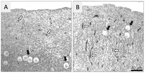
In humans, blood vessels are scarce at young ages (e.g. the 20s), but with an increase in age, the blood vessel density increases along with a decrease in the number of resting oocytes (Delgado-Rosas et al. Citation2009) (). Thus, one may raise a question: Are resting oocytes in humans also protected from radiation injury due to hypoxia? To make the cells resistant to radiation exposure, oxygen levels need to be below 1% (e.g. Ling et al. Citation1981). Studies on tumor biology indicate that the oxygen levels of approximately 85–100 μm from tumor blood vessels, which corresponds to 10 or more cell layers, decrease to 1% or less which may cause effective protection from radiation damage (Kizaka-Kondoh et al. Citation2009, Harada et al. Citation2012, Macklin et al. Citation2020). Since the cortex contains so few blood vessels (especially when young), it is possible that the oxygen level is sufficiently low to protect cells from radiation damage in humans as well.
Figure 2. Cortical region of an ovary at an age of 23 (A) and 37 (B) years. Note blood vessels are scarce when young but increased its density with the increase of age (open arrows). Black arrows indicate resting oocytes. Bar represents 80 µm. (Delgado-Rosas et al. Citation2009).
Observations in mouse resting oocytes are likely to be applicable to humans
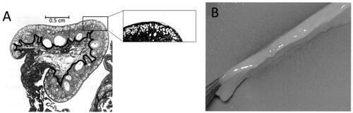
As mentioned earlier, there exists a gradient in stroma density in the mouse ovarian cortex. The question then is whether this observation is applicable to humans. The human ovary is much larger than the mouse ovary (the mouse ovary is 1 to 2 mm in diameter, while the human ovary is several cm in diameter), and the cortical thickness also largely differs. As seen in , most human resting oocytes are located within 2 mm of the ovarian surface. Indeed, an oocyte preservation method is to surgically remove strips from the ovarian cortex and then freeze them in liquid nitrogen () (Silber Citation2016, Kallen et al. Citation2018). Note that the cortex contains few blood vessels and is whitish. Furthermore, the cortex contains a low density of blood vessels () as in mice, but the proportion of the cortex region is larger in the human ovary (e.g. 80%: Forabosco et al. Citation1991, Perven et al. Citation2015). In contrast to humans, the mouse ovarian cortex appears thin as indicated in (Kerr et al. 2006, Niu and Spradling Citation2020).
Figure 3. A) A cross section of an ovary of young woman. The small white dots located close to the ovarian surface are the resting oocytes. A line was added to the photo to indicate the boundary between the cortex and stroma. Original photo is colored (Silber Citation2015). B) Fibrous ovarian cortex removed for cryopreservation of resting oocytes (Silber Citation2016).
In short, it is likely that also in the human ovary, a low oxygen microenvironment is required to maintain a resting state, which can well contribute to radioresistance. It is suggested that the mouse ovary appears exceptional among mammals because ovarian regionalization (differentiation into cortex and medulla regions) is weak (Jiménez Citation2009), so it seems likely that the human ovarian cortex is equally or tougher than that in the mouse, and thus equally or even more protected from radiation injury. In addition, a recent study on mitochondria in resting oocytes of Xenopus and humans showed a surprising fact that Complex-I (Cx-I), one of the five complexes or Cxs which are involved in the electron transport chain and produce ROS as byproducts, is lacking, which could indicate that ROS species are maintained at low levels to protect the oocyte genome from ROS for nearly 50 years, from birth to menopause, in humans (Rodríguez-Nuevo et al. Citation2022). It is also interesting to note that mitochondria in stage I oocytes (resting stage) of Xenopus are clustered near the nuclear membrane, called Balbiani body, and are short (round shape) with poor cristae and no activity of cytochrome c oxidase (COX), which indicates low respiratory activity. The COX activity starts to increase at stage II along with the elongation of mitochondrial length. Thus, the round shape indicates an inactive form of mitochondria (Hertig and Adams Citation1967, Kogo et al. Citation2011). In this regard, it is important to note that, although the Rodríguez-Nuevo paper mentioned that ‘early oocytes are the first and only physiological cell type in animals that exist without a functional mitochondrial complex I’, spermatogonia stem cells in a resting stage are also indicated to contain immature mitochondria and a high-level expression of glycolytic enzymes (Lord and Nixon Citation2020). Thus, it is possible that the two types of germline stem cells are equipped with similar strategic plans to realize long-term survival under the low-level conditions of oxygen and ROS.
From a viewpoint of radiation protection
In the 2001 UNSCEAR report, the risk of transgenerational effects of radiation in humans was expressed as a doubling dose which used the spontaneous frequency of human de novo mutations and the mutation induction rate per Gy following irradiation of mouse spermatogonia. In that report, while it was recognized that mouse immature oocytes are resistant to mutation induction, human resting oocytes were regarded to be equally sensitive to radiation as spermatogonia because there was no reason to expect that the same radio-resistance occurs in human immature oocytes (UNSCEAR (United Nations Scientific Committee on the Effects of Atomic Radiation) Citation2001, section 426). However, if it could be shown that the microenvironment of resting oocytes is hypoxic in both humans and mice, the possible radiation response in terms of mutagenesis would also be equal in both mice and humans. The remaining task is to verify low oxygen conditions in the cortex. Since direct physical measurement of pO2 is difficult, a biological approach would be indispensable.
Estimation of the mean mutation induction rate in spermatogonia
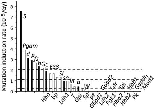
The mouse specific-locus tests (SLT) that were used extensively by Russell’s group for more than 10 years utilized morphologic markers which included coat color genes. The total number of mice used is said to have exceeded 4 million. The results showed that the mutation induction rate varied widely among the 7 genes; for example, the s gene mutated 15 times more frequently than the a gene. The mean mutation induction rate for the 7 genes was about 2 ∼ 3 × 10⁻⁵/Gy per locus, while the mean of 6 genes used in the Medical Research Council in Harwell, England was lower at 0.78 × 10⁻⁵/Gy per locus. Later, more information on additional genes was collected, and the mean mutation rate for 34 genes turned out to be about 1 × 10⁻⁵/Gy per locus (UNSCEAR Citation2001) (). The reasons for the large inter-gene differences in the mutation induction rate are not understood. The data of mouse genome sequence revealed that large genes do not necessarily mutate more often than small ones. Specifically, the s gene is small (about 30 kb) but is highly mutable, while the a and se genes are eight times larger (about 250 kb) but show the lowest mutability among the 7 genes.
Figure 4. Different mutation induction rate in different genes (UNSCEAR Citation2001). The black bars indicate 7 genes used for the initial SLT studies and white bars represent genes examined in later studies. The three dotted lines indicate, starting from the top, the mean mutation induction rate for 7 genes, 34 genes, and 1,190 DNA fragments.
Since the inter-gene difference in mutation induction rate is so large, it was felt necessary to obtain a brief idea of the mean mutation induction rate for many more genes. To study this problem, Asakawa and his coworkers improved the two-dimensional gel electrophoretic method (2DE method) which allows us to examine over 1000 genomic DNA fragments which were end-labeled with 32P at NotI sites and observed as autoradiographic spots by using computer-assisted scanner. Any deletion mutation at an autosomal DNA fragment is expected to give rise to a reduced spot intensity when transitioning from a 2-copy intensity spot to a one-copy intensity spot (a 50% reduction), and characterization of the deletion could then be achieved by cloning DNA from the remaining normal spot and using it as a DNA probe.
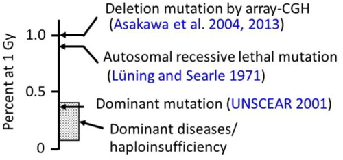
Two rounds of large-scale studies were conducted which tested over 1500 offspring and screened nearly 1.7 million spots, and the results indicated that the mean induction rate of deletion mutations over 1000 DNA fragments was considerably lower than that observed in the SLT studies, probably the order of 10−6/Gy per DNA fragment () (Asakawa et al. Citation2004, Citation2013), which was close to 1.1 × 10⁻⁶/Gy per gene for cataract mutations in mice (where 30 genes are estimated to be involved in cataract genesis) (Graw et al. Citation1986).
Table 2. Results of the mean induction rate of deletion mutations for over one thousand genomic DNA fragments that were end-labeled with 32P at NotI sites (Asakawa et al. Citation2004, Citation2013).
In a subsequent study, the microarray-based comparative genomic hybridization (array-CGH) method was used to detect deletion mutations in offspring derived from irradiated spermatogonia (Asakawa et al. Citation2016). Microarray slides containing 0.3 million to 1 million probes were used to find deletion mutations that had lost multiple probe sites in the genome. In this study, one deletion-bearing individual was found among 100 offspring in the control group (1/100 or 1%) and 5 deletion-bearing mutants were found among 100 offspring derived from spermatogonia irradiated with 4 Gy of X-rays (5/100 or 5%). Since the total number of the mutants is small, the estimated mutation induction rate contains a wide uncertainty, but if we take the results at face value, a 1 Gy exposure is expected to cause one deletion-bearing genome among 100 offspring. It is interesting to note that this value, 1% per genome per Gy, is very close to 0.9%, the estimated induction rate of autosomal recessive lethal mutations in mice (Lüning and Searle Citation1971).
Genes employed in the SLT were hypermutable and do not represent many other genes
It would be worthwhile to think why the mutation induction rates were generally high in the SLT studies. In the 1950s, for the initiation of the studies, it was necessary to prepare homozygous mutants for the seven marker genes, including the coat color genes, to create tester mice. These mutants were derived from pet mice and originated from spontaneous mutations which arose in mouse colonies in genes whose mutations were easily recognized. Evolutionarily, coat color genes in small animals are critically important to help escape attention from predators’ eyes, and hence selective force could have happened to increase the variability in their expression (e.g. Barrett et al. Citation2019), so that a sudden change in the environment would still allow a fraction of the cohort better odds to survive. For example, the mutant alleles of the d and s genes that were used for the tester mice in the SLTstudies had long been thought to be nonfunctional but later turned out to be hypo-morphic (i.e. exhibiting decreased functioning) caused by the insertion of a viral genome (Jenkins et al. Citation1981, Yamada et al. Citation2006). Further, it was found that genes that exhibit a high spontaneous mutation frequency tended to show a high mutation induction rate from radiation in fruit flies and mice (Shukla et al. Citation1979). Therefore, such genes with spontaneous mutation rate of an order of 10−5 would not represent many other genes in the genome.
Haploid insufficient genes as targets of dominant mutations in humans
In the mouse, in addition to the SLT studies, dominant mutations are another tool used to measure the genetic effects of radiation in offspring. Specifically, measurements are made of lethality during developmental processes (dominant lethal), abnormalities in organogenesis (malformations), and hereditary cataracts. Since it is not necessary to use specific genetic markers which are necessary for the SLT study, some of the dominant mutations are also applicable to studies in humans.
In this regard, haploid insufficiency which leads an organism to express a dominant hereditary disease is an important issue to consider (UNSCEAR Citation2001). Dominant mutations may arise from several different mechanisms. The simplest form is caused by a 50% decrease in the gene product, termed loss of function (LOF) mutations. They are caused not only by deletions of one allele of autosomal genes (hemizygosity), but also by the production of nonfunctional polypeptides due to amino acid substitutions. On the other hand, the nonfunctional polypeptides may occasionally cause a decrease in cellular function to 25% or less following the formation of dimers or other forms of the complex. In this case, although the mutation affected only one allele, it looks as if both alleles were affected. The mutation is called dominant negative (DN). The third mechanism is when a mutant protein gains a new function (GOF), and this new mechanism is concerned with genes that code for transcription factors. The former LOF mutations are more relevant for radiation mutagenesis because deletions are the predominant type of mutations induced by radiation. Consequently, when we try to identify genes that may cause dominant hereditary diseases following the exposure of germ cells to radiation, it is crucial to know that LOF mutations of the gene in concern can cause the disease. If it were possible to find patients who are hemizygous for the gene in concern (such as the 5q − syndrome), it is easy to annotate the gene as a haploinsufficient gene. However, if only new base sequences that were not found in the parents were detected in the patient (offspring), it is difficult to determine whether it was caused by LOF mutation or DN or GOF. This occurs commonly in hereditary diseases with only a small number of patients. In addition, it is confusing to find that mutations arising at different locations within the same gene may cause different disease phenotypes (see OMIM data described below).
Estimating the number of haploid insufficient genes
How many genes do we have which may cause haploid insufficient syndromes when one of the two alleles is inactivated? To try to answer this question in the mouse, one needs to knock out thousands of coding genes one by one by using the Crispr/Cas9 technique and observe the abnormal phenotype of the deletion heterozygotes, which in practice is unrealistic. Consequently, it appeared of interest to examine known dominant inherited diseases in humans. For this purpose, one can visit the online version of ‘Mendelian Inheritance in Man’ (OMIM) (https://www.omim.org/) which is an encyclopedia of human hereditary disorders and was first compiled in the 1960s by Dr. Victor A. McKusick. Today, it is updated every day and thus it is not easy to determine the total number of human hereditary diseases.
As a first step, autosomal dominant (AD) disorders were sorted out using a combination of key words; dominant inheritance AND bone malformation, or any other term among the following: mental retardation, birth defect, ribosomal protein mutation, heart malformation, microdeletion, cataract, or intellectual disability. Subsequently, descriptions of each AD disease were scrutinized to see if any description such as ‘probably caused by haploinsufficiency’ can be found. If no such description could be found, the disease cannot be judged as being caused by a mutation in a haploid-sensitive gene.
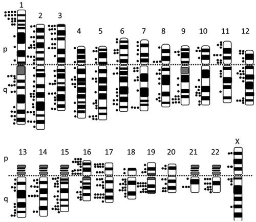
In this way, a total of about 400 genes were identified (Supplementary Table) and shows a chromosomal map of these genes. The karyotype used here represents a G-banding picture in which the negative bands characteristically contain more genes than the positive bands. One can see from the Figure that when a small deletion is detectable with the G-banding method, it would contain multiple haplo-sensitive genes. Under such occasions, each haplo-sensitive gene is thought to act additively and therefore it is anticipated that such fetuses with these conditions are subjected to a strong selective force against live birth.
Figure 5. Mapping of haplo-sensitive genes on human chromosomes.
Estimating the induced frequency of dominant mutations in human spermatogonia
Although it seems certain that we have about 400 haplo-sensitive genes in our genome, there is an additional number of genes that we cannot determine to be haploid sensitive due to the small number of patients. Therefore, we assume here that there are 400 to 2000 haploid sensitive genes. The value of 2000 is derived from observations that the number of genes responsible for recessive mutations in chromosome 1 was nearly 10 times or larger than that for dominant mutations, and our genome seems to contain 20,000 or more protein-coding genes. Thus, it seems that the total number of haploid-sensitive genes would not largely exceed one-tenth of 20,000 or 2000.
Here, assuming that the mean induction rate of deletion mutations per DNA segment (gene) is 1.9 × 10⁻⁶/Gy () in mouse spermatogonia, the estimated frequency of dominant mutations which appear in the offspring of 1 Gy-exposed spermatogonia is 0.8 to 4 per 1000 (0.1 to 0.4%). It is noted that the mutation induction rate used here is derived from mouse data but without considering the possible decreased viability of hemizygous mutants. The estimated frequency is not likely to be very different from 0.34% which was estimated as the frequency of dominant mutations in the F1 mouse derived from irradiated spermatogonia, and which include bone malformations, cataracts, and congenital malformations (UNSCEAR Citation2001). Furthermore, as mentioned earlier, it is interesting to note that mouse data which was acquired by using the array-CGH method showed 1 deletion out of 100 F1 mice in the control group and 5 deletions out of 100 in the 4 Gy-irradiated group, or 1% at 1 Gy. Also, the frequency of autosomal recessive lethal mutations was reported to be 0.9% per Gy (Lüning and Searle Citation1971). summarizes these data. Although each point estimate would involve a wide confidence interval due to the small number of mutants obtained, if we take the results at the face value, then one could summarize the data as indicating that a 1 Gy exposure to spermatogonia might induce deletion mutations in about 1% of the offspring, and a fraction of the deletion mutations may cause autosomal recessive mutations (which is phenotypically hidden and unseen) or dominant mutation which is seen as haploid insufficient disorders.
Figure 6. Estimated frequency of dominant diseases/haploinsufficiency disorders in offspring derived from 1 Gy-exposed spermatogonia but without considering possible decreased viability. The frequencies of other genetic changes are also shown for comparisons. Dominant mutations include bone malformations, cataracts, and congenital malformations.
Similarities and differences in male gametogenesis between mice and humans
Basic structure of the testis
Spermatogonia consist of several types of cells; spermatogonia stem cells (SSCs), undifferentiated progenitor cells, and differentiated cells which lead toward the development of spermatocytes, etc. (). Male germ cells are separated from circulating blood (the blood-testis barrier, or more correctly, the Sertoli cell barrier), since sperm-forming cells located in the lumen of the seminiferous tubules, whereas blood vessels are located primarily outside the seminiferous tubules, i.e. in the interstitial space ().
Figure 7. A) Schematic view which indicates development of male germ cells in the seminiferous tubules (Jung et al. Citation2019). B) A proposed model for the location of mouse spermatogonia stem cells (SSCs) close to the basement membrane in the seminiferous tubules, and away from blood vessels (BV) (Lord and Nixon Citation2020).
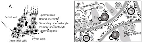
Although the SSCs attach to the basement membrane, their location in the tubules relative to the blood vessels was not known until recently, primarily because of the lack of SSC-specific surface markers. It was found recently in mice that ID4 can be used for such a marker, and over 95% of ID4-labeled SSCs reside in the basement areas where two seminiferous tubules adhere closely to each other (i.e. no blood vessels), or where the tubules face interstitial space which does not contain blood vessels (Chan et al. Citation2014). Lord and Nixon proposed the model shown in based on the most recent information, including single-cell gene expression profiling (Lord and Nixon Citation2020). Because a hypoxic microenvironment is common for tissue stem cells, e.g. hematopoietic stem cells and other tissue stem cells (e.g. Jež et al. Citation2015, Huang et al. Citation2018), it is reasonable to think that SSCs reside in avascular regions, whereas their progenies move laterally along the circumference of the tubule closer to the blood vessels so that oxygen and nutrients, including hormones, may be available.
In support of this, it is reported that in mice, Sertoli cells exhibit a high glycolytic flux (Voigt et al. Citation2021), and Sertoli, Leydig and interstitial cells stain positively for the presence of hypoxia-inducible factor-1α (HIF-1α) (Lysiak et al. Citation2009, Gruber et al. Citation2010), and also positive with pimonidazole staining which indicates that the O2 level is lower than 1.5% (Gruber et al. Citation2010). It is interesting to note that pigment epithelium-derived factor (PEDF), a multi-functional glycoprotein, has anti-angiogenic activity and is secreted by peritubular cells of the testis (Windschüttl et al. Citation2015, Bagdadi et al. Citation2021) and also granulosa cells in the ovary (Chuderland et al. Citation2013), indicating that an anti-angiogenic microenvironment is present in both the testis and ovary. Further, recent studies showed that SSCs contain immature round-shaped mitochondria and express high levels of glycolysis enzymes, whereas in progenitor cells, the mitochondria start to mature and initiate oxidative phosphorylation (Lord and Nixon Citation2020). Such metabolic conditions are very similar to that of immature oocytes as mentioned earlier (Kogo et al. Citation2011, Rogríguez-Nuevo et al. 2022).
Differences in spermatogenesis between mice and humans
There is a big difference between mouse and human testes in the width of interstitial space. Specifically, mouse seminiferous tubules are tightly packed and share the boundaries along their circumference () whereas human tubules are loosely packed and there are wide inter-tubular spaces (). Namely, interstitial space composes 10% of the testicular cross-section in mice but 40% in humans (Hess and Renato de Franca Citation2008). Blood vessels run through the interstitial space and also the basement membrane of the seminiferous tubules (Ergün et al. Citation1994), but the density is low (indicated by the arrows in ), which supports the notion of a low O2 microenvironment in testes.
Figure 8. A) Mouse testis at 12 weeks of age (hematoxylin-eosin staining). Sg stands for spermatogonia, SC for Sertoli cells, and L for Leydig cells. Bar represents 20 µm. (Guillermet-Guibert et al. Citation2015). B) Human testis stained with an anti-MYH11 (a smooth muscle myosin) antibody. Arrow heads indicate blood vessels, and the insert indicates negative results with a control antibody. Bar represents 50 µm (Welter et al. Citation2013). C) A schematic presentation of stem cell pools and sperm output in mice and humans (Fayomi and Orwig Citation2018).
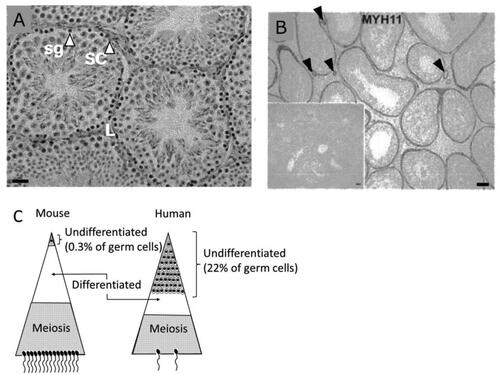
Another difference between mouse and human testes is clone formation by SSCs. In mice, a single SSC can produce about 4000 spermatozoa following 12 cell divisions while about 50% of them are lost by apoptosis. The dead cells seem to be used as an energy source by Sertoli cells (Xiong et al. Citation2009). In contrast, a human SSC produces only 64 sperm following 6 cell divisions in which 75% of the cells are lost through apoptosis. Consequently, only 25% of the cells produced can participate in the sperm pool. Furthermore, about half of the sperm are morphologically abnormal, and so probably only 12% of the sperm produced are thought to contribute to fertilization (Fayomi and Orwig Citation2018). In mice, only 0.3% of germ cells are undifferentiated while 22% are undifferentiated in humans (). As a result, human spermatogenic efficiency is poor when compared to laboratory animals (Holstein et al. Citation2003), but the relatively small number of mitotic cell divisions during human spermatogenesis is probably beneficial toward maintaining a low spontaneous mutation rate. This difference is shown schematically in ( (Fayomi and Orwig Citation2018).
In the mouse, Type A spermatogonia were thought to be similar to renewable stem cells (As) and give rise to differentiated spermatogonia (Type A1 to A4), followed by Type B spermatogonia and then spermatocytes. However, later studies showed that as cells are further divided into three types, a single-cell type (Ais: isolated), a connected two-cell type (Apr: paired), and a connected 4-, 8- or even 16-cell type (Acl: clonal). Currently, Ais cells are regarded as being SSCs while Apr and Acl cells are undifferentiated progenitors, and the number of Acl cells is 2 to 50 times larger than Ais or Apr cells depending on the epithelial stages of the seminiferous tubules (Erickson Citation1981). Dose survival data which was determined microscopically in toto (whole mount) 1 to14 days after irradiation showed that Acl cells are radiosensitive, especially at doses below 1 Gy, whereas Ais cells are quite resistant (). The results are remarkable because many other studies which used cross-sectional approaches repeatedly indicated that mouse spermatogonia were highly sensitive to the killing effect of radiation and that the dose-survival responses had no shoulder. Such observations were probably made primarily on Acl cells.
Figure 9. Dose survival responses of spermatogonia in mice (A) (Erickson Citation1981) and humans (B) (Clifton and Bremner Citation1983). Ais and Acl stand for isolated and clonally aligned A spermatogonia, respectively, in mice.
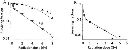
Regarding human spermatogenesis, Type A spermatogonia are classified into only two subtypes, Adark and Apale, based on differences in nuclear morphology and on staining intensity with hematoxylin, and it is thought that Adark cells are stem cells in the G0 stage, and Apale cells are cycling (Fayomi and Orwig Citation2018). There is only one human study in which testes of volunteers in prison were biopsied before and after irradiation (but the interval varied among individuals) in order to determine the dose survival response (it was human experimentation) (Clifton and Bremner Citation1983). The results showed that human spermatogonia cells are highly sensitive to the killing effect of radiation, especially at doses below 1 Gy (), which is consistent with the observations that the sperm count may decrease to nearly zero in 3 months after exposure to 1 Gy. Although no separate counting of Adark and Apale cells was made, it seems that the low-dose response was largely caused by the apoptotic death of Apale cells (Fayomi and Orwig Citation2018).
Spermatogonia stages at which the mutagenesis data were collected
In mouse studies, the mutation frequency increases linearly with the X- or gamma-ray dose until 6 Gy, and then declines (). The decline is more extreme when mice were exposed to fission neutrons. The mutation frequency (15 × 10−5) at about 1 Gy declined to about 1/5 when the dose was increased to about 2 Gy () (Searle Citation1974). The decline in mutation frequency was thought to occur when the radiosensitive fraction of the spermatogonia pool to both mutation induction and cell killing, was selectively killed at low doses, resulting in only radioresistant cells surviving to be exposed at higher doses. It was also suggested that different stages in the spermatogonia cell cycle were involved in this response (Searle Citation1974). However, since the biological effects of high LET radiations are known to be less dependent on the cell cycle stages when compared to the effects of low LET radiations, it appears difficult to attribute the appearance of a very radioresistant fraction of cells as being an effect of the specific stages in a cell cycle, and only hypoxic cells would explain the observations. In addition, as repeatedly mentioned, SSCs are under hypoxic conditions.
Figure 10. A) Results of specific locus tests of mouse spermatogonia after exposures of acute X- or gamma-rays (□) or 500 R + 500 R fractionated exposures given 1 day apart (■). B) Results after acute (■) or chronic (□) exposures of fission neutrons (Searle Citation1974).
These results support the notion that SSCs are in a quiescent stage (G0) and produce Ais cells which subsequently produce Apr to Acl cells. Since SSCs are under hypoxic conditions, they are resistant to radiation-induced cell killing (Searle Citation1974 review) and probably also to mutagenesis, whereas Apr and Acl cells are mutable. However, since the inter-cellular connection of Apr and Acl cells may break and the cells revert to Ais cells (Potter and DeFalco Citation2017), mutated Apr or Acl spermatogonia would not be gradually replaced with undamaged SSCs but may persist in the stem cell niche of the testes.
In contrast, in humans, it takes on an average of 74 days (Amann Citation2008) for Apale spermatogonia cells to become sperm. In atomic-bomb survivor studies, observations were made of births that occurred 3 years or more after exposure. In the studies on the offspring of ex-patients of cancer, it was recommended that adult male patients wait for 6 to 18 months before trying to father their own children (Sabanegh and Ragheb Citation2009). This time interval between radiotherapy and pregnancy becomes naturally even longer (years or decades) for childhood cancer survivors. Thus, in human cases, there is little doubt that it is the effects on SSCs that were (and will be) examined.
Different sensitivities for male fertility in mice and humans
In mouse experiments, single exposures of males to 3 to 6 Gy were often used, whereas, in humans, a fraction of the patients became permanently sterile, even after exposure to 2 Gy or less (e.g. Nygaard et al. Citation1991). For example, sperm count drops to nearly zero at 3 or 6 months after exposure to 1 Gy or 0.5 Gy, respectively (ICRP Citation1984) and it takes years for the recovery. In addition, mouse data showed that a full recovery may not occur at higher doses (Meistrich Citation1986), and hence, in human males, although recovery from radiation-induced injuries may take place to some extent, sub-fertility can easily develop. In addition, when the number of sperm is low, a sufficient number of sperm may not be able to survive in the epididymis, and hence may not appear in the semen (Silber et al. Citation1997).
In summary, recent studies indicate that both human and mouse SSCs express genes that are found to be characteristic of cells under hypoxic conditions (Lord and Nixon Citation2020 and references cited in it). Because the reproductive lifespan is much longer in humans than in mice, it can be hypothesized that human SSCs are equally or better protected than mouse SSCs from DNA damage inflicted by spontaneously arising reactive oxygen species, which implies also from radiation injuries. Such biological and other factors such as radiation sensitivity, the structure of the stem cell pool, and the time interval between radiation exposure and conception, are likely to contribute to differences in the transgenerational effects of radiation between mice and humans.
Species differences after fertilization
Malformed human fetuses tend to be lost
One of the differences between humans and mice is that humans are essentially single-delivery animals whereas mice are multi-delivery animals. Thus, miscarriage is common in humans while mice cannot abort abnormal fetuses selectively (dead fetuses can be absorbed in utero). It is difficult to become pregnant in humans as only 30% of fertilized eggs are thought to give rise to living births. Specifically, 30% of fertilized eggs are lost before implantation, another 30% after implantation, and 10% after medical confirmation of pregnancy (Larsen et al. Citation2013). Among the so-called ‘missed abortions’ (when there is no sign of abortion in the mother), 85% carry malformations and 75% carry an abnormal chromosome constitution/number (XX/XY mosaic, 45XO, trisomy 15, 16, 21, etc.) (Philipp et al. Citation2003).
The human placenta consists of complex structures with maternal and fetal blood vessels, and thus the probability of a life-threatening risk of bleeding is relatively high at the time of childbirth. Thus, the hypothesis has been raised that the endometrial cells can mark or identify fertilized eggs so that low-quality embryos may avoid implantation or be aborted after implantation (Weimar et al. Citation2012). This hypothesis is derived from the observation that patients who experience recurrent miscarriages tend to be pregnant more often than healthy women. An in vitro study indicated that cultured endometrial cells from patients appear to attract even low-quality embryos (fertilized eggs that had three pronuclei or those which do not develop beyond the morula stage, etc.) which are avoided by cultured normal endometrial cells. Furthermore, it is also suggested that when low-quality embryos were unfortunately implanted, they could still be discarded through menstruation (Brosens et al. Citation2014).
In line with these observations, it was reported that aborted fetuses which were generated for medical reasons contained external malformations in as many as 9% of the fetuses among the early-stage embryos (5 weeks of age) but decreased to 1% at a full-term pregnancy (Shiota Citation2021).
Were malformed newborns of atomic-bomb survivors hidden from being formally recorded?
Among personal notes written by atomic-bomb survivors, a midwife wrote that malformed newborns were deliberately hidden to avoid a formal record (in those days, home delivery was the most common form of childbirth). If such activity was common, it threatens the validity of the genetic study on birth defects which was started in 1948, three years after the end of the War, by the Atomic Bomb Casualty Commission (ABCC). However, this possibility is likely to be low because the ABCC’s study scheme could diagnose as many as 93% of the births that were registered at 5 months of pregnancy when mothers visited the city office to receive a special ration of food (Neel and Schull Citation1991).
On the other hand, births within 9 months after the exposure (prior to May 1946) are in a different situation because they are derived from irradiated fetuses in utero, and it is possible that some of the malformed newborns might not be properly recorded but recorded as stillbirths. This is because, at that time, Japanese society was suffering under chaotic conditions, and newborns derived from irradiated fetuses are at much higher risk to develop malformations than those derived from irradiated germ cells; for example, an exposure to 0.5 Gy delivered to fetuses at 8-15 weeks of age could cause mental retardation in 20% of the newborns (Otake et al. Citation1991).
It is mentioned here that no study could be conducted then to collect information on malformations observed among in-utero exposed babies.
Only a small fraction of malformations is genetic
The frequency of congenital malformations varies from about 1% (serious cases) to 3% (including subtle cases) but the frequency does not change greatly by changing time. Importantly, not all malformations are caused by genetic alterations; for example, it is estimated that 65 to 75% of malformations are of unknown origin, 15 to 25% are genetic, and 10% are environmental (Brent Citation2004). This means that even if the genetic component were doubled, the total frequency would still increase only 1.2-fold. On the other hand, the estimated frequency of dominant mutations in mice which includes congenital malformations, bone malformations, and cataracts is 0.34% in the offspring derived from 1 Gy-exposed spermatogonia (UNSCEAR Citation2001), which is on the same order of the estimated frequency for dominant mutations (0.1 to 0.4%) caused by haploinsufficiency in human offspring (it is noted that the induction rate used here is derived from mouse studies). If exposure to 1 Gy, which is a substantial dose, induces congenital malformations at a frequency that is lower than the spontaneous frequency, it would be a difficult task to find evidence for the mutagenic effects of radiation, if any effect were present, in human offspring. A recent whole genome sequencing study showed that the frequency of de novo mutations in developmental disorders is about 0.3% (Deciphering Developmental Disorder Study 2017).
Evidence for the high radio-resistance of human spermatogonia stem cells
In this section, epidemiologic data on cancer survivors will be presented to understand the unusual nature of human spermatogonia stem cells and resting-stage oocytes. This discussion will not consider the epidemiologic reports on atomic-bomb survivors and their offspring. For readers who are interested in those reports, please refer to Grant et al. (Citation2015) for cancer risk, Yamada et al. (Citation2021) for birth defects, and Tatsukawa et al. (Citation2013) for the prevalence of multifactorial diseases.
A study on malformations in the offspring of survivors from Wilms tumors (Green et al. Citation2010) showed that in the case of male survivors, no malformation was observed at total doses below 25 Gy (). It is fascinating to see that some SSCs can survive after exposure to such large doses of radiation, especially when referring to the dose-survival response of spermatogonia (). The secret probably lies in the fractionated exposures where surviving cells can repopulate during the intervals between exposures. At doses higher than 25 Gy, excessive numbers of malformations were observed whereas the authors stated that the observation might be a chance observation due to a small number of the cases and hence further studies are necessary. In the case of female survivors, the frequencies of premature birth and low birth weight tended to increase with the dose (results not included in this table), but no dose-related increase was observed in malformation frequencies (). The generally elevated frequency of malformations in the offspring of female survivors when compared with that in male survivors is most likely attributable to the effects of chemotherapy on the uterus.
Table 3. Epidemiologic data in the offspring of Wilms tumor survivors (Green et al. Citation2010).
In another epidemiologic study of cancer survivors, no increased frequencies for chromosome aberrations, Mendelian diseases, and malformations were observed (Green et al. Citation2009). The mean dose was 1.26 Gy to the ovary and 0.46 Gy to the testis. As this study recruited over as many as 6000 offspring born to the survivors, which means that if the frequency of malformation had increased by 1% above the baseline level of 3.1% (4.1/3.1 = 1.3-fold increase), the increase could have been detected with a high probability with p < .05. One might raise a question that the increase was smaller than 1% and did not reach statistical significance, but the observed frequency (2.2%) was lower than that of the control group.
In short, the epidemiologic data from cancer survivors show that, although the total exposure doses (fractionated doses) to the gonads were much larger than in the cases of atomic-bomb survivors, normal children are born. This fact indicates that the lack of evidence is probably not totally due to a problem in detecting the effect of radiation exposures in humans such as a lack of radiosensitive marker genes, but contributions of biological factors could be more important which makes it difficult for the irradiated germ cells to give rise to abnormal offspring. Such factors include a low oxygen microenvironment and low mutation induction rate following the irradiation of human spermatogonia, and the miscarriage-prone nature of human pregnancy.
Future directions
Microarray comparative genomic hybridization (array-CGH) and whole genome sequencing (WGS) of mouse trios (irradiated sire and non-irradiated dam or the reverse combination, and the offspring) indicated there was a small increase in the frequency of long deletions (Asakawa et al. Citation2016) and short insertions or deletions (indels) in offspring born to irradiated spermatogonia or mature oocytes (Adewoye et al. Citation2015, Satoh et al. Citation2020). In the case of humans, the WGS study was conducted for families whose fathers or mothers were exposed to radiation following the Chernobyl accident (which includes 130 children) (Yeager et al. Citation2021), and three families in Nagasaki whose fathers were exposed to large doses of radiation (Horai et al. Citation2018), but no increased frequency of mutations was observed.
At the Radiation Effects Research Foundation, blood samples of trios (father, mother, and offspring) from 1000 survivor families are stored under liquid nitrogen for future WGS studies. In this regard, it happened once in the past that atomic-bomb survivors and their offspring were discriminated against in obtaining employment or getting married. Therefore, ethical guidelines need to be followed so that such discrimination may not happen again. A summary from an international meeting on this ethical issue was reported by Noda et al. (Citation2021).
Finally, future descriptions of the genetic risks of radiation will be better described as the probability of giving rise to dominant diseases in the offspring. For this purpose, information on both the number of haploid-sensitive genes and the mutation induction rate per gene would be required. The latter information could be obtained in the future by WGS studies while complete knowledge of the former will continue to be difficult to acquire.
Supplemental Material
Download MS Word (172.9 KB)Acknowledgement
The authors are grateful to Dr. L. Kapp for his careful reading of the manuscript.
Disclosure statement
No potential conflict of interest was reported by the author(s).
Additional information
Funding
Notes on contributors
Nori Nakamura
Nori Nakamura, PhD, is a radiation biologist and interested in various aspects of radiation effects in humans.
Noriaki Yoshida
Noriaki Yoshida, MD, PhD, is a molecular scientist working at the RERF in Hiroshima. His current interest is to elucidate pathogenesis of hematopoietic malignancies in atomic-bomb survivors from a molecular perspective. He is also interested in the prevention of the effect of radiation on the development of cancer and other diseases.
Tatsuya Suwa
Tatsuya Suwa, MD, PhD, is a radiation oncologist working at the Department of Radiation Oncology and Image-applied Therapy, Kyoto University, Kyoto, Japan. His current research interest is to elucidate the effects of hypoxic regions in normal and tumor tissues during radiotherapy. He is also interested in the role of innate immunity in these regions.
References
- Adewoye AB, Lindsay SJ, Dubrova YE, Hurles ME. 2015. The genome-wide effects of ionizing radiation on mutation induction in the mammalian germline. Nat Commun. 6:6684.
- Amann RP. 2008. The cycle of the seminiferous epithelium in humans: a need to revisit? J Androl. 29(5):469–487.
- Asakawa J, Kuick R, Kodaira M, Nakamura N, Katayama H, Pierce D, Funamoto S, Preston D, Satoh C, Neel JV, et al. 2004. A genome scanning approach to assess the genetic effects of radiation in mice and humans. Radiat Res. 161(4):380–390.
- Asakawa J, Kodaira M, Cullings HM, Katayama H, Nakamura N. 2013. The genetic risk in mice from radiation: an estimate of the mutation induction rate per genome. Radiat Res. 179(3):293–303.
- Asakawa JI, Kodaira M, Miura A, Tsuji T, Nakamoto Y, Imanaka M, Kitamura J, Cullings H, Nishimura M, Shimada Y, et al. 2016. Genome-wide deletion screening with the array CGH method in mouse offspring derived from irradiated spermatogonia indicates that mutagenic responses are highly variable among genes. Radiat Res. 186(6):568–576.
- Bagdadi N, Sawaied A, AbuMadighem A, Lunenfeld E, Huleihel M. 2021. The expression levels and cellular localization of pigment epithelium derived factor (PEDF) in mouse testis: Its possible involvement in the differentiation of spermatogonial cells. IJMS. 22(3):1147.
- Baker TG. 1971. Comparative aspects of the effects of radiation during oogenesis. Mutat Res. 11(1):9–22.
- Barrett RDH, Laurent S, Mallarino R, Pfeifer SP, Xu CCY, Foll M, Wakamatsu K, Duke-Cohan JS, Jensen JD, Hoekstra HE. 2019. Linking a mutation to survival in wild mice. Science. 363(6426):499–504.
- BEIR (Committee on the Biological Effects of Ionizing Radiation). 1980. The Effects on Populations of Exposure to Low Levels of Ionizing Radiation: 1980 (BEIR-III Report). Washington, D.C: National Academy Press..
- Brent RL. 2004. Environmental causes of human congenital malformations: the pediatrician’s role in dealing with these complex clinical problems caused by a multiplicity of environmental and genetic factors. Pediatrics. 113(Supplement_3):957–968.
- Brosens JJ, Salker MS, Teklenburg G, Nautiyal J, Salter S, Lucas ES, Steel JH, Christian M, Chan YW, Boomsma CM, et al. 2014. Uterine selection of human embryos at implantation. Sci Rep. 4(1):3894.
- Chan F, Oatley MJ, Kaucher AV, Yang QE, Bieberich CJ, Shashikant CS, Oatley JM. 2014. Functional and molecular features of the Id4+ germline stem cell population in mouse testes. Genes Dev. 28:1351–1362.
- Choi Y, Yuan D, Rajkovic A. 2008. Germ cell-specific transcriptional regulator sohlh2 is essential for early mouse folliculogenesis and oocyte-specific gene expression. Biol Reprod. 79:1176–1182.
- Chuderland D, Ben-Ami I, Kaplan-Kraicer R, Grossman H, Komsky A, Satchi-Fainaro R, Eldar-Boock A, Ron-El R, Shalgi R. 2013. Hormonal regulation of pigment epithelium-derived factor (PEDF) in granulosa cells. Mol Hum Reprod. 19(2):72–81.
- Clifton DK, Bremner WJ. 1983. The effect of testicular X-irradiation on spermatogenesis in man. J Androl. 4(6):387–392.
- Deciphering Developmental Disorders Study 2017. Prevalence and architecture of de novo mutations in developmental disorders. Nature. 542:433–438.
- Delgado-Rosas F, Gaytán M, Morales C, Gómez R, Gaytán F. 2009. Superficial ovarian cortex vascularization is inversely related to the follicle reserve in normal cycling ovaries and is increased in polycystic ovary syndrome. Hum Reprod. 24(5):1142–1151.
- Dobson RL, Kwan TC. 1977. The tritium RBE at low-level exposure—Variation with dose, dose rate, and exposure duration. Curr Top Radiat Res Q. 12:44–62.
- Dobson RL, Clifford GK, Felton JS, Kwan TC, Wuebbles BJ, Jones DCL. 1978. Vulnerability of female germ cells in developing mice and monkeys to tritium, gamma rays, and polycyclic aromatic hydrocarbons. In Developmental Toxicology of Energy-Related Pollutants, DD Mahlum, MR Sikov, PL Hackett, FD Andrew (Eds.), D.O.E. Symposium Series 47, CONF-771017, National Technical Information Service, US Department of Commerce, Springfield, Virginia, USA.
- Ergün S, Stingl J, Holstein AF. 1994. Microvasculature of the human testis in correlation to Leydig cells and seminiferous tubules. Andrologia. 26(5):255–262.
- Erickson BH. 1981. Survival and renewal of murine stem spermatogonia following 60Co gamma radiation. Radiat Res. 86(1):34–51.
- Fayomi AP, Orwig KE. 2018. Spermatogonial stem cells and spermatogenesis in mice, monkeys and men. Stem Cell Res. 29:207–214.
- Forabosco A, Sforza C, De Pol A, Vizzotto L, Marzona L, Ferrario VF. 1991. Morphometric study of the human neonatal ovary. Anat Rec. 231(2):201–208.
- Gougeon A. 1986. Dynamics of follicular growth in the human: a model from preliminary results. Hum Reprod. 1(2):81–87.
- Grant EJ, Furukawa K, Sakata R, Sugiyama H, Sadakane A, Takahashi I, Utada M, Shimizu Y, Ozasa K. 2015. Risk of death among children of atomic bomb survivors after 62 years of follow-up: a cohort study. Lancet Oncol. 16:1316–1323.
- Graw J, Favor J, Neuhäuser-Klaus A, Ehling UH. 1986. Dominant cataract and recessive specific locus mutations in offspring of X-irradiated male mice. Mutat Res. 159:47–54.
- Grdina DJ, Nagy B, Hill CK, Sigdestad CP. 1989. Protection against radiation-induced mutagenesis in V79 cells by 2-[(aminopropyl)amino] ethanethiol under conditions of acute hypoxia. Radiat Res. 117(2):251–258.
- Green DM, Sklar CA, Boice JD, Jr, Mulvihill JJ, Whitton JA, Stovall M, Yasui Y. 2009. Ovarian failure and reproductive outcomes after childhood cancer treatment: results from the childhood cancer survivor study. J Clin Oncol. 27:2374–2381.
- Green DM, Lange JM, Peabody EM, Grigorieva NN, Peterson SM, Kalapurakal JA, Breslow NE. 2010. Pregnancy outcome after treatment for Wilms tumor: a report from the national Wilms tumor long-term follow-up study. J Clin Oncol. 28:2824–2830.
- Gruber M, Mathew LK, Runge AC, Garcia JA, Simon MC. 2010. EPAS1 is required for spermatogenesis in the postnatal mouse testis. Biol Reprod. 82:1227–1236.
- Guillermet-Guibert J, Smith LB, Halet G, Whitehead MA, Pearce W, Rebourcet D, León K, Crépieux P, Nock G, Strömstedt M, et al. 2015. Novel role for p110β PI 3-kinase in male fertility through regulation of androgen receptor activity in Sertoli cells. PLOS Genet. 11(7):e1005304.
- Harada H, Inoue M, Itasaka S, Hirota K, Morinibu A, Shinomiya K, Zeng L, Ou G, Zhu Y, Yoshimura M, et al. 2012. Cancer cells that survive radiation therapy acquire HIF-1 activity and translocate towards tumour blood vessels. Nat Commun. 3(1):783.
- Hayashi K, Shimamoto S, Nagamatsu G. 2020. Environmental factors for establishment of the dormant state in oocytes. Develop Growth Differ. 62(3):150–157.
- Hertig AT, Adams EC. 1967. Studies on the human oocyte and its follicle. I. Ultrastructural and histochemical observations on the primordial follicle stage. J Cell Biol. 34(2):647–675.
- Hess RA, Renato de Franca L. 2008. Spermatogenesis and cycle of the seminiferous epithelium. Adv Exp Med Biol. 636:1–15.
- Holstein AF, Schulze W, Davidoff M. 2003. Understanding spermatogenesis is a prerequisite for treatment. Reprod Biol Endocrinol. 1(1):107.
- Horai M, Mishima H, Hayashida C, Kinoshita A, Nakane Y, Matsuo T, Tsuruda K, Yanagihara K, Sato S, Imanishi D, et al. 2018. Detection of de novo single nucleotide variants in offspring of atomic-bomb survivors close to the hypocenter by whole-genome sequencing. J Hum Genet. 63(3):357–363.
- Huang X, Trinh T, Aljoufi A, Broxmeyer HE. 2018. Hypoxia signaling pathway in stem cell regulation: Good and evil. Curr Stem Cell Rep. 4(2):149–157.
- ICRP, 1984. Non-stochastic effects of irradiation. ICRP Publication 41. Ann. ICRP 14, p. 19–21.
- Jenkins NA, Copeland NG, Taylor BA, Lee BK. 1981. Dilute (d) coat colour mutation of DBA/2J mice is associated with the site of integration of an ecotropic MuLV genome. Nature. 293(5831):370–374.
- Jež M, Rožman P, Ivanović Z, Bas T. 2015. Concise review: the role of oxygen in hematopoietic stem cell physiology. J Cell Physiol. 230(9):1999–2005.
- Jiménez R. 2009. Ovarian organogenesis in mammals: mice cannot tell us everything. Sex Dev. 3(6):291–301.
- Jung M, Wells D, Rusch J, Ahmad S, Marchini J, Myers SR, Conrad DF. 2019. Unified single-cell analysis of testis gene regulation and pathology in five mouse strains. Elife. 8:e43966.
- Kallen A, Polotsky AJ, Johnson J. 2018. Untapped reserves: controlling primordial follicle growth activation. Trends Mol Med. 24(3):319–331.
- Kerr JB, Duckett R, Myers M, Britt KL, Mladenovska T, Findlay JK. 2006. Quantification of healthy follicles in the neonatal and adult mouse ovary: evidence for maintenance of primordial follicle supply. Reproduction. 132:95–109.
- Kim DA, Suh EK. 2014. Defying DNA double-strand break-induced death during prophase I meiosis by temporal TAp63α phosphorylation regulation in developing mouse oocytes. Mol Cell Biol. 34(8):1460–1473.
- Kizaka-Kondoh S, Tanaka S, Harada H, Hiraoka M. 2009. The HIF-1-active microenvironment: an environmental target for cancer therapy. Adv Drug Deliv Rev. 61(7-8):623–632.
- Kogo N, Tazaki A, Kashino Y, Morichika K, Orii H, Mochii M, Watanabe K. 2011. Germ-line mitochondria exhibit suppressed respiratory activity to support their accurate transmission to the next generation. Dev Biol. 349(2):462–469.
- Larsen EC, Christiansen OB, Kolte AM, Macklon N. 2013. New insights into mechanisms behind miscarriage. BMC Med. 11(1):154.
- Ling CC, Michaels HB, Gerweck LE, Epp ER, Peterson EC. 1981. Oxygen sensitization of mammalian cells under different irradiation conditions. Radiat Res. 86(2):325–340.
- Lord T, Nixon B. 2020. Metabolic changes accompanying spermatogonial stem cell differentiation. Dev Cell. 52(4):399–411.
- Luan Y, Xu P, Yu SY, Kim SY. 2021. The Role of mutant p63 in female fertility. IJMS. 22(16):8968.
- Lüning KG, Searle AG. 1971. Estimates of the genetic risks from ionizing radiation. Mutat Res. 12:291–304.
- Lysiak JJ, Kirby JL, Tremblay JJ, Woodson RI, Reardon MA, Palmer LA, Turner TT. 2009. Hypoxia-inducible factor-1alpha is constitutively expressed in murine Leydig cells and regulates 3beta-hydroxysteroid dehydrogenase type 1 promoter activity. J Androl. 30(2):146–156.
- Macklin PS, Yamamoto A, Browning L, Hofer M, Adam J, Pugh CW. 2020. Recent advances in the biology of tumour hypoxia with relevance to diagnostic practice and tissue-based research. J Pathol. 250(5):593–611.
- Meistrich ML. 1986. Relationship between spermatogonial stem cell survival and testis function after cytotoxic therapy. Br J Cancer. 53 (Suppl. VII):89–101.
- Muller HJ. 1927. Artificial transmutation of the gene. Science. LXVI:84–87.
- Nakamura N. 2006. Genetic effects of radiation in atomic-bomb survivors and their children: past, present and future. JRR. 47(SupplementB):B67–B73.
- Nakamura N, Suyama A, Noda A, Kodama Y. 2013. Radiation effects on human heredity. Annu Rev Genet. 47(1):33–50.
- Nakamura N. 2018. Why genetic effects of radiation are observed in mice but not in humans. Radiat Res. 189(2):117–127.
- Nakamura N. 2019. History of radiation genetics: light and darkness. Int J Radiat Biol. 95(7):999–1014.
- Neel JV, Schull WJ. 1991. The children of atomic bomb survivors. Washington DC :National Academy Press, , pp. 35.
- Niu W, Spradling AC. 2020. Two distinct pathways of pregranulosa cell differentiation support follicle formation in the mouse ovary. Proc Natl Acad Sci U S A. 117(33):20015–20026.
- Noda A, Kato K, Tamura C, Biesecker LG, Imaizumi M, Inoue Y, Henderson GE, Wilfond B, Muto K, Naito M, et al. 2021. Ethical, legal and social implications of human genome studies in radiation research: a workshop report for studies on atomic bomb survivors at the radiation effects research foundation. J Radiat Res. 62(4):656–661.
- Nygaard R, Clausen N, Siimes MA, Márky I, Skjeldestad FE, Kristinsson JR, Vuoristo A, Wegelius R, Moe PJ. 1991. Reproduction following treatment for childhood leukemia: a population-based prospective cohort study of fertility and offspring. Med Pediatr Oncol. 19(6):459–466.
- Otake M, Schull WJ, Yoshimaru H. 1991. Brain damage among the prenatally exposed. JRR. 32(SUPPLEMENT):249–264.
- Perven HA, Nurunnabi ASM, Siddiqua D, Johora F, Afroz H, Ara S. 2015. Histological study of proportion of cortex and medulla of the ovary in Bangladesh women. Bangladesh Med J. 44(1):8–10.
- Philipp T, Philipp K, Reiner A, Beer F, Kalousek DK. 2003. Embryoscopic and cytogenetic analysis of 233 missed abortions: factors involved in the pathogenesis of developmental defects of early failed pregnancies. Hum Reprod. 18:1724–1732.
- Potter SJ, DeFalco T. 2017. Role of the testis interstitial compartment in spermatogonial stem cell function. Reproduction. 153:R151–R162.
- Rodríguez-Nuevo A, Torres-Sanchez A, Duran JM, De Guirior C, Martínez-Zamora MA, Böke E. 2022. Oocytes maintain ROS-free mitochondrial metabolism by suppressing complex I. Nature. 607(7920):756–761.
- Russell WL. 1977. Mutation frequencies in female mice and the estimation of genetic hazard of radiation in women. Proc Natl Acad Sci USA. 74:3523–3527.
- Russell WL. 1972. The genetic effects of radiation., In Peaceful Use of Atomic Energy, Vol. 13, IAEA Publ. STU/PUB/300. Vienna, p. 487–500.
- Russell LB, Russell WL. 1992. Frequency and nature of specific-locus mutations induced in female mice by radiations and chemicals: a review. Mutat Res. 296(1-2):107–127.
- Sabanegh ES, Jr, Ragheb AM. 2009. Male fertility after cancer. Urology. 73(2):225–231.
- Satoh Y, Asakawa JI, Nishimura M, Kuo T, Shinkai N, Cullings HM, Minakuchi Y, Sese J, Toyoda A, Shimada Y, et al. 2020. Characteristics of induced mutations in offspring derived from irradiated mouse spermatogonia and mature oocytes. Sci Rep. 10(1):37.
- Searle AG. 1974. Mutation induction in mice. Adv Radiat Biol. 4:131–207.
- Selby PB, Lee SS, Kelley EM, Bangham JW, Raymer GD, Hunsicker PR. 1991. Specific-locus experiments show that female mice exposed near the time of birth to low-LET ionizing radiation exhibit both a low mutational response and a dose-rate effect. Mutat Res. 249(2):351–367.
- Shimamoto S, Nishimura Y, Nagamatsu G, Hamada N, Kita H, Hikabe O, Hamazaki N, Hayashi K. 2019. Hypoxia induces the dormant state in oocytes through expression of Foxo3. Proc Natl Acad Sci USA. 116(25):12321–12326.
- Shiota K. 2021. A life-table analysis of the intrauterine fate of malformed human embryos and fetuses. Birth Defects Res. 113(8):623–632.
- Shukla PT, Sankaranarayanan K, Sobels FH. 1979. Is there a proportionality between the spontaneous and the X-ray-induction rates of mutations? Experiments with mutations at 13 X-chromosome loci in Drosophila melanogaster. Mutat Res. 61(2):229–247.
- Silber S. 2015. Unifying theory of adult resting follicle recruitment and fetal oocyte arrest. Reprod Biomed Online. 31(4):472–475.
- Silber S. 2016. Ovarian tissue cryopreservation and transplantation: scientific implications. J Assist Reprod Genet. 33(12):1595–1603.
- Silber SJ, Nagy Z, Devroey P, Tournaye H, Van Steirteghem AC. 1997. Distribution of spermatogenesis in the testicles of azoospermic men: the presence or absence of spermatids in the testes of men with germinal failure. Hum Reprod. 12:2422–2428.
- Stadler LJ. 1928a. Mutations in barley induced by X-rays and radium. Science. 68(1756):186–187.
- Stadler LJ. 1928b. Genetic effects of X-rays in maize. Proc Natl Acad Sci USA. 14(1):69–75.
- Suh EK, Yang A, Kettenbach A, Bamberger C, Michaelis AH, Zhu Z, Elvin JA, Bronson RT, Crum CP, McKeon F. 2006. p63 protects the female germ line during meiotic arrest. Nature. 444:624–628.
- Suwa T, Kobayashi M, Shirai Y, Nam J-M, Tabuchi Y, Takeda N, Akamatsu S, Ogawa O, Mizowaki T, Hammond EM, et al. 2021. SPINK1 as a plasma marker for tumor hypoxia and a therapeutic target for radiosensitization. JCI Insight. 6(21):e148135.
- Tatsukawa Y, Cologne JB, Hsu WL, Yamada M, Ohishi W, Hida A, Furukawa K, Takahashi N, Nakamura N, Suyama A, et al. 2013. Radiation risk of individual multifactorial diseases in offspring of the atomic-bomb survivors: a clinical health study. J Radiol Prot. 33(2):281–293.
- UNSCEAR (United Nations Scientific Committee on the Effects of Atomic Radiation). 2001. Hereditary effects of radiation 2001 Report. New York (NY) :United Nations.
- Voigt AL, Thiageswaran S, de Lima E Martins Lara N, Dobrinski I. 2021. Metabolic requirements for spermatogonial stem cell establishment and maintenance in vivo and in vitro. IJMS. 22(4):1998.
- Wallace WH, Thomson AB, Kelsey TW. 2003. The radiosensitivity of the human oocyte. Hum Reprod. 18(1):117–121.
- Weimar CH, Kavelaars A, Brosens JJ, Gellersen B, de Vreeden-Elbertse JM, Heijnen CJ, Macklon NS. 2012. Endometrial stromal cells of women with recurrent miscarriage fail to discriminate between high- and low-quality human embryos. PLOS One. 7(7):e41424.
- Welter H, Kampfer C, Lauf S, Feil R, Schwarzer JU, Köhn FM, Mayerhofer A. 2013. Partial loss of contractile marker proteins in human testicular peritubular cells in infertility patients. Andrology. 1(2):318–324.
- Windschüttl S, Kampfer C, Mayer C, Flenkenthaler F, Fröhlich T, Schwarzer JU, Köhn FM, Urbanski H, Arnold GJ, Mayerhofer A. 2015. Human testicular peritubular cells secrete pigment epithelium-derived factor (PEDF), which may be responsible for the avascularity of the seminiferous tubules. Sci Rep. 5:12820.
- Xiong W, Wang H, Wu H, Chen Y, Han D. 2009. Apoptotic spermatogenic cells can be energy sources for Sertoli cells. Reproduction. 137(3):469–479.
- Yamada M, Furukawa K, Tatsukawa Y, Marumo K, Funamoto S, Sakata R, Ozasa K, Cullings HM, Preston DL, Kurttio P. 2021. Congenital malformations and perinatal deaths among the children of atomic bomb survivors: a reappraisal. Am J Epidemiol. 190:2323–2333.
- Yamada T, Ohtani S, Sakurai T, Tsuji T, Kunieda T, Yanagisawa M. 2006. Reduced expression of the endothelin receptor type B gene in piebald mice caused by insertion of a retroposon-like element in intron 1. J Biol Chem. 281:10799–10807.
- Yeager M, Machiela MJ, Kothiyal P, Dean M, Bodelon C, Suman S, Wang M, Mirabello L, Nelson CW, Zhou W, et al. 2021. Lack of transgenerational effects of ionizing radiation exposure from the Chernobyl accident. Science. 372(6543):725–729.
- Zheng W, Zhang H, Gorre N, Risal S, Shen Y, Liu K. 2014a. Two classes of ovarian primordial follicles exhibit distinct developmental dynamics and physiological functions. Hum Mol Genet. 23(4):920–928.
- Zheng W, Zhang H, Liu K. 2014b. The two classes of primordial follicles in the mouse ovary: their development, physiological functions and implications for future research. Mol Hum Reprod. 20(4):286–292.