
ABSTRACT
The rapid development of 5G network technology has gained much popularity as well as concerns about its adverse effects. In this study, we investigated the effects of 4.9 GHz (one of working frequencies of 5G communication) radiofrequency (RF) field on emotional behaviours and spatial memory in adult male mice. Open field test (OFT), tail suspension test (TST) and Y maze were used to evaluate anxiety, depression-like behaviour and spatial memory ability, respectively. It was found that the anxiety-like behaviour and spatial memory ability of mice did not change, but the depression-like behaviour was induced in mice after 4.9 GHz RF exposure. In addition, the number of neurons significantly reduced and the level of pyroptosis obviously increased in amygdala rather than hippocampus. These results suggested that 4.9 GHz RF exposure could induce depression-like behaviour, which might be associated with the neuronal pyroptosis in amygdala.
Introduction
Radiofrequency electromagnetic radiation (RF-EMR) refers to the electromagnetic waves with frequencies ranging from 3 kHz to 300 GHz, which is widely used in the living environment since the increasing number of radios, televisions and wireless telecommunications, including mobile phones. Recently, 5 G (the fifth generation) network technology has gained much popularity for its faster data transmission rate and higher communication quality. According to the Ministry of Industry and Information Technology of China, the number of mobile phone users in China has reached 1.642 billion by November 2021, among which 496 million were 5 G users. One the one hand, 5 G communication has brought great convenience to humans, on the other hand, it raises the concerns of its potential health hazards.
It was reported that RF field emitted by 2 G-4 G communication network could cause adverse health effects to humans, such as headache, fatigue, sleep disturbance and even cancer (Durusoy et al. Citation2017; Gupta et al. Citation2022; Pareja-Peña et al. Citation2022). In 2011, the International Agency for Research on Cancer (IARC) classified RF as a “possible carcinogenic to humans” (Group 2B) (IARC Citation2013). The brain is one of sensitive organs to RF (Hu et al. Citation2021), and head also receives much more RF energy than other parts of body, whether RF exposure contributes to brain dysfunction has become the focus of investigations. Nittby et al (Nittby et al. Citation2008) reported that exposure to Global System for Mobile Communication-900 MHz (GSM-900) radiation with SAR levels of 0.6 and 60 mW/kg significantly reduced memory function in rats. Narayanan et al (Narayanan et al. Citation2013) revealed that exposure to GSM-900 GHz mobile phone radiation with a peak power density of 146.60 μW/cm2 for 28 days could affect the emotionality of rats. Another study (Shehu et al. Citation2016) found that exposure to mobile phone RF field, vibration, ringtone or both for 4-week significantly increased anxiety-like behaviour in rats. However, the results from studies relating to the effect of RF-EMR on brain functions are inconsistent. Some scholars (Dubreuil et al. Citation2003; Zhang et al. Citation2017b) found that exposure to RF could not affect the emotional behaviour and spatial memory of rats or mice. Until today, no studies have been able to theoretically and practically prove whether RF exposure is hazardous or not.
4.9 GHz RF is commonly used in 5 G network communication technology in China, but its biological effects on brain function have not been clarified. Therefore, in the present study, we aim to investigate the effects of 4.9 GHz RF on the spatial memory and emotionality in mice, so as to provide theoretical and experimental basis for the evaluation and prevention of potential health hazards caused by RF from 5 G communications.
Materials and methods
Animals
Healthy adult C57BL/6 mice (8 weeks, 22.6 ± 2.7 g) were purchased from the Laboratory Animal Centre of Air Force Medical University (Xi’an, China) and housed in the clean room (12-h light/dark cycle) with controlled temperature 20-26°C, and humidity 45–65% with free access to water and food. All mice were randomly divided into sham exposure group (Sham) and RF exposure group (4.9 GHz RF), with 12 mice in each group. All animal operations were conducted in accordance with the ethical guidelines of the Animal Welfare Committee of the Air Force Medical University (SYXK-2019-001). The body weight of mice was measured daily in the first 3 days, and then recorded every 3 days.
Exposure system
The exposure system consists of a signal generator, an amplifier, and a radiating antenna. The mice in the 4.9 GHz RF group were placed individually and freely in a plexiglass box (32 mm × 42 mm × 80 mm) with small ventilation holes on the walls, and then exposed to 4.9 GHz RF for 21 d, 1 h/d. The power density (PD) was 50 W/m2, and the distance from the radiation antenna to the target animals was 70 cm. The animals in Sham group were treated in the same way as that of in 4.9 GHz RF group, but the exposure control system was turned off. After exposure, the mice were returned to original cages, and the general health status was observed every day.
Open field test (OFT)
OFT is used to evaluate spontaneous movement and emotion of animals in a novel environment which was reported previously (Kraeuter et al. Citation2018a). Briefly, the mice were separately placed in the centre of open field (50 cm × 50 cm × 40 cm) and allowed to explore freely for 5 min. After the exploration, the open field arena was wiped with 75% alcohol, the feces and urine were cleaned to prevent the odor traces. The total locomotion distance, number of central area entries and time spent in central squares were recorded and measured by the video camera tracking system (Ethovision XT 15.0 Noldus software, Holland).
Y-maze test
The Y-maze behavioural test was performed based on the Kraeuter’s report (Kraeuter et al. Citation2018b) with proper modifications. Briefly, the experiment consisted of two stages. During the first stage, one arm of Y-maze was closed and mice were gently put into one of two arms and allowed to explore the maze undisturbed for 10 min. All arms were opened 1 h after the first stage and mice were placed into the same arm that what used in the first stage, and allowed to freely explore the maze for 5 min. After each test, the feces and urine were cleaned and all walls of the maze were wiped with 75% alcohol to remove the odor in the maze. Video tracking system (Ethovision XT 15.0 Noldus software, Holland) was used to record the track of mice and analyse the percentage of time in novel arm and total number of novel arm entries.
Tail suspension test (TST)
The TST procedure was adapted from Can et al (Can et al. Citation2012) with proper modifications. Briefly, the mouse was suspended with adhesive tape and fixed in a 20 cm × 20 cm × 30 cm box, the head of mouse was about 50 cm above the bottom of box. In order to prevent the mice from climbing their tails, a small plastic cylinder was placed around the tails prior to applying the tape. All mice were suspended for 6 min, and digital video-camera was applied to record the movement of mice for 6 min. After each test, mice were returned to their home cages and the tapes from the tails were carefully removed, the feces and urine were cleaned with 75% alcohol before the next session. The accumulated immobility time of mice within 6 min was measured, mice were considered to be immobile when they remained motionless and gave up struggling.
Hematoxylin-eosin (HE) and Nissl staining
After the behavioural tests, the mice were deeply anesthetized with 1% sodium pentobarbital (60 mg/kg) and perfused with 4% paraformaldehyde (PFA). Then the brain was harvested on the ice and fixed in 4% PFA. After routine dehydrated and embedded, the brain tissue was sliced at a thickness of 4 μm with a Leica RM2135 rotary microtome (Leica, Germany). Subsequently, the brain slices were stained with HE and Nissl solution. The morphology and histology of hippocampus and amygdaloid nucleus were observed with Leica DMI400B microscope (Leica, Germany).
Enzyme-linked immunosorbent assay (ELISA)
Mice were deeply anesthetized with 1% sodium pentobarbital and blood samples were collected from the left ventricle of the heart and centrifuged at 3000 r/min for 15 min at 4°C after 2 h sufficient clotting. The serum (n = 10 for each group) was collected to determine the levels of neuron specific enolase (NSE) and S100 calcium binding protein B (S100B) with ELISA kit (HY306 and HY1017, Huaying, China) according to the manuals.
TUNEL staining
The apoptosis in hippocampal cell and amygdala were evaluated by terminal deoxynucleotidyl transferase (TdT) enzymaticated dUTP nick end labelling (TUNEL) assay using Cell Death Detection Kit (Roche, Basel, Switzerland) according to the manuals. After initial deparaffinization and antigen recovery, the sections were permeabilized with Triton X-100 (ST795, Beyotime, Shanghai, China), followed by 30 μl TUNEL reaction mixture for 60 min at 37°C. Finally, 6 random fields for each group were chosen for analysis with a fluorescence microscope (Leica, Germany).
Immunofluorescence staining
The brain tissues were carefully dissected out from the mice , fixed in 4% PFA for 24 h, and then dehydrated in 30% sucrose solution for at least 48 h. Hippocampal coronal slices were sliced at 20 μm thickness and were treated with 1% Bovine serum albumin (BSA) for 1 h, then the sections were incubated with GSDMD (1:200, rabbit polyclonal antibody, Affinity, USA) and Cleaved caspase-1 (1:200, rabbit polyclonal antibody, Affinity, USA) at 4°C overnight. After washing 3 times with PBS, the sections were incubated in a dark place with fluorescent secondary antibodies: Alexa Fluor 594-labeled goat anti-rabbit IgG (1:200) for 1 h at room temperature. All sections were later counterstained with DAPI to label nuclei at room temperature. Digital images were obtained with a Leica DMI4000B microscope (Leica, Germany).
Western blotting analysis
Total hippocampal protein was extracted and quantified as previously described (Liu et al. Citation2022). Briefly, approximately 30 μg hippocampus samples were separated by 12% SDS-PAGE gel electrophoresis and transferred to polyvinylidene fluoride immunoblot (PVDF) membranes (Millipore, USA). Then the membranes were blocked with 5% non-fat milk for 2 h at room temperature, and incubated with primary antibodies: GSDMD (1:1000, rabbit polyclonal antibody, Affinity, USA) and Cleaved caspase 1 (1:1000, rabbit polyclonal antibody, Affinity, USA) overnight at 4°C. The membranes were then incubated with species-matched HRP-conjugated secondary antibodies (1:5000, CWBIO, Beijing, China) for 2 h at room temperature. Quantity One 4.6.2 software (Bio-Rad, CA, USA) was used to analyse the optical density of each target band.
Statistical analysis
The data were presented as the mean and standard error (mean ± SEM), and analysed by SPSS 22.0 software (SPSS Inc., Chicago, IL, USA). Two-tailed student’s t-test was used to compare two independent groups. The graphs were performed with GraphPad Prism 8.0 software (San Diego, CA, USA), the difference was considered statistically significant when P < 0.05.
Results
The effects of 4.9 GHz RF exposure on the spatial memory and emotional behaviour in mice
shows the timeline of RF exposure. During the whole experiment, the mice in the Sham group and 4.9 GHz RF group were raised in general conditions. The results () showed that the body weight of mice in 4.9 GHz RF group increased gradually, and there was no obvious difference in body weight between two groups.
Figure 1. The effects of 4.9 GHz RF on spatial memory and emotional behaviours in mice. (a) The experimental procedure. (b) Body weight of mice during exposure. (c) Representative activity track in OFT. The red and blue dots in open field indicate the beginning and ending points, respectively. (d) Total distance travelled in OFT. (e) Time spent in central area. (f) Number of central entries. (g) Representative activity track in Y-maze. The red and blue dots in Y-maze indicate the beginning and ending points, respectively. (h) Percentage of time in novel arm. (i) Number of novel arm entries. (j) The movement status of mice in TST. (k) Immobility time in TST. n = 12 for each group. All data are presented as mean ± SEM. *P <0.05.
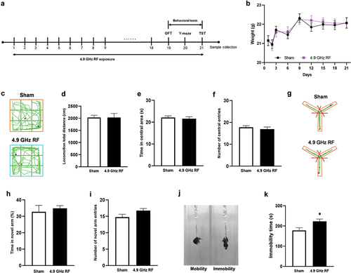
The anxiety-like behaviour of mice was evaluated by OFT (). The results showed that compared with Sham group, the accumulative total distance (), total time spent in central area () and central area entries () remained unchanged in 4.9 GHz RF group (P = 0.998, P = 0.631, P = 0.517, respectively), which indicated that 4.9 GHz RF exposure had no obvious effect on anxiety-like behaviour in mice.
The spatial memory ability of mice was evaluated by Y-maze (). The results showed that compared with Sham group, the time spending in novel arm (%) () and number of novel arm entries did not change in 4.9 GHz RF group (, P = 0.627, P = 0.080, respectively), which indicated that 4.9 GHz RF exposure could not affect spatial learning and memory ability of mice.
The immobility status was recorded () in TST. The results showed that compared with Sham group, 4.9 GHz RF exposure significantly increased the immobility time of mice (, P = 0.018), suggesting that 4.9 GHz RF exposure could induce depression-like behaviour of mice.
The effects of 4.9 GHz RF on the morphology and histology of brain in mice
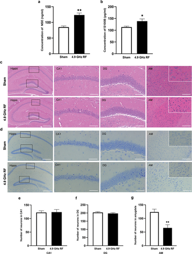
The results of ELISA showed that compared with Sham group, 4.9 GHz RF exposure significantly increased the levels of NSE and S100B concentrations in mice (, B, P < 0.01 and P < 0.05, respectively). As is shown in , no obvious morphological change was found in hippocampus between Sham group and RF group. However, karyopyknosis, disordered cells arrangement and necrosis were found in the amygdala of 4.9 GHz RF group. The results of Nissl staining () showed that the number of neurons in hippocampal CA1 and DG regions did not change (, P = 0.916 and P = 0.497, respectively) compared with Sham group. While the number of neurons in amygdala of 4.9 GHz RF exposure group significantly reduced (, P < 0.01). The above results indicated that 4.9 GHz RF exposure had no obvious effects on the histology and morphology of hippocampus, while it could induce the structural damage and neuronal loss in amygdala.
Figure 2. The level of brain injury after exposure of 4.9 GHz RF. (a) The level of NSE concentration in serum. (b) The level of S100B concentration in serum. n = 10 for each group. (c) Representative images of HE staining in hippocampus (Hippo) and amygdala (AM). (d) Representative images of Nissl staining in Hippo and AM. Scale bar = 100 μm for Hippo; scale bar = 50 μm for CA1 and DG regions; scale bar = 200 μm for AM. (e) The number of neurons in CA1. (f) The number of neurons in DG. (g) The number of neurons in amygdala. All data are presented as mean ± SEM. *P <0.05, **P <0.01.
The effects of 4.9 GHz RF on cell apoptosis in the hippocampus and amygdala
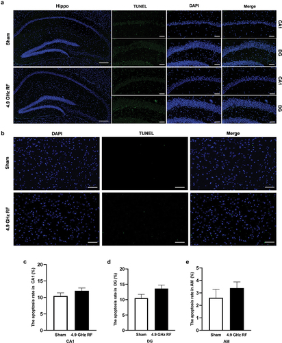
The results of TUNEL staining in hippocampus and amygdala were shown in . The apoptosis rate in hippocampus and amygdala slightly increased in 4.9 GHz RF group, while the differences were not statistically significant, compared with Sham group (, P = 0.236 and 0.082 for CA1 and DG, respectively; P = 0.410 for AM). The results suggested that 4.9 GHz RF exposure did not induce cell apoptosis in hippocampus and amygdala.
Figure 3. The effects of 4.9 GHz RF on the levels of cell apoptosis in hippocampus (Hippo) and amygdala (AM). (a) Representative images of TUNEL staining in hippocampus. Scale bar = 100 μm for Hippo, scale bar = 50 μm for CA1 and DG regions. (b) Representative images of TUNEL staining in amygdala. Scale bar = 100 μm. (c) The apoptosis rate in hippocampal CA1 region. (d) The apoptosis rate in hippocampal DG region. (e) The apoptosis rate in amygdala. All data are presented as mean ± SEM.
The effects of 4.9 GHz RF on the levels of cell pyroptosis in hippocampus and amygdala
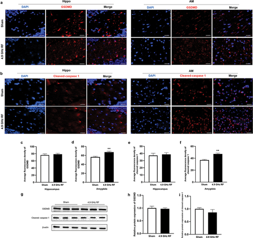
We next examined the level of cell pyroptosis by immunofluorescence staining in hippocampus and amygdala. The immunoreactivity of Gasdermin D (GSDMD) and Cleaved caspase-1 were shown in . Compared with Sham group, the average fluorescence density of GSDMD and Cleaved caspase-1 in hippocampus did not change in 4.9 GHz RF group (, P = 0.370 and P = 0.736, respectively), which was consistent with the results of western blot (, P = 0.205 and P = 0.135, respectively). These results indicated that 4.9 GHz exposure could not induce pyroptosis in hippocampus. In addition, the average fluorescence density of GSDMD and Cleaved caspase-1 in amygdala significantly increased in 4.9 GHz RF group, compared with Sham group ( P < 0.01). These results above suggested that 4.9 GHz RF exposure for 21 d may induce neuronal death in amygdala through cell pyroptosis.
Figure 4. The effects of exposure to 4.9 GHz RF on pyroptosis in hippocampus (Hippo) and amygdala (AM). (a) Representative images of immunofluorescence staining of GSDMD in Hippo and AM. (b) Representative images of immunofluorescence staining of Cleaved caspase-1 in Hippo and AM. Scale bar = 50 μm for Hippo, scale bar = 100 μm for AM. (c, e) Average fluorescence density of GSDMD and Cleaved caspase-1 in hippocampus. (d, f) Average fluorescence density of GSDMD and Cleaved caspase-1 in amygdala. n = 3 for each group. (g) Representative western blot results of GSDMD and Cleaved caspase 1 in hippocampus. n = 3 for each group. (h) Relative protein level of GSDMD. (i) Relative protein level of Cleaved caspase-1. All data are presented as mean ± SEM. **P <0.01.
Discussion
With the popularity of 5 G network communication, the effects of RF-EMR exposure from 5 G telecommunication on brain function has become a major public concern (Zhang et al. Citation2017a; Russell Citation2018; Simkó and Mattsson Citation2019; Kostoff et al. Citation2020). 4.9 GHz RF is one of commonly used 5 G communication frequencies in China, in this study, based on literature and mobile phone usage reality, we investigated long-term 4.9 GHz RF exposure (1 hour/day for 21 d consecutively) on emotional behaviours and spatial memory in mice.
The results of OFT showed that after exposure to 4.9 GHz RF for 21 d, the cumulative time and number of entries in central area did not change, which indicated that under this experimental condition, 4.9 GHz RF exposure could not induce anxiety-like behaviour. This result was consistent with Júnior’s result (Júnior et al. Citation2014), they found that exposure to 1.8 GHz RF emitted by mobile phone for 3 days (the RF originated from 25-second-long phone calls, every 2 min), no anxiety patterns were observed in exposed rats. Another study (Petitdant et al. Citation2016) also revealed that after exposing young rats to 900 MHz RF (SAR: 0, 1.5, and 6 W/kg, 45 min/d) for 30 days, no significant changes were found in novelty perception and anxiety-like behaviour compared with Sham groups. However, Obajuluwa et al (Obajuluwa et al. Citation2017) reported that there was an increase in anxiety-like behaviours of rats after exposure to 2.5 GHz RF for a period of 4, 6 and 8 weeks. Similarly, Varghese et al (Varghese et al. Citation2018) found that adult female rats showed increased anxiety-like behaviours after exposure to 2.45 GHz RF (PD: 7.88 W/m2) for 4 h/d for 45 days. Although our results showed that 4.9 GHz RF exposure had no obvious effects on anxiety-like behaviours, it could induce depression-like behaviour in mice. Previously, Zhang et al (Zhang et al. Citation2017b) reported that depression-like behaviour of mice did not change after 4-week exposure to 1.8 GHz RF field (SAR: 2.7 W/kg for whole body and 2.2 W/kg for brain; PD: 530 µW/cm2). Regarding the inconsistency, it is noted that the present study and those studies used discrepant species of animals with different parameters of RF, which may give rise to diverse outcomes.
Y-maze is a hippocampal-dependent task which is widely used to evaluate spatial memory in mice. Our results showed that long-term 4.9 GHz RF exposure had no adverse effect on spatial memory in mice, which was consistent with Daniels and Keleş et al report (Daniels et al. Citation2009; Keleş et al. Citation2018). However, inconsistent findings were also reported. Gupta et al (Gupta et al. Citation2018) found that exposure to 2450 MHz RF for 28 consecutive days caused cognitive deficiency in male rats. Interestingly, Tafakori et al (Tafakori et al. Citation2020) revealed that short-term RF could increase working memory in rats, while it was temporary and would reduce by one-week rest, and gradually decreased to initial level. Therefore, based on the discussion above, the effects of RF on spatial learning and memory remain inconsistent, further investigations are needed.
S100B and NSE are two important biomarkers for central nervous system (CNS) (Missler et al. Citation1997). When neuronal cells are damaged, the concentrations of S100B and NSE are detected to be increased in serum (Yu et al. Citation2020). To explore the potential mechanism of 4.9 GHz RF induced emotional behaviour change, the levels of S100B and NSE in serum were detected. It was found that 4.9 GHz RF exposure significantly increased the levels of S100B and NSE in mice serum, which indicated that 4.9 GHz RF exposure could cause brain injury. It was proved that hippocampus and amygdala are associated with emotionality in mice (Janak and Tye Citation2015). Therefore, we next observed the histology of hippocampus and amygdala. It was found that 4.9 GHz RF resulted in the alteration of morphology of amygdala, rather than hippocampus, meanwhile, the number of neurons in amygdala significantly reduced, which suggested that amygdala was involved in 4.9 GHz RF induced emotional behaviour change. Previously, Hong et al (Hong et al. Citation2020) reported that exposure to 800–1900 MHz RF for 19 days did not affect hippocampal architecture in pregnancy rats. However, Narayanan et al (Narayanan et al. Citation2018) revealed that after exposing rats to 900 MHz radiation (1 h/day, SAR: 1.15 W/kg) for 28 d, a decrease of neurons in amygdala was found, which was consistent to this study.
To investigate the manner of 4.9 GHz RF induced neuronal death in amygdala, we then detected the level of apoptosis and pyroptosis in amygdala and hippocampus. It was found that 4.9 GHz RF exposure had no obvious effects on cell apoptosis in hippocampus and amygdala. Pyroptosis, differs from necrosis and apoptosis, is an inflammasome-mediated and caspase-1-dependent programmed cell death (Fu et al. Citation2019). GSDMD and Cleaved caspase-1 are widely used as biomarkers of pyroptosis. Our results showed that the fluorescence density of GSDMD and Cleaved caspase-1 increased obviously in amygdala after 4.9 GHz RF exposure, which suggested that 4.9 GHz RF could induce pyroptosis rather than apoptosis in amygdala. Furthermore, the expression levels of pyroptosis-related proteins showed no obvious difference in hippocampus. Combined the results of HE and Nissl staining, we speculated that the morphology damage and neuronal loss in amygdala were, at least partly, due to 4.9 GHz RF induced pyroptosis.
In this study, we first investigated the effects of 4.9 GHz RF from 5 G Communications on spatial memory and emotionality, and found that 4.9 GHz RF induced depression-like behaviour in mice, which might be associated with the pyroptosis in amygdala. However, this study has some limitations. For example, we did not observe whether 4.9 GHz RF induced depression-like behaviour was reversible; besides pyroptosis, is there other ways of neuronal death initiated by 4.9 GHz RF in amygdala? These remain to be further studied.
Conclusion
Our study reveals that 21-d exposure to 4.9 GHz RF may induce depression-like behaviour in mice and the pyroptosis in amygdala may be involved in it.
Disclosure statement
No potential conflict of interest was reported by the author(s).
Additional information
Funding
References
- Can A, Dao DT, Terrillion CE, Piantadosi SC, Bhat S, Gould TD. 2012. The tail suspension test. J Vis Exp. (59):e3769. doi:10.3791/3769.
- Daniels WM, Pitout IL, Afullo TJ, Mabandla MV. 2009. The effect of electromagnetic radiation in the mobile phone range on the behaviour of the rat. Metab Brain Dis. 24(4):629–641. doi:10.1007/s11011-009-9164-3.
- Dubreuil D, Jay T, Edeline JM. 2003. Head-only exposure to GSM 900-MHz electromagnetic fields does not alter rat’s memory in spatial and non-spatial tasks. Behav Brain Res. 145(1–2):51–61. doi:10.1016/S0166-4328(03)00100-1.
- Durusoy R, Hassoy H, Özkurt A, Karababa AO. 2017. Mobile phone use, school electromagnetic field levels and related symptoms: a cross-sectional survey among 2150 high school students in Izmir. Environ Health-Glob. 16(1):51. doi:10.1186/s12940-017-0257-x.
- Fu Q, Wu J, Zhou XY, Ji MH, Mao QH, Li Q, Zong MM, Zhou ZQ, Yang JJ. 2019. Nlrp3/caspase-1 pathway-induced pyroptosis mediated cognitive deficits in a mouse model of sepsis-associated encephalopathy. Inflammation. 42(1):306–318. doi:10.1007/s10753-018-0894-4.
- Gupta SK, Mesharam MK, Krishnamurthy S. 2018. Electromagnetic radiation 2450 MHz exposure causes cognition deficit with mitochondrial dysfunction and activation of intrinsic pathway of apoptosis in rats. J Biosci. 43(2):263–276. doi:10.1007/s12038-018-9744-7.
- Gupta S, Sharma RS, Singh R. 2022. Non-ionizing radiation as possible carcinogen. Int J Environ Health Res. 32(4):916–940. doi:10.1080/09603123.2020.1806212.
- Hong S, Huang H, Yang M, Wu H, Wang L. 2020. Enriched environment decreases cognitive impairment in elderly rats with prenatal mobile phone exposure. Front Aging Neurosci. 12:162. doi:10.3389/fnagi.2020.00162.
- Hu C, Zuo H, Li Y. 2021. Effects of radiofrequency electromagnetic radiation on neurotransmitters in the brain. Front Public Health. 9:691880. doi:10.3389/fpubh.2021.691880.
- IARC. 2013. Non-ionizing radiation, part 2: radiofrequency electromagnetic fields. IARC Monogr Eval Carcinog Risks Hum. 102(Pt 2):1–460.
- Janak PH, Tye KM. 2015. From circuits to behaviour in the amygdala. Nature. 517(7534):284–292. doi:10.1038/nature14188.
- Júnior LC, Guimarães ES, Musso CM, Stabler CT, Garcia RM, Mourão-Júnior CA, Andreazzi AE. 2014. Behaviour and memory evaluation of Wistar rats exposed to 1·8 GHz radiofrequency electromagnetic radiation. Neurol Res. 36(9):800–803. doi:10.1179/1743132813Y.0000000276.
- Keleş A0, Yıldırım M, Ö G, ÇolakoğluS, Kaya H, Baş O, Sönmez OF, Odacı E. 2018. The effects of a continuous 1-h a day 900-MHz electromagnetic field applied throughout early and mid-adolescence on hippocampus morphology and learning behaviour in late adolescent male rats. J Chem Neuroanat. 94:46–53. doi:10.1016/j.jchemneu.2018.08.006.
- Kostoff RN, Heroux P, Aschner M, Tsatsakis A. 2020. Adverse health effects of 5G mobile networking technology under real-life conditions. Toxicol Lett. 323:35–40. doi:10.1016/j.toxlet.2020.01.020.
- Kraeuter A, Guest PC, Sarnyai Z. 2018a. The open field test for measuring locomotor activity and anxiety-like behaviour. New York, NY: Springer New York; pp. 99–103. doi:10.1007/978-1-4939-8994-2_9.
- Kraeuter A, Guest PC, Sarnyai Z. 2018b. The Y-maze for assessment of spatial working and reference memory in mice. New York, NY: Springer New York; pp. 105–111. doi:10.1007/978-1-4939-8994-2_10.
- Liu LY, Qin TZ, Guo L, Rong-Rong H, Jing YT, Lai PP, Xue YZ, Ding GR. 2022. The preventive and therapeutic effect of repetitive transcranial magnetic stimulation on radiation-induced brain injury in mice. Int J Radiat Biol. 98(8):1–14. doi:10.1080/09553002.2022.2038806.
- Missler U, Wiesmann M, Friedrich C, Kaps M. 1997. S-100 protein and neuron-specific enolase concentrations in blood as indicators of infarction volume and prognosis in acute ischemic stroke. Stroke. 28(10):1956–1960. doi:10.1161/01.STR.28.10.1956.
- Narayanan SN, Kumar RS, Paval J, Kedage V, Bhat MS, Nayak S, Bhat PG. 2013. Analysis of emotionality and locomotion in radio-frequency electromagnetic radiation exposed rats. Neurol Sci. 34(7):1117–1124. doi:10.1007/s10072-012-1189-4.
- Narayanan SN, Mohapatra N, John P, N K, Kumar RS, Nayak SB, Bhat PG. 2018. Radiofrequency electromagnetic radiation exposure effects on amygdala morphology, place preference behaviour and brain caspase-3 activity in rats. Environ Toxicol Pharmacol. 58:220–229. doi:10.1016/j.etap.2018.01.009.
- Nittby H, Grafström G, Tian DP, Malmgren L, Brun A, Persson BR, Salford LG, Eberhardt J. 2008. Cognitive impairment in rats after long-term exposure to GSM-900 mobile phone radiation. Bioelectromagnetics. 29(3):219–232. doi:10.1002/bem.20386.
- Obajuluwa AO, Akinyemi AJ, Afolabi OB, Adekoya K, Sanya JO, Ishola AO. 2017. Exposure to radio-frequency electromagnetic waves alters acetylcholinesterase gene expression, exploratory and motor coordination-linked behaviour in male rats. Toxicol Rep. 4:530–534. doi:10.1016/j.toxrep.2017.09.007.
- Pareja-Peña F, Burgos-Molina AM, Sendra-Portero F, Ruiz-Gómez MJ. 2022. Evidences of the (400 MHz - 3 GHz) radiofrequency electromagnetic field influence on brain tumor induction. Int J Environ Health Res. 32(1):121–130. doi:10.1080/09603123.2020.1738352.
- Petitdant N, Lecomte A, Robidel F, Gamez C, Blazy K, Villégier AS. 2016. Cerebral radiofrequency exposures during adolescence: impact on astrocytes and brain functions in healthy and pathologic rat models. Bioelectromagnetics. 37(5):338–350. doi:10.1002/bem.21986.
- Russell CL. 2018. 5 G wireless telecommunications expansion: public health and environmental implications. Environ Res. 165:484–495. doi:10.1016/j.envres.2018.01.016.
- Shehu A, Mohammed A, Magaji RA, Muhammad MS. 2016. Exposure to mobile phone electromagnetic field radiation, ringtone and vibration affects anxiety-like behaviour and oxidative stress biomarkers in albino Wistar rats. Metab Brain Dis. 31(2):355–362. doi:10.1007/s11011-015-9758-x.
- Simkó M, Mattsson MO. 2019. 5G wireless communication and health effects—a pragmatic review based on available studies regarding 6 to 100 GHz. Int J Environ Res Public Health. 16(18):3406. doi:10.3390/ijerph16183406.
- Tafakori S, Farrokhi A, Shalchyan V, Daliri MR. 2020. Investigating the impact of mobile range electromagnetic radiation on the medial prefrontal cortex of the rat during working memory. Behav Brain Res. 391:112703. doi:10.1016/j.bbr.2020.112703.
- Varghese R, Majumdar A, Kumar G, Shukla A. 2018. Rats exposed to 2.45 GHz of non-ionizing radiation exhibit behavioural changes with increased brain expression of apoptotic caspase 3. Pathophysiology. 25(1):19–30. doi:10.1016/j.pathophys.2017.11.001.
- Yu D, Liu B, Jiang G, Pei S, Pan H. 2020. Correlation of changes in serum S100β, NSE and inflammatory factor levels with MMSE and MoCA in intracranial tumor patients with cognitive impairment. Oncol Lett. 20(2):1968–1972. doi:10.3892/ol.2020.11751.
- Zhang J, Sumich A, Wang GY. 2017a. Acute effects of radiofrequency electromagnetic field emitted by mobile phone on brain function. Bioelectromagnetics. 38(5):329–338. doi:10.1002/bem.22052.
- Zhang JP, Zhang KY, Guo L, Chen QL, Gao P, Wang T, Li J, Guo GZ, Ding GR. 2017b. Effects of 1.8 GHz radiofrequency fields on the emotional behaviour and spatial memory of adolescent mice. Int J Environ Res Public Health. 14(11):1344. doi:10.3390/ijerph14111344.