Abstract
The freshwater microalga Haematococcus pluvialis exhibits a unique morphological response to environmental stress, accumulating carotenoid pigment during encystment. The complexity of characterizing the different cell stages and monitoring the pigment cell content during the life cycle of this microalga is one of the main problems reported when assessing astaxanthin accumulation and degradation. Therefore, with the aim of studying the potential encystment response in this microalga by means of flow cytometry (FCM), we induced oxidative stress in cultures of vegetative growing cells by treating them with paraquat, a known generator of superoxide anion radicals. Two flow cytometric approaches were successfully used to monitor the effect of oxidative stress on morphological changes and genesis of carotenoids in H. pluvialis: (1) a cytometric characterization of different cell types based on analysis of the fluorescence of chlorophyll a vs the fluorescence of astaxanthin, and (2) staining with the fluorochromes hydroethidium (HE) and dihydrorhodamine 123 (DHR), in order to measure the in vivo intracellular levels of reactive oxygen species (ROS). FCM data showed that astaxanthin accumulation during encystment hampers the production of ROS. Furthermore, the cell content of astaxanthin seems to be a good indicator of the extent to which H. pluvialis cells undergo oxidative stress, and also of how the cells defend themselves under stress conditions.
Introduction
Astaxanthin (3,3′-dihydroxy-β,β-carotene-4,4′-dione), a secondary carotenoid, is commonly used as a pigmentation source for marine fish aquaculture and has potential pharmaceutical, cosmetic, food, and feed applications (Jyonouchi et al., Citation1991; Guerin et al., Citation2003) due to its strong antioxidant activity, which is ten times higher than β-carotene and over 500 times more effective than α-tocopherol (Miki, Citation1991; Palozza & Krinsky, Citation1992). Furthermore, astaxanthin has been found to have many essential biological functions, including protection against lipid-membrane peroxidation of essential polyunsaturated fatty acids and proteins, DNA damage and ultraviolet light effects (Savoure et al., Citation1995). The search for natural sources for the production of the astaxanthin has been focused mainly on the freshwater unicellular alga Haematococcus pluvialis and the yeast Phaffia rhodozyma (Johnson & An, Citation1991).
Haematococcus pluvialis exhibits a morphological response to environmental stress, stimulating the synthesis of astaxanthin during encystment (Goodwin & Jamikorn, Citation1954; Droop, Citation1955). The complexity of characterizing the different cell stages and monitoring the pigment cell content during the life cycle of this microalga is one of the main problems reported when assessing astaxanthin accumulation and degradation, especially when cultures are not fully synchronized and flagellated and cyst cells are present simultaneously in the same culture (Fábregas et al., Citation2003). Although individual cells can be examined microscopically, there is no efficient method to facilitate the analysis of controversial points in astaxanthin biosynthesis, such as the phases of the cell cycle of H. pluvialis when astaxanthin is accumulated or the relationship between cell size and astaxanthin content in the cyst cells of this microalga. Previous studies have reported the successful implementation of flow cytometry (FCM) to study cell parameters in microalgae, such as size or chlorophyll a fluorescence (Cunningham & Buonnacorsi, Citation1992; Cid et al., Citation1995; González-Barreiro et al., Citation2004; Rioboo et al., Citation2009).
The aim of the present work was to develop a new FCM method to analyse changes in the cell content of chlorophyll a and astaxanthin pigments of H. pluvialis cultures, identifying the main cell types of this microalga: vegetative cells, immature cysts and mature cysts. Moreover, FCM allows the independent analysis of the different cell types and their evolution, rather than whole populations. Since reactive oxygen species (ROS) are considered to be involved in the regulation of astaxanthin synthesis in H. pluvialis (Kobayashi et al., Citation1993, Citation2001; Hu et al., Citation2008), we induced oxidative stress in cultures of vegetative growing cells by treating them with paraquat, a herbicide known to produce intracellular superoxide anion radicals, in order to study the encystment response in this microalga by means of FCM. We also determined the intracellular production of both superoxide anion and hydrogen peroxide radicals in vivo in vegetative and cyst cells of H. pluvialis.
Materials and methods
Microalgal cultures
Haematococcus pluvialis Flotow (Chlorophyceae) obtained from the Culture Collection of Algae and Protozoa of the Institute of Freshwater Ecology (Cumbria, UK) (strain CCAP-34/7) was grown on sterile Bristol medium (Brown et al., Citation1967).
All cultures were grown in sterilized Pyrex glass bottles containing 400 ml of medium. For the assays, cells growing in logarithmic phase were used as inoculum. Microalgal cultures were maintained at 18 ± 1°C, with a photon flux of 80 µmol m−2 s−1 photosynthetically active radiation and a light/dark cycle of 12:12 h; they were continuously aerated for 96 h. The initial cell density of experimental cultures was 0.10 × 10⁶ cells ml−1. Haematococcus pluvialis cultures were checked daily using an Eclipse E400 light microscope (Nikon, Japan). Photographs of cultures were taken with a cooled Nikon DS-5Mc high-definition colour camera and images were manipulated with the imaging software Nis-Elements (Nikon, Japan).
Paraquat treatment
At time zero, paraquat was added to H. pluvialis cultures at three final concentrations, 50 nM, 300 nM and 600 nM. These concentrations were selected on the basis of the effect observed on growth in previous studies carried out for 96 h, which showed that 50 nM is lower than the EC50, 300 nM is close to the EC50, and 600 nM is c. twice the EC50. Paraquat was the Riedel-de Häen Pestanal standard for environmental analysis (>99.5% purity: RdH Laborchemikalien, Seelze, Germany). Stock solutions (1 mM) were prepared by dissolving the granulated paraquat in distilled and sterilized water and filtering through 0.2 µm membrane filters. Cultures without herbicide were included as control in all experiments.
Starting from time 0 of the assays, samples were taken from each culture every 12 h for analysis, at the beginning of the period of light and at the beginning of the period of darkness. Samples were taken up to 96 h of the assay.
Flow cytometric analysis of microalgal cells
Flow cytometric analysis of H. pluvialis cells was performed using a Coulter Epics XL4 flow cytometer (Beckman Coulter, Fullerton, California, USA) equipped with an argon-ion excitation laser (blue light, 488 nm), two light scatter detectors that measured forward (FS) and side (SS) light scatter, and four fluorescence detectors that detected filtered light at 525 ± 20 nm (FL1), 575 ± 15 nm (FL2), 620 ± 15 nm (FL3) and 675 ± 25 nm (FL4) wavelengths. For each parameter, analysed data were recorded in a logarithmic scale and results were expressed as mean values obtained from histograms in arbitrary units (a.u.). At least 10 000 cells per culture were analysed. The flow rate was kept at the lowest setting (data rate, 200–300 events s−1) to avoid cell coincidence. Data were collected using listmode files and statistically analysed off-line using the SYSTEM II software (Beckman Coulter). Fluorescent staining of the microalgal cells was always verified at 200× magnification using an Eclipse E400 epifluorescence microscope (Nikon, Japan), with excitation by blue light provided from a mercury lamp.
Analysis of H. pluvialis subpopulations: chlorophyll-rich and astaxanthin-rich cells
Photosynthetic single-cell microorganisms generally show a very strong natural fluorescence due to the presence of chlorophyll and/or other pigments, such as carotenoids (Cunningham, Citation1993). In H. pluvialis cells, FCM signals gathered in the FL4 and FL2 detectors correspond to red (FL4, 675 ± 25 nm) and yellow (FL2, 575 ± 15 nm) fluorescences, emitted by chlorophyll a and astaxanthin, respectively, after being excited at 488 nm. Based on this fact, chlorophyll a and astaxanthin fluorescences were represented as biparametric histograms in order to identify chlorophyll-rich from astaxanthin-rich cell subpopulations, allowing the direct analysis of other cytometric parameters of both subpopulations separately.
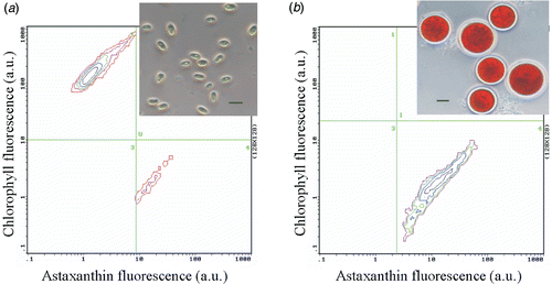
Before each experiment, vegetative cells growing in logarithmic phase and cysts from nitrogen-free cultures were used to define the acquisition settings and the regions corresponding to each subpopulation in the biparametric histograms. The instrument was set to threshold on either astaxanthin or chlorophyll regions. Cells collected in the first logarithmic decade of the FL4 detector and also in channels greater than channel 10 of the FL2 detector were considered astaxanthin-rich cells. In contrast, cells collected in the first logarithmic decade of the FL2 detector and also in channels greater than channel 10 of the FL4 detector were considered chlorophyll-rich cells ().
Fig. 1. Biparametric histograms of H. pluvialis cultures: astaxanthin fluorescence (FL2, 575 ± 15 nm; arbitrary units) vs chlorophyll a fluorescence (FL4, 675 ± 25 nm; arbitrary units). Analysis of flow cytometric data of H. pluvialis cultures after 96 h exposure to 0 nM (a) and 600 nM (b) of paraquat using chlorophyll a fluorescence to gate non-microalgal particles. Scale bar: 10 µm.
Chlorophyll a fluorescence (FL4, 675 ± 25 nm) was used as a FCM gate to ensure exclusive analysis of intact microalgal cells and exclude non-microalgal particles. This gate does not exclude microalgal cells with a low chlorophyll a content, because these cells always maintain a basal level, detectable by FCM.
Cell size
Since forward light scatter (FS) is correlated with the size or volume of a cell or particle (Shapiro, Citation1995), cell suspensions [0.10 × 10⁶ cells ml−1 in phosphate buffered saline (PBS)] were analysed by FCM to study alterations in the cell size of H. pluvialis cultures treated with paraquat.
Production of reactive oxygen species (ROS): superoxide anion radical and hydrogen peroxide
Oxidative stress was assessed from intracellular levels of ROS, including the superoxide anion radical () and hydrogen peroxide (H2O2). Intracellular ROS detection in microalgal cells was performed by FCM using dihydroethidium and dihydrorhodamine 123 probes (both from Molecular Probes, Eugene, Oregon, USA). Dihydroethidium, also called hydroethidine (HE), is able to enter into the cell, where it is selectively oxidized by superoxides into fluorescent ethidium, which is trapped by intercalating with DNA, resulting in a fluorescent signal that can be quantified by FCM. Dihydrorhodamine 123 (DHR) can be oxidized by hydrogen peroxide (H2O2) and peroxynitrite into green fluorescent compound rhodamine 123 (R123), which binds selectively to the inner mitochondrial membrane (Shapiro, Citation1995). Cell suspensions (0.10 × 10⁶ cells ml−1 in PBS) were loaded with HE (1.6 µM at final concentration) or with DHR (1.45 µM at final concentration) for 20 min at room temperature and protected from direct light. Afterwards, H. pluvialis cells were washed and fluorescence emissions from the oxidized products of HE and DHR – ethidium and rhodamine 123 – were quantified at 620 ± 15 nm (FL3) and 525 ± 20 nm (FL1), respectively. Fluorescence values were corrected by subtracting the autofluorescence of cells. In each experiment, a positive control for ethidium and rhodamine 123 detection was obtained by incubating microalgal cells for 15 min with menadione (100 µM) or H2O2 (5 mM), respectively.
Data analysis
All experiments were carried out in triplicate. The three replicates per assayed concentration of paraquat and data were analysed statistically by one-way analysis of variance (ANOVA) using the SPSS 14.0 software. When significant differences were observed, means were compared by the multiple-range Duncan test. P values ≤ 0.05 were considered statistically significant. Data are given as means ± standard errors (SE).
Results
FCM analysis allowed stages of the cell cycle of H. pluvialis cultures to be monitored at the cell level
Based on the analysis of the red fluorescence of chlorophyll a (FL4, 675 ± 25 nm) vs the yellow fluorescence of astaxanthin (FL2, 575 ± 15 nm), the development of a new cytometric protocol allowed discrimination of two cell subpopulations in all H. pluvialis cultures: a chlorophyll-rich (and astaxanthin-poor) subpopulation () and an astaxanthin-rich (and chlorophyll-poor) subpopulation (). These corresponded to flagellated vegetative cells and cyst cells, respectively, as revealed by microscopical observation.
Paraquat exposure induced the formation of astaxanthin-rich cells
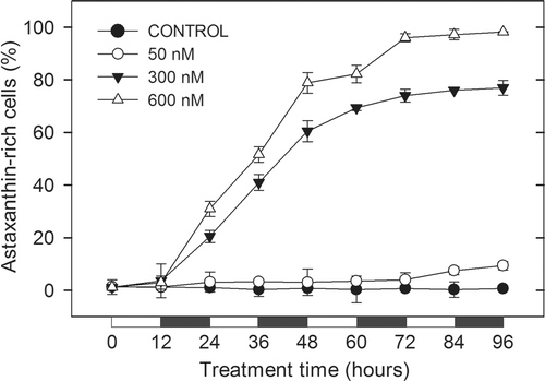
Paraquat exposure provoked an increase in the percentage of astaxanthin-rich cells, which was more acute as the concentration of the herbicide increased in the culture medium (). In control cultures, the percentage of astaxanthin-rich cells remained lower than 1% of the microalgal population throughout the 96 h of culture. Low paraquat concentration cultures, 50 nM, showed no differences in astaxanthin-rich subpopulation with respect to control cultures until 84 h of experiment, when the percentage of this cell type increased slightly to become nearly 10% at the end of the assay (). After 12 h of exposure to the medium and high paraquat assayed concentrations (300 and 600 nM), the percentage of astaxanthin-rich cells of H. pluvialis cultures increased continuously and astaxanthin-rich cells became the main subpopulation present in these cultures from 48 h to the end of the study, reaching the maximum value (99%) after 96 h of exposure to 600 nM of paraquat ().
Fig. 2. Evolution of astaxanthin-rich subpopulation analysed by FCM in cultures of H. pluvialis exposed to different concentrations of paraquat. Results are expressed as the percentage of astaxanthin-rich cells in each culture with respect to microalgal population. Black and white bars indicate dark and light periods, respectively.
Paraquat exposure provoked an accumulation of astaxanthin, an enlargement of size, and a loss of chlorophyll a content in both chlorophyll-rich and astaxanthin-rich cells
Chlorophyll-rich cells
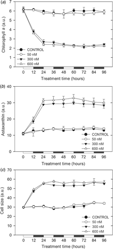
The cytometric results showed that paraquat affected both the content of chlorophyll a and astaxanthin and the cell size of chlorophyll-rich H. pluvialis (). In cultures without paraquat and those exposed to 50 nM paraquat, the cell size and the content of both chlorophyll a and astaxanthin followed a marked light/dark cycle, showing an increase in the values of the three parameters in the microalgal cells throughout the periods of light, whereas during the dark periods, the values diminished, returning to the initial basal levels ().
Fig. 3. Chlorophyll a fluorescence (a), astaxanthin fluorescence (b) and size (c), analysed by FCM of chlorophyll-rich cells in cultures of H. pluvialis exposed to different concentrations of paraquat. Black and white bars indicate dark and light periods, respectively. a.u., arbitrary units.
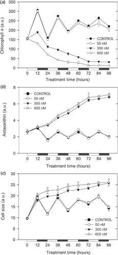
In contrast, exposure to medium and high concentrations of paraquat (300 and 600 nM) provoked a rapid drop in chlorophyll a content, as analysed by FCM (). The decrease was observed after 12 h of treatment, when 300 and 600 nM paraquat-exposed cells showed statistically lower chlorophyll a values than those observed in control cultures (P < 0.05).
Addition of medium and high concentrations of paraquat to the medium induced the accumulation of astaxanthin and an increase in size of chlorophyll-rich cells during the 96 h of exposure (, ). These changes were detected after 24 h of exposure, when 300 and 600 nM paraquat-treated cells showed statistically higher values than those observed in control cultures (P < 0.05). The maximum accumulation of astaxanthin observed in the chlorophyll-rich cells occurred after 96 h of exposure to the highest assayed concentration of paraquat.
Astaxanthin-rich cells
In control cultures and cultures exposed to a low paraquat concentration (50 nM), no significant differences were recorded, either in the cell content of chlorophyll a and astaxanthin or in the cell size of astaxanthin-rich cells during the 96 h experimental period (). However, in cultures exposed to medium and high paraquat concentrations (300 and 600 nM), the chlorophyll content of the astaxanthin-rich cells showed a rapid three-fold decrease, whereas the astaxanthin content and cell size quickly tripled and doubled, respectively. After 24 h exposure, the astaxanthin-rich cells exposed to 300 and 600 nM of paraquat showed the minimum recorded value of chlorophyll a content, while the astaxanthin content and the size of these cells reached the maximum values displayed in the present study. Beyond this point, these three parameters remained constant throughout the exposure period.
Paraquat induced oxidative stress in chlorophyll-rich H. pluvialis cells but not in astaxanthin-rich ones
Chlorophyll-rich cells
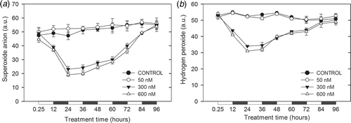
Addition of paraquat to H. pluvialis cultures affected the intracellular levels of superoxide radical and hydrogen peroxide in a similar pattern (). Intracellular levels of both ROS analysed showed a marked light/dark cycle in chlorophyll-rich cells during the 96 h of exposure, except in cultures exposed to medium and high paraquat assayed concentrations, 300 and 600 nM (). This light/dark cycle was characterized by an increase in the generation of both ROS at the beginning of the light period and a decrease in both at the beginning of the dark period. A rapid increase in the production of both ROS was recorded in chlorophyll-rich cells after only 15 min of exposure to paraquat, when all the herbicide-treated cells showed statistically higher values than those observed in control cells (P < 0.05) ().
Fig. 5. Intracellular generation of superoxide radical (a) and hydrogen peroxide (b), analysed by FCM of chlorophyll-rich cells in cultures of H. pluvialis exposed to different concentrations of paraquat. Black and white bars indicate dark and light periods, respectively. a.u., arbitrary units.
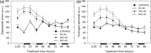
The maximum levels of oxidative stress observed in the present study were detected (for both ROS) after 12 h of exposure in chlorophyll-rich cells exposed to 600 nM of paraquat (). During the subsequent 12 h of treatment, oxidative stress exhibited by chlorophyll-rich cells exposed to medium and high paraquat concentrations remained constant. Nevertheless, after 24 h of paraquat exposure, a significant decrease in the intracellular production of both ROS was observed in microalgal cells treated with 300 and 600 nM concentrations of paraquat, compared with control chlorophyll-rich cells at the end of the treatment period (P < 0.05).
Astaxanthin-rich cells
After 15 min exposure, no significant oxidative stress alterations were detected in astaxanthin-rich cells as compared with control cultures, even at the high concentration of paraquat (600 nM) (; P > 0.05). In fact, in both non-exposed astaxanthin-rich cells and those exposed to 50 nM paraquat, no significant differences (P > 0.05) were observed in the production of ROS during the 96 h of treatment. However, the intracellular generation of ROS in astaxanthin-rich cells exposed to the medium and high assayed concentrations of paraquat, 300 and 600 nM, decreased significantly during the first 24 h of exposure (P < 0.05); this drop was especially marked in the levels of superoxide radical (). Then the ROS production increased until the end of the assay; after 96 h exposure to paraquat, astaxanthin-rich cells exhibited no significant differences in the levels of ROS with respect to control cells (P > 0.05).
Discussion
The results reported here demonstrate a new cytometric protocol to identify the cell types of H. pluvialis cultures present during the encystment process, based on the analysis of a set of intrinsic cellular parameters such as size and complexity, and the chlorophyll and astaxanthin fluorescences. Two main subpopulations were detected by FCM in microalgal cultures (). Cultures grown under favourable conditions predominantly comprised chlorophyll-rich cells, with just a few astaxanthin-rich cells, corresponding to a situation of fast and active cell growth and division (Del Río et al., Citation2005). When paraquat was added to growing vegetative cultures, chlorophyll-rich cells stopped dividing and underwent remarkable morphological and biochemical changes, leading to the formation of red cyst cells (), as several previous studies have reported (see review by Kobayashi, Citation2003). Since, in most of the cases, astaxanthin accumulation was accompanied by inhibition of cell division, it is a general belief that a decrease or cessation of cell division is imperative for astaxanthin accumulation in Haematococcus (Droop, Citation1955; Borowitzka et al., Citation1991; Boussiba, Citation2000; Orosa et al., Citation2001), but the accumulation of astaxanthin in actively growing cells has also been demonstrated (Lee & Soh, Citation1991; Grünewald et al., Citation1997; Del Río et al., Citation2005). In the present work, no astaxanthin-rich growing cells were observed, probably due to the strong and continued oxidative stress induced by paraquat in H. pluvialis cells.
The higher the assayed concentration of paraquat, the higher the percentage of vegetative cells converted to cyst cells. Nevertheless, no significant differences in the rates of chlorophyll-rich subpopulation transformation were detected when microalgal cells were exposed to 50 nM of paraquat. It is well known that ROS can cause damage to cellular constituents or metabolic processes, e.g. through destruction of membranes, inhibition of enzyme activity or oxidation of amino acids from proteins (e.g. Apostol et al., Citation1989; Asada, Citation1999). In order to counter the deleterious effects of ROS, cells have evolved a number of enzymatic and non-enzymatic antioxidant defence systems that repair the various types of damage and scavenge toxic ROS (Asada, Citation1994). The activity of enzymes such as superoxide dismutase or catalase as the first line of defence against ROS in green Haematococcus cells has been demonstrated (Kobayashi et al., Citation1997; Wang et al., Citation2004); this, together with the presence of low molecular-mass antioxidants, can explain the behaviour of chlorophyll-rich cells exposed to a low paraquat concentration. Nevertheless, in cells exposed to the two highest paraquat assayed concentrations, this primary defence response seems to be overwhelmed by the overproduction of ROS, leading to the active formation of cyst cells ().
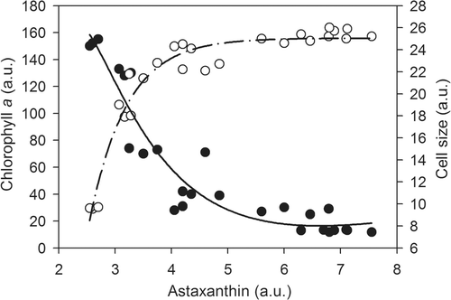
Diel oscillations in size and chlorophyll content have been described for other microalgal species in normal conditions (Rioboo et al., Citation2009). In control and 50 nM paraquat-treated H. pluvialis cells, the size, chlorophyll and astaxanthin content of vegetative cells exhibited a marked light/dark cycle (). This cycle reflects the temporal separation between growth in cell volume and cell division of vegetative cells: the growth of chlorophyll-rich mother cells was produced during the light periods (), whereas the liberation of daughter cells took place during the periods of darkness, as is generally described for microalgae (Tamiya, Citation1966; Edmunds, Citation1988; Rioboo et al., Citation2009). Cultures treated with medium and high concentrations of paraquat did not show this diel oscillation when analysed by flow cytometry (), as observed also for Chlorella vulgaris cells stressed by the herbicide terbutryn (Rioboo et al., Citation2009). Since chlorophyll is one of the main photosynthetic components, the reduction in its synthesis detected in vegetative cultures exposed to 300 and 600 nM of paraquat concentrations may lead to the overall suppression of photosynthetic activity, as reported by other authors (Borowitzka et al., Citation1991; Yong & Lee, Citation1991; Zlotnik et al., Citation1993; Lu et al., Citation1994; Tan et al., Citation1995). Furthermore, FCM analysis revealed pigment and size changes during encystment (, ). shows a correlation of both chlorophyll a content and cell size parameters with intracellular astaxanthin accumulation in chlorophyll-rich cells during the 96 h exposure to 600 nM of paraquat.
Fig. 7. Correlation between chlorophyll a content (•) or cell size (○) parameters and accumulated intracellular astaxanthin in chlorophyll-rich cells during 96 h of exposure to 600 nM of paraquat. The relation between chlorophyll content and astaxanthin content was characterized by an exponential decay function (solid line; R 2 = 0.92), whereas the relation between cell size and astaxanthin content was asymptotic, characterized by an exponential rise to maximum function (dash-dot line, R 2 = 0.95). a.u., arbitrary units.
With regard to the cyst cell subpopulation, FCM analysis of astaxanthin-rich cells allowed monitoring of the encystment process on the basis of astaxanthin accumulation, as indicated previously. At time zero of the experiments, H. pluvialis cultures showed a small subpopulation of astaxanthin-rich cells (). Exposure to medium and high concentrations of paraquat provoked the maturation of these cyst cells, which was accompanied by a three-fold increase in their cellular astaxanthin content (). Mature cysts consisted mainly of enlarged red cells, which had the highest astaxanthin content recorded in this work (). Therefore, paraquat treatment of H. pluvialis could induce not only morphogenesis from vegetative to cyst cells but also enhanced carotenogenesis in the cyst cells. In addition, since the highest astaxanthin content of mature cyst cells cannot be explained by the observed increase in cellular size alone (), astaxanthin seems to be more densely accumulated in the cytoplasm of mature cyst than in immature cyst cells, as has been described previously (Kobayashi et al., Citation2001).
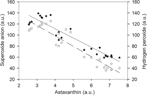
In both control and 50 nM paraquat-exposed cultures, the intracellular ROS levels of vegetative cells increased at the beginning of the light period and decreased at the beginning of the dark period (). Since it has been reported that the production of ROS from mitochondrial activity is relatively constant and independent of light/dark cycle, the diel oscillations detected in the production of ROS could be related to the photosynthetic metabolism of microalgal cells (Rhoads et al., Citation2006). Although paraquat was mostly effective in increasing superoxide anion radical levels, the two highest paraquat-assayed concentrations also provoked a severe increase in production of hydrogen peroxide (), since the superoxide anion radical can be sequentially converted to H2O2 (Elstner, Citation1982). Nevertheless, the production of both ROS in these vegetative cells began to revert to basal levels or lower after the first 24 h of exposure (); this decrease in ROS levels was closely associated with the increase in intracellular astaxanthin content and with the formation of cyst cells in these H. pluvialis cultures. Particularly, a plot of the ROS levels (generation of superoxide anion and hydrogen peroxide) against cellular astaxanthin content in chlorophyll-rich cells exposed to 600 nM of paraquat revealed a negative linear relationship (R 2 = 0.94 and R 2 = 0.89 respectively; ), indicating that astaxanthin could function as a protective agent to cope with oxidative stress. In fact, the ROS levels data in cyst cells showed that tolerance to paraquat was higher in this cell type than in vegetative cells, the maximum levels of ROS always being lower in astaxanthin-rich cells than in chlorophyll-rich ones (, ). As in chlorophyll-rich cells, the higher the induction of astaxanthin synthesis, the lower levels of ROS were detected in cyst cells exposed to paraquat. Interestingly, the ROS levels in astaxanthin-rich cells began to increase, reaching control values, when astaxanthin accumulation had ceased. This could suggest that the astaxanthin is not itself the protective antioxidant agent; rather it is the intermediates in the process of astaxanthin biosynthesis that scavenge ROS (Li et al., Citation2008).
Fig. 8. Correlation between superoxide anion (•) or hydrogen peroxide (○) levels and accumulated intracellular astaxanthin in chlorophyll-rich cells during 96 h of exposure to 600 nM of paraquat. Both are linear regressions, represented by a solid line for the superoxide anion (R 2 = 0.94) and dash-dot line for hydrogen peroxide (R 2 = 0.89). a.u., arbitrary units.
Our results indicate that astaxanthin could play a critical role in protecting cells from oxidative stress, as a defence response involving cellular accumulation of this pigment and cyst formation, as many authors have suggested before. Thus, the cell content of astaxanthin may be a good indicator of the extent to which H. pluvialis cells undergo oxidative stress, and also of how the cells defend themselves under stress conditions.
With regard to FCM methods, the approaches assayed in this work provide a rapid and precise method of monitoring the encystment process, based on quantitative measurement of the responses of individual algal cells, rather than determination of average values for whole populations. Finally, our results confirm that HE and DHR dyes are indicators of oxidative stress in H. pluvialis cells, i.e. the net formation of ROS when the capacity of cellular defence mechanisms becomes overwhelmed. Based on this fact, these fluorochromes are a powerful tool for the study of oxidative stress in microalgae under a wide range of environmental conditions.
Acknowledgements
This work was carried out with the financial support of the Consellería de Innovación e Industria, Xunta de Galicia (INCITE08ENA103032ES). The authors wish to thank the anonymous reviewers who helped improve this work.
References
- Apostol , I , Heinstein , PF and Low , PS . 1989 . Rapid stimulation of an oxidative burst during elicitation of cultured plant cells. Role in defense and signal transduction . Plant Physiol. , 90 : 109 – 116 .
- Asada , K . 1994 . “ Production and action of active oxygen species in photosynthetic tissues ” . In Causes of Photooxidative Stress and Amelioration of Defense Systems in Plants , Edited by: Foyer , CH and Mullineaux , PM . 77 – 104 . Boca Raton : CRC Press .
- Asada , K . 1999 . The water-water cycle in chloroplasts: scavenging of active oxygens and dissipation of excess photons . Ann. Rev. Plant Physiol. Plant Mol. Biol. , 50 : 601 – 639 .
- Borowitzka , MA , Huisman , JM and Osborn , A . 1991 . Cultures of the astaxanthin producing green alga Haematococcus pluvialis. 1. Effects of nutrients on growth and cell type . J. Appl. Phycol. , 3 : 295 – 304 .
- Boussiba , S . 2000 . Carotenogenesis in the green alga Haematococcus pluvialis: Cellular physiology and stress response . Physiol. Plant. , 108 : 111 – 117 .
- Brown , TE , Richardson , FL and Vaughn , ML . 1967 . Development of red pigmentation in Chlorococcum wimmeri (Chlorophyta: Chlorococcales) . Phycologia , 6 : 167 – 184 .
- Cid , A , Fidalgo , P , Herrero , C and Abalde , J . 1995 . Flow cytometry determination of acute physiological changes in a marine diatom stressed by copper . Microbiologia , 11 : 455 – 460 .
- Cunningham , A . 1993 . “ Analysis of microalgae and cyanobacteria by flow cytometry ” . In Flow Cytometry in Microbiology , Edited by: Lloyd , D . 131 – 142 . London : Springer-Verlag .
- Cunningham , A and Buonnacorsi , GA . 1992 . Narrow-angle forward light scattering from individual algal cells: implications for size and shape discrimination in flow cytometry . J. Plankton Res. , 14 : 223 – 234 .
- Del Río , E , Acién , FG , García-Malea , MC , Rivas , J , Molina-Grima , E and Guerrero , MG . 2005 . Efficient one-step production of astaxanthin by the microalga Haematococcus pluvialis in continuous culture . Biotechnol. Bioeng. , 91 : 808 – 815 .
- Droop , MR . 1955 . Carotenogenesis in Haematococcus pluvialis . Nature , 175 : 42
- Edmunds , LM Jr . 1988 . Cellular and Molecular Bases of Biological Clocks , New York : Springer-Verlag .
- Elstner , EF . 1982 . Oxygen activation and oxygen toxicity . Ann. Rev. Plant Physiol. , 33 : 73 – 96 .
- Fábregas , J , Domínguez , A , Maseda , A and Otero , A . 2003 . Interactions between irradiance and nutrient availability during astaxanthin accumulation and degradation in Haematococcus pluvialis . Appl. Microbiol. Biotechnol. , 61 : 545 – 551 .
- González-Barreiro , O , Rioboo , C , Cid , A and Herrero , C . 2004 . Atrazine-induced chlorosis in Synechococcus elongatus cells . Arch. Environ. Contam. Toxicol. , 46 : 301 – 307 .
- Goodwin , TW and Jamikorn , M . 1954 . Studies in carotenogenesis 11. Carotenoid biosynthesis in the alga Haematococcus pluvialis . Biochem. J. , 57 : 376 – 381 .
- Grünewald , K , Hagen , C and Braune , W . 1997 . Secondary carotenoid accumulation in flagellates of the green alga Haematococcus lacustris . Eur. J. Phycol. , 32 : 387 – 392 .
- Guerin , M , Huntley , ME and Olaizola , M . 2003 . Haematococcus astaxanthin: applications for human health and nutrition . Trends Biotechnol. , 21 : 210 – 216 .
- Hu , Z , Li , Y , Sommerfeld , M , Chen , F and Hu , Q . 2008 . Enhanced protection against oxidative stress in an astaxanthin-overproduction Haematococcus mutant (Chlorophyceae) . Eur. J. Phycol. , 43 : 365 – 378 .
- Johnson , EA and An , GH . 1991 . Astaxanthin from microbial sources . Crit. Rev. Biotechnol. , 11 : 297 – 326 .
- Jyonouchi , H , Hill , RJ , Tomita , Y and Good , RA . 1991 . Studies of immunomodulating actions of carotenoids. I. Effects of beta-carotene and astaxanthin on murine lymphocyte functions and cell surface marker expression in in vitro culture system . Nutr. Cancer , 16 : 93 – 105 .
- Kobayashi , M . 2003 . Astaxanthin biosynthesis enhanced by reactive oxygen species in the green alga Haematococcus pluvialis . Biotechnol. Bioproc. Eng. , 8 : 322 – 330 .
- Kobayashi , M , Kakizono , T and Nagai , S . 1993 . Enhanced carotenoid biosynthesis by oxidative stress in acetate-induced cyst cells of a green unicellular alga, Haematococcus pluvialis . Appl. Environ. Microbiol. , 59 : 867 – 873 .
- Kobayashi , M , Kakizono , T , Nishio , N , Kurimura , Y and Tsuji , T . 1997 . Antioxidant role of astaxanthin in the green alga Haematococcus pluvialis . Appl. Microbiol. Biotechnol. , 48 : 351 – 356 .
- Kobayashi , M , Katsuragi , T and Tani , Y . 2001 . Enlarged and astaxanthin-accumulating cyst cells of the green alga Haematococcus pluvialis . J. Biosci. Bioeng. , 92 : 565 – 568 .
- Lee , YK and Soh , CW . 1991 . Accumulation of astaxanthin in Haematococcus lacustris (Chlorophyta) . J. Phycol. , 27 : 575 – 577 .
- Li , Y , Sommerfeld , M , Chen , F and Hu , Q . 2008 . Consumption of oxygen by astaxanthin biosynthesis: A protective mechanism against oxidative stress in Haematococcus pluvialis (Chlorophyceae) . J. Plant Physiol. , 165 : 1783 – 1797 .
- Lu , F , Vonshak , A and Boussiba , S . 1994 . Effect of temperature and irradiance on growth of Haematococcus pluvialis (Chlorophyceae) . J. Phycol. , 30 : 829 – 833 .
- Miki , W . 1991 . Biological functions and activities of animal carotenoids . Pure Appl. Chem. , 63 : 141 – 146 .
- Orosa , M , Franqueira , D , Cid , A and Abalde , J . 2001 . Carotenoid accumulation in Haematococcus pluvialis in mixotrophic growth . Biotechnol. Lett. , 23 : 373 – 378 .
- Palozza , P and Krinsky , NI . 1992 . Astaxanthin and canthaxanthin are potent antioxidants in a membrane model . Arch. Biochem. Biophys. , 297 : 291 – 295 .
- Rhoads , DM , Umbach , AL , Subbaiah , CC and Siedow , JN . 2006 . Mitochondrial reactive oxygen species. Contribution to oxidative stress and interorganellar signaling . Plant Physiol. , 141 : 357 – 366 .
- Rioboo , C , O′Connor , JE , Prado , R , Herrero , C and Cid , A . 2009 . Cell proliferation alterations in Chlorella cells under stress conditions . Aquat. Toxicol. , 94 : 229 – 237 .
- Savoure , N , Briand , G , Amory-Touz , MC , Combre , A , Maudet , M and Nicol , M . 1995 . Vitamin A status and metabolism of cutaneous polyamines in the hairless mouse after UV irradiation: action of beta-carotene and astaxanthin . Int. J. Vitam. Nutr. Res. , 65 : 79 – 86 .
- Shapiro , HM . 1995 . Practical Flow Cytometry , New York : Wiley-Liss Inc. .
- Tamiya , H . 1966 . Synchronous cultures of algae . Annu. Rev. Plant Physiol. , 17 : 1 – 26 .
- Tan , S , Cunningham , FX Jr , Youmans , M , Grabowski , B , Sun , Z and Gantt , E . 1995 . Cytochrome f loss in astaxanthin accumulating red cells of Haematococcus pluvialis (Chlorophyceae): Comparison of photosynthetic activity, photosynthetic enzymes and thylakoid membrane polypeptides in red and green cells . J. Phycol. , 31 : 897 – 905 .
- Wang , S-B , Chen , F , Sommerfeld , M and Hu , Q . 2004 . Proteomic analysis of molecular response to oxidative stress by the green alga Haematococcus pluvialis (Chlorophyceae) . Planta , 220 : 17 – 29 .
- Yong , YYR and Lee , YK . 1991 . Do carotenoids play a photoprotective role in the cytoplasm of Haematococcus lacustris (Chlorophyta)? . J. Phycol. , 30 : 257 – 261 .
- Zlotnik , IS , Sukenik , A and Dubinsky , Z . 1993 . Physiological and photosynthetic changes during the formation of red aplanospores in the chlorophyte Haematococcus pluvialis . J. Phycol. , 29 : 463 – 469 .
