Abstract
Proline residues in transmembrane helices have been found to have important roles in the functioning of membrane proteins. Moreover, Pro residues occur with high frequency in transmembrane α-helices, as compared to α-helices for soluble proteins. Here, we report several properties of the bacteriorhodopsin mutants P50A (helix B), P91A (helix C) and P186A (helix F). Compared to wild type, strongly perturbed behaviour has been found for these mutants. In the resting state, increased hydroxylamine accessibility and altered Asp-85 pKa and light-dark adaptation were observed. On light activation, hydroxylamine accessibility was increased and proton transport activity, M formation kinetics and FTIR difference spectra of M and N intermediates showed clear distortions. On the basis of these alterations and the near identity of the crystalline structures of mutants with that of wild type, we conclude that the transmembrane proline residues of bacteriorhodopsin fulfil a dynamic role in both the resting and the light-activated states. Our results are consistent with the notion that mutation of Pro to Ala allows the helix to increase its flexibility towards the direction originally hindered by the steric clash between the ring Cγ and the carbonyl O of the i-4 residue, at the same time decreasing the mobility towards the opposite direction. Due to their properties, transmembrane Pro residues may serve as transmission elements of conformational changes during the transport process. We propose that these concepts can be extended to other transmembrane proteins.
Introduction
The role of prolines in transmembrane α helices has been a challenge for a long time Citation[1], Citation[2]. Prolines occur relatively frequently in transmembrane helices, where they are involved usually in a change of the orientation of the helix Citation[3], Citation[4]. The introduction of a steric clash with the backbone carbonyl oxygen at position i-4 and the loss of the backbone hydrogen bond may give rise to a kink or bend in the helix and to an anisotropic distribution of its flexibility (a, b) Citation[5–8]. On the other hand, due to the influence of the nitrogen atom, the ring Cδ is able to make non-conventional, low-energy C–H· · ·O hydrogen bonds with the carbonyl oxygen of the i-3 and/or i-4 residues Citation[9]. This feature may compensate in part for the lack of a conventional hydrogen bond, and may play a significant role in the transmission of conformational changes along the helix. These properties explain that Pro residues can help the helix to adopt the optimal disposition to establish important helix-helix packing interactions Citation[10]. Another possible role of Pro side chains may come from the cis-trans isomerisation of the peptidyl prolyl bonds, facilitating the movement of a part of the helix Citation[11].
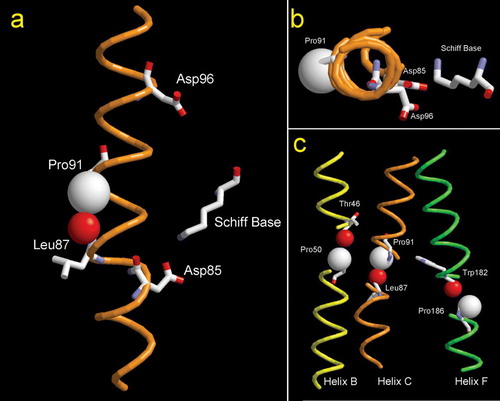
Figure 1. (A) Plot of helix C of WT bacteriorhodopsin. The two proton transporting residues (Asp85 and Asp96) are indicated, as well as the location of the Schiff base. The steric clash between the ring Cγ of Pro91 and the carbonyl O of Leu87 is evidenced by using van der Waals spheres. (B) Cytoplasmic-to-extracellular view of helix C of bacteriorhodopsin. (C) Location of transmembrane prolines in bacteriorhodopsin. Van der Wals radii of the atoms involved in the steric clash are displayed. Cγ of proline residues are in white and the peptydil O of the i-4 residue is in red (PDB code 1PY6).
Thus, proline can act as a hinge, changing the path of the transmembrane helix, and becoming an efficient mechanism in switching the conformation in transport proteins. However, at present there is still no definite answer about the relative importance of the different structural/functional roles in which Pro residues in transmembrane helices can participate: static or dynamic or both? Conflicting reports have been published in this respect. According to Nilsson et al. Citation[12], proline residues appear to have no gross conformational effects when placed centrally in the transmembrane helix. Other works emphasize a role for helix–helix interactions Citation[13], or give importance to the particular nature of the side chain Citation[14]. Recently, studies have revealed the modulation of proline kinks in α helices through Ser and Thr side chains, enhancing or attenuating the bend of the helix Citation[15].
Bacteriorhodopsin (BR) constitutes a suitable model for the analysis of the role of transmembrane proline residues for several reasons, but especially because the 3D structures of the resting state of wild type and the three transmembrane Pro mutants are known Citation[14], Citation[16]. Additionally, it is possible to perform structural and functional analyses under native conditions (purple membrane). To provide experimental evidence about the role of Pro side chains we analyze the structural and functional consequences of changing separately each of the three transmembrane Pro residues of BR to Ala. As shown in c, these residues are located in helix B (Pro-50), helix C (Pro-91) and helix F (Pro-186) and may act as hinges, facilitating the protein mobility necessary for the proton transfers. Pro-91 and Pro-186 are fully conserved among the 25 known archaeal rhodopsins Citation[17]. Some reports have already appeared studying the proline residues of BR in particular Citation[18–20]. Movements have been detected in the helices where these prolines are embedded Citation[21–23] and conformational changes during the photocycle involving prolines have been described Citation[24]. Folding experiments Citation[13] suggest that all three prolines participate in the formation of the retinal binding pocket, as deduced from altered regeneration dynamics of the apoprotein with retinal. Mutagenesis studies on E. coli-reconstituted systems showed altered proton pumping rates, particularly for Pro-186 mutants Citation[19], Citation[20]. Even though this system has been recognized to yield results that are not reproducible in the native system Citation[25], these data already suggested a contribution of transmembrane prolines to bacteriorhodopsin structure and/or function. By using the BR mutants expressed in the homologous organism we now extend the earlier studies made on the E. coli system Citation[19], Citation[20], Citation[26], Citation[27]. We show that Pro to Ala mutations induce strong reduction of transport and cause other important alterations, pointing towards a dynamic role of proline residues in transmembrane helices of BR.
Materials and methods
The construction of BR mutants P50A, P91A and P186A was performed through transformation of the plasmid pXL-NovR containing the mutated bop gene in the L33 strain of Halobacterium salinarum Citation[28]. The expression and isolation of the WT and mutant proteins were done as described in Oesterhelt and Stoeckenius Citation[29]. Protein concentration was determined spectrophotometrically at the maximum wavelength of the visible band, taking for the mutants the same extinction coefficient as wild type (WT). This was considered to be correct, since the ratio of intensities between the UV band and the visible band was the same for all four samples.
Dark-light adaptation spectra of purple membrane suspensions (1.5×10⁻⁵ M) were recorded with a Cary Bio3 spectrophotometer, using an integrating sphere when necessary. The difference spectra were obtained by subtracting dark-adapted from light-adapted samples.
pH titrations to obtain pKa values were carried out by adding micro-volumes of HCl or NaOH solutions to membrane suspensions (1.5×10⁻⁵ M BR). Absorption spectra were taken using an integrative sphere device (to minimize the loss of signal caused by light scattering) placed in a Varian Cary 3 spectrophotometer. Absorbance changes at 615 nm as a function of pH were used to monitor the purple-to-blue transition. Experimental data were normalized to the largest value at 615 nm and fitted to the Henderson-Hasselbalch equation.
Membrane suspensions (1.5×10⁻⁵ M BR) were reacted with 1 M hydroxylamine in a medium containing 150 mM sodium phosphate (pH 7.0). Reactions under light were done using white light of 300 lux of luminance.
Liposome preparation for proton pumping determination was done as described Citation[30]. After stabilization of the sample pH under dim red light at room temperature, proton pumping was initiated by illumination of the sample with yellow-filtered light of a luminance of about 8×10⁴ lux. The initial rate of proton pumping was determined by a linear fit of the pH changes within the first 15 s of illumination.
Transient pH changes in the bulk medium were followed by measuring the absorbance changes of 50 µM pyranine at 460 nm in a purple membrane suspension, in 1M KCl pH 7.2. To obtain the net absorbance changes of pyranine, the traces of samples in the absence of the dye were subtracted from those in its presence. The negative signal of ΔΔA indicates the release of protons by BR (pyranine protonation), whereas the positive signal indicates BR proton uptake. Kinetics of the M intermediate of PM suspensions in 1M KCl were followed by the acquisition of absorbance changes at 410 nm as a function of time, at pH 6.5 and pH 10.0. Time constants of M rise (τave) were obtained from the fitting of the M rise kinetics to a single or a double exponential.
FTIR experiments were performed at 277 K and 243 K with wet and dry samples. The temperature was controlled and maintained using a homemade cell holder and a cryostat. Membrane samples were suspended in 150 mM KCl and either 3 mM carbonate-bicarbonate for pH 10.0 or 3 mM sodium phosphate for pH 7.0. Preparation of membrane films and spectra acquisition was done as described Citation[31] with a Bio-Rad FTS6000 spectrometer at 2 cm−1 resolution. At least 3 cycles of 350 scans were averaged (i.e., at least 1050 interferograms were accumulated per spectrum). Difference spectra were calculated by subtracting unphotolysed BR from the corresponding photointermediate.
Distances between the Proline ring Cδ and the carbonyl oxygen of the i-3 or i-4 residue were measured on the crystal structures indicated in the Tables using the RasTop software (http://www.geneinfinity.org/rastop).
Results
Initial dark state of bacteriorhodopsin
As described in references 14 and 16, the resting state structures of P50A, P91A and P186A were found to be nearly identical to that of wild type (WT). Therefore, it is of interest to monitor the initial state and find out possible alterations of bacteriorhodopsin upon mutation of transmembrane Pro to Ala. With this objective, we compared the behaviour of the mutants with WT in the dark state. Some alterations were found for the visible absorption spectrum and their pH dependence (the purple-to-blue transition). Absorbance maxima of the three Pro mutants in the light- or dark-adapted states are somewhat shifted (see ), and show altered light-dark adaptation. Upon light adaptation, P50A shows an increase of its absorption coefficient of about 8%, whereas P186A shows 12% increase. The light-dark adaptation of P91A depends on ionic strength as evidenced by the increase of the absorption coefficient, ranging from 0% in water to about 12% in 1 M KCl (not shown). This behaviour parallels that described for the T90A mutant Citation[32].
Table I. Spectral features of bacteriorhodopsin WT and proline mutants.
The Asp85 pKa, obtained from the purple-to-blue transition, is altered by an amount depending on the mutant (). In 150 mM KCl, the P186A mutation exerts the most pronounced modification of the Asp85 pKa, 1.3 pH units, while there is an increase of one pH unit for P50A and of 0.4 pH units for P91A.
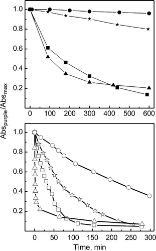
As a means to evaluate changes in the path leading to the Schiff base (SB) of BR, hydroxylamine accessibility to the Schiff base was measured in the dark. It was found to be greatly increased (see a and ). Both P91A and P50A show a fast reaction with a t1/2 of 1.3 and 3 h, respectively, whereas P186A has a t1/2 of 8 h and WT 160 h.
Figure 2. Hydroxylamine reactions. (Top) Rates of Schiff-base reaction with hydroxylamine in the absence of light recorded for WT (•) and mutants P50A (▪), P91A (▴) and P186A (★). (Bottam) Rates of Schiff-base reaction with hydroxylamine performed under illumination for WT (○), P50A (□), P91A (▵) and P186A (☆), using light of 300 lux of luminance. Plots correspond to changes of the intensity of the visible absorption band as a function of time.
The light-activated bacteriorhodopsin
The accessibility of hydroxylamine to the Schiff base was monitored under light-induced conditions (b, ). Upon illumination, P50A shows a t1/2 of 27 min, P91A 35 min and P186A 46 min, as compared to WT that has a t1/2 of 180 min. Therefore, Pro mutations increase hydroxylamine accessibility under illumination, as was observed in darkness.
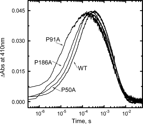
To determine possible alterations of the photocycle of the BR Pro mutants, M rise and decay kinetics were acquired by flash photolysis, at pH 6.5, 1 M KCl and room temperature. In all three mutants a rate of M formation faster than WT is apparent (), indicating a faster SB deprotonation. This is in keeping with the reported faster rate of light-induced SB deprotonation for these mutants expressed in E. coli and reconstituted into liposomes Citation[20]. Virtually no changes were observed for the M decay kinetics.
Figure 3. Kinetics of M formation and decay. Changes in absorbance at 410 nm of WT, P50A, P91A and P186A suspensions in 1M KCl, pH 6.5 at room temperature after a ns laser flash were averaged and plotted as a function of time. The time constants of M rise (τave) were: WT 60 µs, P50A 45 µs, P91A 7 µs and P186A 35 µs.
Proton uptake and release upon illumination of WT and mutant pigments in membrane sheets was estimated by using the pH indicator pyranine. shows the pyranine signal obtained at pH 7.2, after a laser flash. Compared to the WT a decrease in the release and uptake for all the three Pro mutants is apparent. P186A is the most affected mutant having about 20% of the amount of proton release and uptake compared to WT, whereas P91A shows about 35% and P50A about 70% (see ).
Figure 4. Light-induced pH changes as measured with the pH-sensitive dye pyranine. Pyranine absorption changes were measured at 460 nm in suspensions of WT, P50A, P91A and P186A in 1M KCl (pH 7.2) at 298 K as a function of time, after laser flash excitation. Proton uptake from BR causes an increase of absorbance, while proton release causes a decrease. Protein concentration, 1.5×10⁻⁵ M.
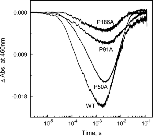
Table II. Bacteriorhodopsin relative proton pumping activity.
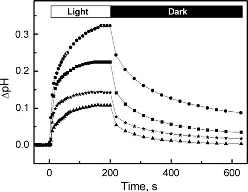
To monitor the proton pumping activity, purple membrane patches were reconstituted into liposomes Citation[19], Citation[30] and the variation of pH in the medium was recorded while illuminating the liposome suspension. Proton pumping activity is altered by the substitution of transmembrane embedded prolines (see and ). The relative proton pumping activity values agree well with pyranine experiments, except in the case of P186A where the pyranine value is about half of that obtained for the liposomes. Irrespective of the causes that may explain this difference, it is clear that the substitution of the proline side chains at the 91 or 186 positions decreases significantly the proton pumping capacity to less than 45% of WT, as measured by both pyranine and light-induced pH changes.
Figure 5. Light-induced pH changes of BR-incorporated liposomes as a function of time. The samples correspond to WT (•), P50A (▪), P91A (▴) and P186A (★). Initial pH 6.5, in KCl 150 mM.
We used Fourier transform infrared (FTIR) spectroscopy to determine differences in conformational changes at the level of the photocycle intermediates that could clarify the role of Pro residues in proton transport. To this end, we analyzed the light-induced difference spectra of membrane films under conditions that are known to drive the WT protein to M intermediate (243 K dry film, pH 10), or to N intermediate (277 K, wet film, pH 10). All three mutants showed diverse alterations in the difference spectra obtained under these conditions, as compared to WT pigment.
presents spectra obtained at 243 K, dry film, pH 10. The WT protein shows a difference spectrum corresponding to the M intermediate. In this state, major events of the transport have occurred: protonation of Asp85 by the SB, which becomes deprotonated, and release of a proton to the extracellular medium. It is characterized, among other features, by the positive peak at 1762 cm-1 due to Asp85 protonation and by a vibration of retinal at 1186 cm−1 below the baseline. Although all three Pro mutants showed the peak at 1761–1763 cm−1 of protonated Asp85, several differences were observed in the rest of the spectrum, compared to WT.
Figure 6. Fourier transform infrared difference spectra corresponding to M intermediate state minus the BR resting state. Spectra for WT, P50A, P91A and P186A were collected under M-yielding conditions (150 mM KCl, dry film, pH 10, 243 K). Spectra shown are the addition of 1050 interferograms taken at a resolution of 2 cm−1.
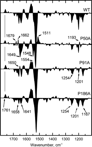
P50A shows changes in the secondary structure, as indicated by changed peaks in the amide region (). New positive peaks at 1679 and at 1662 cm−1 appear and a broad negative peak at 1649 cm−1 replaces the peaks at 1658 and 1641 cm−1. In the amide II region, new peaks appear at 1548 and 1511 cm−1. These differences indicate variations in the conformational changes accompanying formation of the M intermediate. In the retinal fingerprint region (), a small positive band at 1193 cm−1 is seen, whereas the negative peak at 1167 cm−1, corresponding to M intermediate C-C stretching vibration of retinal, is absent. Compared to WT, these changes are indicative of a severely altered retinal pocket, which in turn affects the retinal structure.
The mutant P91A shows, in the amide region, a positive peak at 1650 cm−1 and a more prominent peak at 1554 cm−1, indicative of alterations in α helices. In the retinal region, several major changes are evident. The C14-C15 all trans vibration at 1201 cm−1 is significantly decreased, the peak at 1186 cm−1 is slightly positioned above the baseline, and the peak at 1254 cm−1 appears distorted. These alterations demonstrate an altered retinal pocket. Together with the variations observed in the amide region, they indicate a changed conformation of the protein in this intermediate.
In P186A, the negative peaks at 1658 and 1641 cm−1 have less intensity, suggesting a lesser amount of protein conformational changes in this M-like intermediate, as compared to WT. The retinal region shows the three peaks at 1254, 1201 and 1167 cm−1 with similar intensities among them, whereas for the WT these peaks have very different intensities. This indicates the presence of distortions in the retinal pocket.
Under conditions yielding N intermediate (wet film, 297 K, pH 10), a state in which the SB has been reprotonated by a proton coming from Asp96, all four spectra show an overall pattern corresponding to N-like intermediate: the Asp85 peak at 1755 cm−1 and a positive peak of retinal at 1186 cm−1 (). However, like for the M intermediate, the mutants show several alterations.
Figure 7. Fourier transform infrared difference spectra corresponding to N intermediate state minus the BR resting state. Spectra for WT, P50A, P91A and P186A were collected under N-yielding conditions (150 mM KCl, wet film, pH 10, 277 K). Spectra shown are the addition of 1050 interferograms taken at a resolution of 2 cm−1.
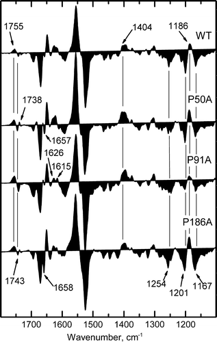
P50A presents the most similar spectrum to WT, although it has the negative band of Asp96 at 1744 cm−1 and the 1738 cm−1 band of Asp115 are more intense. In the amide region, a peak at 1657 cm−1 is seen. The retinal region shows only minor changes as compared to WT.
The spectrum of P91A shows the absence of the positive band at 1737 cm−1, corresponding to Asp115. In the amide I, the bands at 1626 cm−1 corresponding to β sheets and at 1615 cm−1 of Pro side chains Citation[24] appear slightly changed. The band at 1404 cm−1 that is decreased or absent, is of more difficult assignment. It can correspond to symmetric -COO− stretching vibrations of Asp96 Citation[33] or to Pro side chains Citation[24]. In the retinal region, a clearly decreased 1201 cm−1 negative band and changed 1186 and 1253 cm−1 bands indicate a strongly distorted retinal environment. As a whole, these changes point to the presence of variations in the protein structure.
In the carboxylic region, P186A presents a shape very similar to WT. In the amide region, an extra peak at 1658 cm−1 is evident, indicating abnormal changes of helices in this intermediate. P186A also shows an increased negative peak at 1640 cm−1. This band has been reported as corresponding in the major part to the C = N stretching vibration of protonated Schiff base. As for P91A, the peak at 1404 cm−1 is absent in P186A. The retinal fingerprint region shows that, like for the M intermediate, the three retinal peaks have similar intensity, indicating some distortions of the retinal environment.
In summary the FTIR difference spectra of M- and N-like intermediate states show important alterations in the retinal fingerprint region, accompanied by changes in the amide I and II regions corresponding mainly to protein conformational changes. Similar trends are seen in the two intermediates studied, being P91A the most affected mutant in the retinal region and P186A the least affected. Because the retinal configuration is shaped by the protein groups making contacts with it, the retinal changes reflect conformational differences of the retinal pocket induced by the Pro mutations.
Discussion
The availability of crystal structures of the Ala mutants of the three intra-helical prolines of BR (P50A Citation[14], P91A and P186A Citation[16]) provides a unique opportunity of gaining insight into the role, static or dynamic, for transmembrane prolines. As shown by Yohannan et al. Citation[16] mutation of any of the three transmembrane prolines does not eliminate the bending of the helices, neither induces noticeable conformational changes in the BR resting state. However, while the mutated proteins do not show more open or closed structures compared to WT, our results demonstrate that replacement of these proline residues cause severe alterations of a number of protein properties in both the resting and in the light-activated states. Therefore, changes in protein flexibility should be invoked to explain these changed properties.
Initial dark state of Bacteriorhodopsin
All three mutated proteins have a different behaviour in the resting state compared to WT. Hydroxylamine experiments performed in the dark argue for an increased accessibility of the Schiff base, whereas Asp85 pKa and dark-light adaptation appear altered in the mutants. How can these differences be explained in spite of the almost unchanged structures revealed by the crystals? As is known, Pro side chains can act as hinges that decouples the pre- and post-proline portions of the helix, due to the lack of a regular hydrogen bond Citation[5], Citation[7]. The resulting flexibility is anisotropic, whereas in an isolated helix, mutation of Pro to Ala will give rise to an isotropic flexibility. Thus, observing closely the environment of the mutated residues in the crystal structures (), the steric clash between a Pro ring and the carbonyl oxygen of the i-4 and/or the i-3 residue Citation[8], Citation[34] of the WT has been changed to a different interaction: Ala50 in P50A may establish a backbone hydrogen bond with Thr46, and Ala186 of P186A may form a hydrogen bond with the i-3 residue. Ala91 in P91A does not seem to be able to form new interactions. Thus, from a structural point of view, mutation of Pro to Ala does not recover a regular α-helix, but will instead change the local interactions and mobility in a helix-dependent manner and may explain the altered behaviour observed experimentally in this initial dark state.
Table III. Distances in Å between Cδi of Pro (50, 91 and 186) and the peptidyl O of the i-3 and i-4 residues of bacteriorhodopsin.
The changes may be important especially for helix C (see ) because Pro91 is oriented in such a way that its substitution with Ala will allow new motions of the helix away from the proton pathway. Because helix C carries Asp85, these fluctuations may entail perturbations of the Asp85 pKa and the SB environment, and may explain the higher accessibility of the SB to hydroxylamine. In the case of helix F, the location of Pro186 inhibits movements toward the retinal ring. Mutation to Ala may permit these movements and increase the retinal motion, especially considering that Trp182 and Trp189, which interact directly with the retinal, will increase their motion. In helix B, Pro50 limits motions towards helices A and G. The increased motions toward these helices upon mutation to Ala may increase the hydroxylamine accessibility and perturb the retinal binding pocket. It is worth to note that the inability of crystallography to detect these changes is related to the fact that crystallography yields information on the mean structures of tightly packed crystal. This tends to favour or induce certain conformations of the protein and obscure others.
The light-activated Bacteriorhodopsin
As described in Results, the decrease of proton transport efficiency of the mutants argues for strong alterations of the conformational changes associated with the transport process. This is in keeping with the increase of the hydroxylamine accessibility that occurs under illumination. Both these altered conformational changes and the higher flexibility in the path of hydroxylamine to the SB during the photocycle can be accounted for by similar reasons as those explained above for the role of Pro residues in the resting state of BR.
To get more precise information on the effects of Pro mutations on the photocycle, we analyzed the FTIR difference spectra at the level of the M- and the N-like intermediates. Results show significant alterations in the retinal fingerprinting region in all three mutants. This observation, along with other discrete changes observed in the amide region and around 1400 cm−1, indicates that the mutated Pro residues affect principally the protein structural changes at the level of the retinal pocket.
On the other hand, the influence of Pro side chains on the dynamics of the retinal pocket and thus on the transport depends on their location in relation to the retinal: Pro91 produces the largest effect whereas Pro50, which is farther, has the least effect. It is also conceivable that Pro residues, especially Pro91 and Pro186 allow transmission of the conformational changes from the retinal to the rest of the protein. Generally speaking, the absence of the transmembrane Pro can cause futile photocycles, in which the coupling between the retinal changes and proton transport is decreased due to the different pattern of mobility of helices B, C and F. This different pattern of mobility may be derived from the absence of the steric clash and the lost C-H···O hydrogen bonds ().
Conclusions
The general consequence of our results, when considering the absence of significant structural changes in the resting state of the BR mutants as compared to WT, is that proline residues in transmembrane helices have a dynamic role. On the basis of infrared Citation[35], electron diffraction Citation[23] and 13C NMR experiments Citation[36], a dynamic role for Pro-186, serving as a hinge residue, has been anticipated. With regards to Pro-91, it is worthwhile to consider the important role of the preceding residue in the helix, Thr-90, in BR function Citation[30], Citation[32]. The results about Thr-90 and Pro-91 mutants taken together emphasize the essential contribution of the middle of helix C, in terms of its precise flexibility and location in the photocycle steps, to the function and dynamics of BR. Moreover, according to crystallographic studies, the deformation of helix C during the photocycle seems to play a central role in setting the stage for proton transfer Citation[21], Citation[37]. Mutation of Pro91 may hamper this conformational change, as in the case of mutants of Thr90, decreasing the proton pumping efficiency.
An important role of transmembrane Pro residues has also been proposed for the family of hepta-helical G-protein coupled receptors, implicating these residues in the functional mechanism of signal transduction Citation[5], Citation[38], Citation[39]. Therefore, our results give strong support to the important role of Pro residues in this receptor family. As for the BR function, the involvement of Pro side chains in the dynamics of the protein segments defining the substrate pocket can be essential.
More generally, considering a protein as a dynamic system in which the major part of interactions are transient in nature, the relevance of a proline located in a transmembrane helix will not be given by the kink it induces in the resting state, but by its environment. That is, a Pro side chain present in a non-kinked transmembrane helix may develop its dynamic role during the conformational changes involved in the function, without the need of a kink to be observed in the resting state.
An important corollary of this work that can be extended to other membrane proteins containing Pro residues in their transmembrane helices is that even that crystal structures of wild type and mutated proteins appear the same, caution should be taken in deriving conclusions about their function. That is, the mutation will alter the helix flexibility or the capacity to establish transient interactions, but the protein may still have the same structure in the initial state. Therefore, direct information about function and structural dynamics should be gained to assess if a Pro replacement leads to functional impairment or not.
This paper was first published online on prEview on 27 January 2006.
This work was supported by grants BMC2003-04941 from the Dirección General de Investigación (MCYT), FP2000-6326 from the Secretaría de Estado de Educación y Universidades, MECYD and 2001SGR-00197 from the Direcció General de Recerca, DURSI. We thank Drs Tzvetana Lazarova, Josep Cladera and Leonardo Pardo for helpful discussions and suggestions, and Elodia Serrano and Alex Fernández for their skilful technical assistance.
References
- Bywater RP, Thomas D, Vriend G. A sequence and structural study of transmembrane helices. J Comput Aided Mol Des 2001; 15: 533–552
- Slepkov ER, Chow S, Lemieux MJ, Fliegel L. Proline residues in transmembrane segment IV are critical for activity, expression and targeting of the Na + /H+ exchanger isoform 1. Biochem J 2004; 379: 31–38
- Woolfson DN, Mortishire-Smith RJ, Williams DH. Conserved positioning of proline residues in membrane-spanning helices of ion-channel proteins. Biochem Biophys Res Commun 1991; 175: 733–737
- Cordes FS, Bright JN, Sansom MSP. Proline-induced distortions of transmembrane helices. J Mol Biol 2002; 323: 951–960
- Sansom MS, Weinstein H. Hinges, swivels and switches: the role of prolines in signalling via transmembrane alpha-helices. Trends Pharmacol Sci 2000; 21: 445–451
- von Heijne G. Proline kinks in transmembrane alpha-helices. J Mol Biol 1991; 218: 499–503
- Reiersen H, Rees AR. The hunchback and its neighbours: proline as an environmental modulator. Trends Biochem Sci 2001; 26: 679–684
- Bright JN, Sansom MSP. The flexing/twirling helix: exploring the flexibility about molecular hinges formed by proline and glycine motifs in transmembrane helices. J Phys Chem B 2003; 107: 627–636
- Chakrabarti P, Chakrabarti S. C–H · · · O hydrogen bond involving proline residues in alpha-helices. J Mol Biol 1998; 284: 867–873
- Orzáez M, Salgado J, Giménez-Giner A, Pérez-Payá E, Mingarro I. Influence of proline residues in transmembrane helix packing. J Mol Biol 2004; 335: 631–640
- Bruns K, Fossen T, Wray V, Henklein P, Tessmer U, Schubert U. Structural characterization of the HIV-1 Vpr N terminus: evidence of cis/trans-proline isomerism. J Biol Chem 2003; 278: 43188–43201
- Nilsson I, von Heijne G. Breaking the camel's back: proline-induced turns in a model transmembrane helix. J Mol Biol 1998; 284: 1185–1189
- Lu H, Marti T, Booth PJ. Proline residues in transmembrane alpha helices affect the folding of bacteriorhodopsin. J Mol Biol 2001; 308: 437–446
- Faham S, Yang D, Bare E, Yohannan S, Whitelegge JP, Bowie JU. Side-chain contributions to membrane protein structure and stability. J Mol Biol 2004; 335: 297–305
- Deupi X, Olivella M, Govaerts C, Ballesteros JA, Campillo M, Pardo L. Ser and Thr residues modulate the conformation of pro-kinked transmembrane alpha-helices. Biophys J 2004; 86: 105–115
- Yohannan S, Faham S, Yang D, Whitelegge JP, Bowie JU. The evolution of transmembrane helix kinks and the structural diversity of G protein-coupled receptors. Proc Natl Acad Sci USA 2004; 101: 959–963
- Ihara K, Umemura T, Katagiri I, Kitajima-Ihara T, Sugiyama Y, Kimura Y, Mukohata Y. Evolution of the archaeal rhodopsins: evolution rate changes by gene duplication and functional differentiation. J Mol Biol 1999; 285: 163–174
- Deber CM, Sorrell BJ, Xu GY. Conformation of proline residues in bacteriorhodopsin. Biochem Biophys Res Commun 1990; 172: 862–869
- Mogi T, Stern LJ, Chao BH, Khorana HG. Structure-function studies on bacteriorhodopsin. 8. Substitutions of the membrane-embedded proline-50, proline-91, and proline-186-the effects are determined by the substituting amino-acids. J Biol Chem 1989; 264: 14192–14196
- Zhang YN, El-Sayed MA, Stern LJ, Marti T, Mogi T, Khorana HG. Effects of mutagenetic substitution of prolines on the rate of deprotonation and reprotonation of the Schiff-base during the photocycle of bacteriorhodopsin. Photochem Photobiol 1993; 57: 1027–1031
- Edman K, Royant A, Larsson G, Jacobson F, Taylor T, van der Spoel D, Landau EM, Pebay-Peyroula E, Neutze R. Deformation of helix C in the low temperature L-intermediate of bacteriorhodopsin. J Biol Chem 2004; 279: 2147–2158
- Kouyama T, Nishikawa T, Tokuhisa T, Okumura H. Crystal structure of the L intermediate of bacteriorhodopsin: evidence for vertical translocation of a water molecule during the proton pumping cycle. J Mol Biol 2004; 335: 531–546
- Subramaniam S, Henderson R. Molecular mechanism of vectorial proton translocation by bacteriorhodopsin. Nature 2000; 406: 653–657
- Gerwert K, Hess B, Engelhard M. Proline residues undergo structural-changes during proton pumping in bacteriorhodopsin. FEBS Lett 1990; 261: 449–454
- Needleman R, Chang M, Ni BF, Varo G, Fornes J, White SH, Lanyi JK. Properties of Asp212→Asn bacteriorhodopsin suggest that Asp212 and Asp85 both participate in a counterion and proton acceptor complex near the Schiff-base. J Biol Chem 1991; 266: 11478–11484
- Ahl PL, Stern LJ, During D, Mogi T, Khorana HG, Rothschild KJ. Effects of amino-acid substitutions in the F-helix of bacteriorhodopsin. Low-temperature ultraviolet – visible difference spectroscopy. J Biol Chem 1988; 263: 13594–13601
- Rothschild KJ, He YW, Mogi T, Marti T, Stern LJ, Khorana HG. Vibrational spectroscopy of bacteriorhodopsin mutants .4. Evidence for the interaction of proline-186 with the retinylidene chromophore. Biochemistry 1990; 29: 5954–5960
- Sanz C, Lazarova T, Sepulcre F, González-Moreno R, Bourdelande JL, Querol E, Padrós E. Opening the Schiff base moiety of bacteriorhodopsin by mutation of the four extracellular Glu side chains. FEBS Lett 1999; 456: 191–195
- Oesterhelt D, Stoeckenius W. Isolation of the cell membrane of Halobacterium halobium and its fractionation into red and purple membrane. Methods Enzymol 1974; 31: 667–678
- Perálvarez A, Barnadas R, Sabés M, Querol E, Padrós E. Thr90 is a key residue of the bacteriorhodopsin proton pumping mechanism. FEBS Lett 2001; 508: 399–402
- Lazarova T, Padrós E. Helical and reverse turn changes in the BR→N transition of bacteriorhodopsin. Biochemistry 1996; 35: 8354–8358
- Perálvarez-Marín A, Márquez M, Bourdelande JL, Querol E, Padrós E. Thr-90 plays a vital role in the structure and function of bacteriorhodopsin. J Biol Chem 2004; 279: 16403–16409
- Dioumaev AK, Brown LS, Needleman R, Lanyi JK. Coupling of the reisomerization of the retinal, proton uptake, and reprotonation of Asp-96 in the N photointermediate of bacteriorhodopsin. Biochemistry 2001; 40: 11308–11317
- Bright JN, Shrivastava IH, Cordes FS, Sansom MSP. Conformational dynamics of helix S6 from Shaker potassium channel: Simulation studies. Biopolymers 2002; 64: 303–313
- Ludlam CFC, Sonar S, Lee CP, Coleman M, Herzfeld J, Rajbhandary UL, Rothschild KJ. Site-directed isotope labeling and ATR-FTIR difference spectroscopy of bacteriorhodopsin: the peptide carbonyl group of Tyr-185 is structurally active during the Br→N-transition. Biochemistry 1995; 34: 2–6
- Kira A, Tanio M, Tuzi S, Saito H. Significance of low-frequency local fluctuation motions in the transmembrane B and C alpha-helices of bacteriorhodopsin, to facilitate efficient proton uptake from the cytoplasmic surface, as revealed by site-directed solid-state 13C NMR. Eur Biophys J 2004; 33: 580–588
- Takeda K, Matsui Y, Kamiya N, Adachi S, Okumura H, Kouyama T. Crystal structure of the M intermediate of bacteriorhodopsin: Allosteric structural changes mediated by sliding movement of a transmembrane helix. J Mol Biol 2004; 341: 1023–1037
- Gether U. Uncovering molecular mechanisms involved in activation of G protein-coupled receptors. Endocr Rev 2000; 21: 90–113
- Stitham J, Martin KA, Hwa J. The critical role of transmembrane prolines in human prostacyclin receptor activation. Mol Pharmacol 2002; 61: 1202–1210