Abstract
Particles or cells suspended in an appropriately designed ultrasound standing wave field can be aggregated at a node to form a single monolayer in a plane that can be interrogated microscopically. The approach is applied here to investigate the temporal development of F-actin and Cx43 distribution and of gap junctional intercellular communication in 2-D chondrocyte aggregates (monolayers) rapidly and synchronously formed and held in suspension in an ultrasound trap. Development of the F-actin cytoskeleton in the confluent single layer of ‘cuboidal’ cells forming the aggregate was completed within 1 h. Chondrocytes levitated in the trap synchronously formed functional gap junctions (as assessed by CMFDA dye transfer assays) in less than 1 h of initiation of cell-cell contact in the trap. It was shown that Cx43 gene expression was retained in isolated chondrocytes in suspension. Preincubation of cells with the protein synthesis inhibitor cycloheximide caused a six-fold decrease in Cx43 accumulation (as assessed by immunofluorescence) at the interfaces of chondrocytes in the aggregate. It is shown that the ultrasound trap provides an approach to studying the early stages of cytoskeletal and gap junction development as cells progress from physical aggregation, through molecular adhesion, to display the intracellular consequences of receptor interactions.
Introduction
The first morphological event in the differentiation of the limb skeleton is the condensation of mesenchymal cells in areas of prospective cartilage and bone formation Citation[1], Citation[2]. Cellular condensation is associated with an increase in cell-cell contacts through cell adhesion molecule interaction involving neural cadherin (N-cadherin) and neural cell adhesion molecule (NCAM) Citation[3]. As the condensations mature however, N-cadherin and NCAM expression decreases. These molecules are not found in differentiated cartilage in chondrocytes Citation[4]. Perturbation of the function of these molecules reduces chondrogenesis both in vivo and in vitro Citation[5].
Gap junctions are also important for the condensation process; they facilitate intercellular communication and the transfer of small molecules between cells. They also organize the newly forming tissue and segregate future cartilaginous elements from the surrounding connective tissues Citation[2], Citation[5–7]. Gap junctional intercellular communication (GJIC) in cartilage has been demonstrated in vivo in whole tissue sections (i.e. without differentiating between zones) of mouse and rat knee joints Citation[10] and femoral condyles of mature rabbits Citation[8]. In vitro, monolayer and micromass chondrocyte cultures have shown widespread occurrence of gap junctions Citation[11–13].
Within the limb, transcripts of the gap junction gene, connexin 43 (Cx43), are strongly expressed in the mesenchyme condensations forming cartilage Citation[1]. Connexins are also expressed in mature cartilage. For example, it has been shown that some cells within the surface zone of adult articular cartilage occur in pairs and thus are able to communicate in vivo Citation[8]. Additionally, chondrocytes re-express Cx43 in cell clusters (chondrones) that are characteristic in osteoarthritis Citation[9].
Within articular cartilage, the chondrocyte cytoskeleton comprises a dynamic three dimensional network consisting principally of the proteins actin, vimentin and tubulin that are organised into microfilaments, intermediate filaments and microtubules respectively Citation[14]. The cytoskeleton plays an important role in the physical interactions between the chondrocyte and its extracellular matrix and is involved in the process of mechanical signal transduction in articular cartilage Citation[15], as well as in the control of the chondrocyte phenotype Citation[16].
A recently developed ultrasound standing wave trap (USWT) has been used to synchronously and rapidly form and levitate 2-D cell aggregates/monolayers in suspension away from the influence of solid substrata. The trap is an ultrasound resonator where the acoustic pathlength in the cell suspension is a single half wavelength. The resonator has a pressure node plane half way through the cell suspension and parallel to the transducer. The cell trap exploits the fact that particles in general experience an axial direct acoustic radiation force when in an ultrasound standing wave field. This force drives them towards a node plane. (Cells normally go to a pressure node). In an appropriately designed field Citation[17] they then move, within that plane, to accumulate at the centre of the field (). The technique has provided insight to the role of particle/cell surface properties in determining aggregate morphology for latex particles and for cells Citation[17], Citation[18]. Application of the trap has been extended to provide data, such as the intracellular temporal progression of F-actin formation in neural cells to be extracted from a large (ca. 104) sample of cells Citation[17].
Figure 1. Schematic diagram of the temporal progression (from time zero (i) to less than one second (ii) to tens of seconds (iii)) of aggregation of suspended cells in a single pressure node half-wavelength ultrasound trap. Reproduced with permission from Citation[17]. This Figure is reproduced in colour in Molecular Membrane Biology online.
![Figure 1. Schematic diagram of the temporal progression (from time zero (i) to less than one second (ii) to tens of seconds (iii)) of aggregation of suspended cells in a single pressure node half-wavelength ultrasound trap. Reproduced with permission from Citation[17]. This Figure is reproduced in colour in Molecular Membrane Biology online.](/cms/asset/96cf7c0f-a841-41e7-99a8-6cf14b0f0ca9/imbc_a_155573_f0001_b.jpg)
In the present report we elucidate cytoskeleton formation, the distribution of Cx43 and gap junction communication in surface zone chondrocyte 2-D aggregates (cell monolayers) levitated in suspension in an ultrasound trap.
Surface zone chondrocytes were selected for study for the practical reason that they are the preferred cells in investigations of approaches to articular cartilage repair. Additionally, from a ‘biology of adhesion’ perspective, surface zone chondrocytes in vivo are least in contact with each other compared to middle and deep zone chondrocytes. They therefore offer a model system to explore cell-cell interactions in vitro in a system where cell-matrix interactions dominate in vivo.
Material and methods
Ultrasound trap
The ultrasound trap employed in the present work had four layers; a transducer (Ferroperm, Kvistgard, Denmark) nominally resonant in the thickness mode at 1.5 MHz and mounted in a radially symmetric housing, a steel layer coupling the ultrasound to a one half wavelength (λ/2 or 0.5 mm depth, where λ is the wavelength of sound in water at 1.5 MHz) aqueous layer and a reflector that provided optical access from above (). The outer diameter of the cylindrical steel body was 35 mm. The ‘sample-containing’ active area had a diameter of 18 mm. The disc transducer (12 mm diameter) was driven at 1.59 MHz. Its back electrode was etched to a 6 mm diameter circle so as to give a single central aggregate in a single half-wavelength chamber Citation[17]. The quartz glass reflector had a thickness of 1 mm (λ/4) so as to locate the single pressure node plane half way through the sample volume. The transducer drive voltage came from a function generator (HM 8138, HAMEG, Germany). The sound pressure amplitude (Po) was estimated as described by Khanna et al. Citation[19]. P0 was 0.54 MPa for the first 30 s of ultrasound initiation and subsequently reduced to 0.27 MPa for 30 s and 0.06 MPa (the level at which the acoustic radiation force just balanced the gravitational force on the aggregate and thus maintained the aggregate in suspension study) for the remaining 55 min of aggregate levitation in the trap.
Figure 2. (a) Schematic diagram of the cylindrical steel trap assembly, epi-microscope, sample loading and ultrasound generation. Its main components were a 1.5 MHz disc transducer attached to a steel acoustic coupling layer, a sample volume and a glass acoustic reflector. (b) Back view of the trap assembly.
Optical system
Observation in the direction of sound propagation (negative z-axis) was performed as described by Bazou et al. Citation[17]. The trap is shown in position on the microscope stage in . The microscope was pre-focused on the trap's pressure nodal plane. Sonication started shortly after the sample was introduced into the trap. Images were captured and processed using the analySIS 3.1 software.
Chondrocyte isolation and preparation
Chondrocytes were isolated from the surface zone (SZ) of articular cartilage of 7-day old calves by sequential digestion in 3.17 U/ml pronase for 3 h (from Streptomyces griseus; Boehnringer Mannheim, Germany) and 0.12 U/ml collagenase for 12 h (Sigma, UK) as previously described Citation[20]. The sample was introduced into the sonication trap at room temperature with a 2 ml sterile syringe (Plastipak, Becton Dickinson, UK).
Cell viability tests
Viability assays for ultrasound exposed cells were performed in situ on cells (106 cells/ml) suspended for 1 h in the trap in serum-free DMEM/Ham's F12 containing Ethidium Homodimer-1 (EthD-1) or MitoTracker Green FM as previously described for a neural cell suspension Citation[17]. The number of cells that had taken up the dye after 1 min of ultrasound exposure was compared to that after 1 h of ultrasound exposure.
Immunolabelling
Chondrocytes prepared as above, were diluted to 5×10⁶ cells/ml. An aggregate of significant size (i.e., ca. 1 mm diameter) was formed within 30 s of ultrasound exposure. Aggregates remained levitated in the trap for 1 or 60 min. They were then slowly removed from the trap using a 2 ml sterile syringe, placed on a HistoBond microscope slide (RA Lamb, UK) and fixed with 90% ethanol. The slides were then rinsed with saline. Cells were dual-labelled for F-actin and Cx43. Samples were blocked at room temperature with goat serum (DAKO, UK) at 1:20 dilution in PBS and incubated with the primary antibody (monoclonal anti-connexin 43, 5 µg/ml, Chemicon, UK) overnight at 4°C Citation[21]. After washing in PBS, samples were incubated with Alexa 594 conjugated anti-mouse IgG (5 µg/ml, Molecular Probes, Inc. Eugene, OR, USA) for 1 h. After further washing (×3), F-actin was labelled by adding 10 u/ml of Phalloidin-Alexa 488 conjugate (Molecular Probes, Inc. Eugene, OR, USA). The slides were incubated at room temperature in the dark, rinsed and mounted in Vectashield (Vector, UK). Dual-labeled slides were observed (Olympus BX61 epi-fluorescent microscope) with different filters, as appropriate for each stain. Each resulting pair of images was merged using the analysis 3.1 software.
Immunolabelling of non-sonicated chondrocytes was performed on cells (106 cells/ml) retained for 1 h in suspension in serum-free DMEM/Ham's F12 medium. A 100 µl cell suspension aliquot was placed on a Histobond slide, fixed and separately stained for Cx43 and F-actin.
RNA extraction and PCR
Slivers of surface and deep zone cartilage were removed from three metacarpal phalangeal joints of three different 7-day immature bovine calves (as for chondrocyte preparation), washed in PBS and then snap frozen in liquid nitrogen. Pooled slivers of frozen cartilage from the same joint were homogenised in the presence of 1 ml frozen TRI Reagent™ (Sigma, UK) using a Mikro-Dismembrator (B. Braun Biotech International, Melsunger, Germany), and total RNA extracted using the manufacturers recommended conditions. RNA was resuspended in DEPC-treated water, treated with DNAse1 (RQ1; Promega) and then quantified by UV spectrophotometry. Total RNA (200 ng) was reverse transcribed using standard molecular biology protocols. Primers used in this study were based on sequences for bovine glyceraldehyde-3-phosphate dehydrogenase (GAPDH; sense 5′- ATT CTG GCA AGT GGA CAT CG-3′; antisense 5′- GGG CCA TCC ACA GTC TTC TG-3′) and bovine connexin 43 (sense 5′- TGA TTT CCC AGA CGA CCA CCA-3′; antisense 5′- TCC CTC TCC ACT CGC CTA TCC-3′). The cDNA was subjected to the following PCR cycles: 94°C for 30 s, 53°C for 30 s and 72°C for 30 s for 35 cycles with each primer pair. PCR products were separated by 1% agarose gel electrophoresis containing 0.1 µg/ml ethidium bromide and visualized by exposure to UV light.
Gap junction function
Chondrocytes were prepared as above and diluted to 106 cells/ml. The sample was then aliquoted into two tubes. One sample was stained with Green CMFDA Cell Tracker Probe according to the manufacturers’ instructions (20 µM; Molecular Probes, Inc. Eugene, OR, USA), whereas the other population remained unstained. (Gap junctions are permeable to the reaction product of Green CMFDA Cell Tracker Probe Citation[22]). The two cell populations were then mixed at a ratio of 1:1 by volume. A sample was then introduced to the resonator and the aggregates formed at the pressure node were retained there for 60 min. Gap junction functionality was assessed by continuous video fluorescence microscopy. In separate experiments, to demonstrate that cell to cell dye transfer occurs via gap junctions, the gap junctional blocker 1-octanol (4 µM, Sigma, UK) was included in the mixture of the two cell populations Citation[7]. An aggregate was formed in the trap and the microscope (objective magnification ×50) was focussed on a randomly selected field of view containing approximately 200 cells (the total number of cells in an aggregate is approximately 10,000). This particular set of cells was monitored over a period of 1 h. Images were captured and processed using the analySIS 3.1 software during the 60 min period. The experiment was performed twice and the number of fluorescent cells after 60 min of ultrasound exposure was compared to the initial number of fluorescent cells in the same field of view (i.e. at 5 min following activation of the trap).
Protein synthesis inhibition
Chondrocytes (5×10⁶ cells/ml) were suspended in serum-free DMEM/Ham's F12 containing 10 µg/ml cycloheximide (CHX) Citation[23] (Sigma, UK) and incubated for 1 h at 37°C/5% CO2. Cells were then washed in fresh serum-free DMEM/Ham's F12 prior to introduction to the resonator. Aggregates were removed from the trap after 1 or 60 min of ultrasound levitation and double labeled for F-actin and Cx43. The integral intensity of F-actin and Cx43 at the cell-cell contact interface was measured at the above times for 10 randomly selected pairs of cells from each of two replicate experiments.
Results
Formation and morphology of cell monolayers
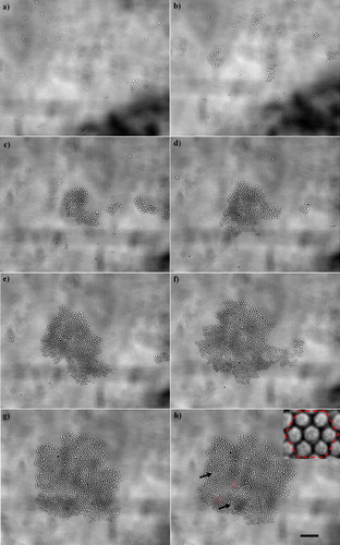
Cells isolated from the surface zone of articular calf cartilage moved into the optically focussed nodal plane of the USWT within 1 s of initiation of sonication. Single cells and some small clusters moved towards the central aggregation area after 10 s of initiation of ultrasound (a). Some small clumps (consisting of approximately 20 cells) were formed outside the field of view and moved towards the central pressure node (20 s) (b). A small aggregate had formed by 30 s (c). Incoming single cells and 2-D closely-packed small clusters rolled along the periphery of the growing aggregate and rearranged to give a closely-packed structure within 1 min of sonication (d). Aggregate growth (e–h) continued with the incorporation of further clusters over the following 4 min. The aggregate growth was complete within 5 min of initiation of ultrasound, as characterized by the absence of free single cells from the field of view. The aggregate morphology was a hexagonally ordered (h, zoom-in image) closely-packed one with only a few voids (h). Cells were exposed to ultrasound for an overall period of 1 h. Cell morphology progressed from a circular to a pentagonal form within 1 h of exposure. In situ viability assays performed with EthD-1 and MitoTracker Green FM showed that cell viability was >99% and was not significantly (p<0.05) different to that of control, non-sonicated, cells.
Figure 3. Development of a 2-D chondrocyte aggregate: (a) 10 s, (b) 20 s, (c) 30 s, (d) 1 min, (e) 2 min, (f) 3 min, (g) 4 min and (h) 5 min after ultrasound initiation. Scale bar is 100 µm. The few voids of the aggregate are shown with arrows in (h), while the hexagonal order of the aggregate is depicted in the zoom-in image shown on the top right-hand part of (h). This Figure is reproduced in colour in Molecular Membrane Biology online.
F-actin localization and Cx43 distribution
Aggregates removed from the trap after 1 min of ultrasound exposure dissociated into small 2-D clumps (typically 10–50 cells) upon deposition on a glass slide. In contrast, 1 h aggregates remained intact when placed on a slide (indicating that the mechanical strength of the aggregate had increased), but subsequently broke up into large fragments of 100 or more cells during the fixation process. Since a 2-D aggregate with a diameter of 1 mm contains approximately 10,000 cells, approximately 100 large fragments (each containing ∼100 cells) were present on every slide. Images were captured from all the fragments of the aggregate, providing guidance on the typical recurring patterns characteristic of a sample, as in and . It is noted that, while samples were removed from the trap after 1 and 60 min of levitation, the immunofluorescent images correspond to the ‘real’ times of 2.5 and 61.5 min due to the time gap (typically 1.5 min) from termination of ultrasound to specimen fixation.
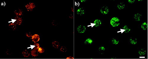
Cx43 in control (non-sonicated) cells was concentrated in finely granulated discrete regions, located mainly in the cytoplasm as shown in a (arrows). The actin cytoskeleton on the other hand, presented as an intricate network throughout the cell (b; arrows).
Figure 4. Distribution of (a) Cx43 and (b) F-actin in non-sonicated chondrocytes 1 h after their preparation. Cx43 (a) appeared highly granulated at the cytoplasm of the cells (arrows), while F-actin (b) consisted of an intricate network (arrows). Scale bar is 10 µm.
Sonicated samples were then assayed. Cx43 was distributed mainly at the periphery of the cells in the form of foci (a; arrows) after 1 min of ultrasound exposure. However, 1 h later (d; arrows) Cx43 had clearly accumulated at the cell-cell contact interface as a nearly continuous line.
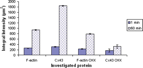
F-actin was distributed in a ring-type pattern at the cell surface following 1 min of ultrasound exposure. A thin and short region of high intensity labelling occurred at the cell-cell contact region. Some internal F-actin organisation was also observed (b; arrows). After 1 h, F-actin had clearly accumulated at the cell-cell contact interface (e; arrows). Comparing the superimposed images (c and 5f) shows that the extent of co-localization of F-actin and Cx43 at 60 min had not yet been established at 1 min. Integral intensity measurements of the phalloidin and Alexa 594 labelling of 20 randomly selected cell-cell interfaces confirmed that the amount of F-actin and Cx43 at the cell-cell contact region increased over a time exposure of 1 h (). The integral intensity of an image is defined for this purpose as the sum of all the intensities within the defined area multiplied by the pixel area Citation[17] and is used here as an indicator of change in the amount of the labeled molecule.
Figure 5. Distribution of peripheral (a) and interfacial (d) Cx43, and short (b) and long (e) interfacial F-actin in aggregates isolated from the trap after 1 (a, b) and 60 (d, e) min of ultrasound exposure respectively: (c, f) superimposed images. (g, h) distribution of Cx43 (intense cytoplasmic Cx43 pool, g) and F-actin (accumulation at the cell-cell interface, h) respectively in chondrocytes pre-incubated with 10 µg/ml CHX and levitated for 1 h in the trap. The superimposed image of g, h is shown in i. Scale bar is 10 µm.
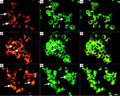
Figure 6. Measurements of the integral intensity of F-actin and Cx43 in control and cycloheximide (10 µg/ml CHX) -pretreated cells, at the cell-cell interface of 20 randomly selected pairs of cells (10 randomly selected pairs of cells from each of two replicate experiments.) The error bars represent one standard error of the mean. This Figure is reproduced in colour in Molecular Membrane Biology online.
Reverse transcriptase-PCR
Reverse transcriptase-PCR was used to detect whether Cx43 mRNA present in RNA isolated from slivers of surface and deep zone chondrocytes is transcribed. A fragment of 327 bp was detected in agarose gel electrophoresis suggestive of the presence of Cx43 mRNA product (). The presence of Cx43 mRNA was confirmed by gene sequencing.
Figure 7. RT-PCR analysis of Cx43 gene expression in immature bovine articular cartilage. cDNA was prepared from cartilage removed from the surface and deep zones of the joint and subjected to PCR using primers specific for housekeeping gene GAPDH (501 bp) and Cx43 (327 bp). The results are representative amplifications from experiments using explants taken from 3 different donors.
Functional gap junction activity
Dye transfer from CMFDA labelled cells to unlabelled cells was used as an index of functional gap junctions coupling. While stained and unstained cell suspension had been prepared on a 1:1 volume ratio fields of view containing a low initial number of stained cells were selected to highlight the spread of dye to the large number of adjacent unstained cells. The results () showed that, for cases where there were 31 and 35 CMFDA stained cells, at time 5 min, in single fields of view from repeated experiments, all (183 and 192 respectively) cells in those fields were stained after 60 min of exposure showing that there was intercellular dye transfer ().
Table I. Counts of labeled cells present in a randomly selected field of view in the presence and absence of the gap junctional blocker 1-octanol over time.
On the other hand, in the presence of the gap junctional blocker 1-octanol, the increase in the number of fluorescently-labeled cells over a time period of 60 min was small. In particular, after 5 min of sonication the number of labeled cells was 40 and 48 (from total cells per field of 197 and 214 respectively), whereas 55 min later that number had increased to 52 and 57 respectively so that most (145 and 157 respectively) of the cells were unstained ().
Protein synthesis inhibition
Chondrocytes were exposed to the protein synthesis inhibitor cycloheximide (10 µg/ml) to examine the distribution of F-actin and Cx43 upon stimulation of cell-cell contact in the ultrasound trap when synthesis of total cellular proteins has been inhibited. The qualitative microscopic patterns showing little accumulation of Cx43 at the cell-cell interface in aggregates of such cells when recovered from the trap after 1 h of ultrasound exposure (g) were quantitatively confirmed by intensity measurements of Alexa 594 staining (). g also shows intense cytoplasmic Cx43 staining (g; arrows). (The mean intensities of Cx43 and F-actin () were derived from measurements of 10 randomly selected pairs of cells from each of two replicate experiments. The variances and means of the intensity distributions of the sets of measurements were not significantly different. The mean intensity was therefore calculated from the combined set of 20 measurements).
After 1 h of levitation in the trap F-actin had accumulated at the cell-cell contact interface (h). i emphasises the co-localisation of F-actin and Cx43. The integral intensity data () show that the amount of F-actin in 1 h samples was reduced by only 1.2-fold in cycloheximide pre-treated cells compared to control non-cycloheximide pretreated cells.
Discussion
Morphology of chondrocyte aggregates formed in a USWT
The gradual change in aggregate form from an initial ordered structure of circular-profile cells to a continuous sheet of quadrilateral and pentagonal cells (typical of confluent monolayers on glass) reflects a freedom to reorganize form rapidly within the suspended monolayer where cells are not constrained by interactions with a substratum Citation[17].
F-actin redistribution
The progression, within 60 min of F-actin accumulation at the cell-cell contact interface (b, e) and increased mechanical robustness of the aggregate during the immunostaining procedures are consistent with previous results on neural cell adhesion, within 30 min, in the trap Citation[17] and with a recent report of Chu et al. Citation[24]. The latter authors showed that; (i) after 30 min F-actin had strongly accumulated at the linear interface of doublets of Sarcoma 180 (S180) cells formed in suspension, and (ii) that the force required to separate cells in a doublet formed by bringing cells, each supported by a micropipette, into contact for a period of 60 min increased rapidly over the first 30 min and more slowly thereafter to the 60 min value.
Cx43 redistribution and formation of gap junctions
The progression of Cx43 distribution from a cytoplasmic location to the plasma membrane and final concentration at the cell-cell contact interface (d) is consistent with reports of Cx43 trafficking where connexin oligomerises into connexons or hemichannels in the endoplasmic reticulum and proceed through the endoplasmic-Golgi-intermediate compartment to completion in the Golgi Citation[25]. The final destination of the connexons, via tubulovesicular networks, is the gap junctions at the cell surface Citation[26].
That formation of functional gap junctions occurred within 60 min in the present work is consistent with the ‘within 30 min’ period from de novo cell-cell contact of mouse sarcoma cells in confluent monolayer cultures Citation[27]. Distinct gap junctional complexes in human gingival fibroblasts were evident by 3 h in a donor-acceptor system Citation[28]. Transportation of Cx43 to the plasma membrane occurred within 10 min in a Cx43-Aeq chimera model in COS-7 cells cultured on coverslips Citation[29], while Lauf et al. Citation[30] reported that the phase of transportation of Cx43-GFP from the Golgi compartment to the plasma membrane occurs within 90 s in HeLa cells grown on cover glasses.
The upper limit (60 min) for F-actin cytoskeletal organization and functional gap junction formation reported here for chondrocytes is of the order of magnitude of the times reported above for these events in different cell systems. Within that agreement, the rather wide range of data for ‘rapid’ Citation[24] intracellular molecular responses to contact may reflect influences of cell type, selection of discrete time points for measurement (as in the ‘within 60 min’ of the present work), inter-laboratory variations in the end point scored, e.g. completion or initial stage of a process, and variations in physical constraints such as attachment of all (monolayer) or some (donor-receptor) interacting cells to a substratum. It is known that substratum properties can influence cell adhesion and even phenotype expression Citation[31]. A general merit of the physical trap employed here is that the interacting cells are free of the consequences of attachment to a solid substratum as they respond to contact. We note also that rapid responses have not been explored in micromass cultures that also provide, at least for many of their cells, a solid-substratum free environment. Micromass cultures are often employed as 3-D models of the prechondrogenic condensation process Citation[13–16]. Times of the order of 60 min are ‘allowed for adhesion to occur’ Citation[6] prior to disturbing the micromass by initiating molecular studies. In the particular case of chondrocytes, data on GJIC or actin cytoskeleton development have been extracted from these systems after at least 1 day in culture Citation[6], Citation[32] and at 3–6 days from the initiation of monolayer cultures Citation[10], Citation[12], Citation[33]. Our data show that functional gap junctions can be assembled in chondrocytes within 1 h. The technique described offers the potential to obtain insights to the trafficking of connexins and its control.
The possibility that ultrasound influences reactions in the trap is now considered. Coakley et al. Citation[34] have shown that the attractive Bjerkness force between cells in suspension in a pressure node plane equals the van der Waals attractive force when the membrane-membrane separation, h, is 24 nm. Since the acoustic interaction is independent of surface separation when h≪r (r is the radius of the cell) and the van der Waals interaction depends on h−2 the latter force dominates at smaller h. The van der Waals force (equals -Ar/h2, where A is the Hamaker constant and is taken as 3×10⁻²¹ J Citation[34]) at a separation of 24 nm is itself 5.8 pN. This force is much smaller that the 60 pN required to disrupt even a single cadherin-cadherin bond Citation[35] and negligible compared to the 20 nN required to separate two sarcoma S180 cells that has been in contact for just 30 s Citation[24]. It is reasonable to conclude that the increased rate of cell adhesion in the trap compared to that in micromass culture reflects increased probability of contact in the trap because the cells are held in close proximity rather than any change in receptor activation energy caused by ultrasound.
It is known that low intensity pulsed ultrasound promotes fracture healing and articular cartilage repair in vivo Citation[36] by mechanisms that remain unclear Citation[37]. Studies of these processes have included assays of chondrocyte properties following exposure to ultrasound. The time scales at which responses were monitored were typically days after ultrasound administration rather than the 1 h upper limit of the present work. For example, daily ultrasound exposures of mesenchymal stem cells in pellet culture increased aggrecan deposition and transforming growth factor beta (TGF-β)- induction of cell differentiation to chondrocytes Citation[38]. Also, chondrocytes in alginate beads exposed to ultrasound showed inhibition of collagen type X expression after 7 days Citation[36]. Since temporal changes in the F-actin distributions, similar to those reported here for chondrocytes and to those summarised above where cell contact occurs in non-ultrasound systems, have been found also in USWT neural cell adhesion Citation[17] and in ongoing USWT work here on prostate epithelial cells, it is concluded that the observed effects follow from engagement of receptors rather than from any currently unknown ultrasound mechanism.
Cx43 protein expression
Chondrocytes retained the ability to transcribe Cx43 mRNA after chondrocyte differentiation (), despite the fact that most chondrocytes within mature articular cartilage exist as individual cells not in physical contact with one another Citation[11], Citation[33]. This retention may, among other things, accommodate the recently reported Citation[8] occurrence of some pairs of chondrocytes in the surface zone of adult human articular knee and ankle cartilage Citation[39].
Blocking the production of new connexin molecules with the protein synthesis inhibitor cycloheximide resulted in a major reduction of Cx43 at the cell-cell interface as shown in g. The six-fold reduction in measured Cx43 fluorescence intensity at the cell-cell interface of cycloheximide-preincubated cells supports the immunofluorescent data. Gap junctions are subject to rapid turnover Citation[27] because of the short half-life (e.g. <1.5 h in the adult rat heart in vivo and cultured cardiac myocytes Citation[40]) of connexin molecules. In contrast to the Cx43 data, cycloheximide pretreatment resulted in only a 1.2-fold reduction in F-actin accumulation at the cell-cell interface (b). F-actin exists in a dynamic equilibrium with monomeric G-actin and has been reported to have a half-life of ∼4.5 h Citation[41]. As a result, the preexisting cytoplasmic pool of actin is sufficient to support reorganisation at the interface of cells in contact in the trap, in contrast to the situation observed for Cx43 molecules.
In conclusion, the results presented in this study showed that Cx43 gene expression is retained in isolated chondrocytes in suspension. The development of the chondrocyte F-actin cytoskeleton occurred within 60 min of initiation of cell-cell contact in the ultrasound field. This time scale is consistent with reports on F-actin redistribution in other cell systems, such as in keratinocytes Citation[42] and neural cells Citation[17]. Functional gap junction formation that had not previously been explored in chondrocytes at times less than 24 h occurred within 1 h of contact formation. It also emerges from this work that the interactions at the plasma membranes of cells in close proximity in the above physical trap transduce signals to the cytoplasm that set in train the reorganisation of F-actin at the cell interfaces, changes in cell and the development of functional gap junctions. The passive physical trap facilitation of cell contact led to accumulation of Cx43 in cells released from protein synthesis inhibition, showing that the biological consequences of the contact proceeded, as in the in vivo situation, to influencing intracellular protein trafficking. The cell-cell interactions and underlying molecular regulatory networks occurring in cartilage cells in the USWT may also be of interest in the area of scaffold-free tissue engineering approaches used to repair cartilage defects Citation[43].
We would like to thank Miss K. Richardson and Miss J. Ni Brugha for their assistance with the PCR technique.
References
- Coleman CM, Tuan RS. Functional role of growth/differentiation factor 5 in chondrogenesis of limb mesenchymal cells. Mech Develop 2003; 120: 823–836
- Hall BK, Miyake T. All for one and one for all: condensations and the initiation of skeletal development. Bioessays 2000; 22: 138–147
- Tavella S, Raffo P, Tacchetti C, Cancedda R, Castagnola P. N-CAM and N-cadherin expression during in vitro chondrogenesis. Exp Cell Res 1994; 215: 354–362
- Widelitz RB, Jiang TX, Murray BA, Chuong CM. Adhesion molecules in skeletogenesis: II. Neural cell adhesion molecules mediate precartilaginous mesenchymal condensations and enhance chondrogenesis. J Cell Physiol 1993; 156: 399–411
- DeLise AM, Tuan RS. Alterations in the spatiotemporal expression pattern and function of N-cadherin inhibit cellular condensation and chondrogenesis of limb mesenchymal cells in vitro. J Cell Biochem 2002; 87: 342–359
- Coelho CND, Kosher RA. Gap junctional communication during limb cartilage differentiation. Dev Biol 1991; 144: 47–53
- Levin M. Isolation and community: a review of the role of gap-junctional communication in embryonic patterning. J Membr Biol 2001; 185: 77–192
- Chi SS, Rattner JB, Matyas JR. Communication between paired chondrocytes in the superficial zone of articular cartilage. J Anat 2004; 205: 363–370
- Hellio LE, Graverand MP, Sciore P, Eggerer J, Rattner JP, Vignon E, Barclay L, Hart DA, Rattner JB. Formation and phenotype of cell clusters in osteoarthritic meniscus. Arthritis Rheum 2001; 44: 1808–1818
- Schwab W, Hofer A, Kasper M. Immunohistochemical distribution of connexin 43 in the cartilage of rats and mice. Histochem J 1998; 30: 413–419
- Donahue HJ, Guilak F, Vander Molen MA, McLeod KJ, Rubin CT, Grande DA, Brink PR. Chondrocytes isolated from mature articular cartilage retain the capacity to form functional gap junctions. J Bone Miner Res 1995; 10: 1359–1364
- D'Andrea P, Vittur F. Gap junctions mediate intercellular calcium signaling in cultured articular chondrocytes. Cell Calcium 1996; 20: 389–397
- Zhang W, Green C, Scott NS. Bone morphogenetic protein-2 modulation of chondrogenic differentiation in vitro involves gap junction-mediated intercellular communication. J Cell Physiol 2002; 193: 233–243
- Trickey WR, Vail TP, Guilak F. The role of the cytoskeleton in the viscoelastic properties of human articular chondrocytes. J Orthop Res 2004; 22: 131–139
- Langelier E, Suetterlin R, Hoemann CD, Aebi U, Buschmann MD. The chondrocyte cytoskeleton in mature articular cartilage: structure and distribution of actin, tubulin and vimentin filaments. J Histochem Cytochem 2000; 48: 1307–1320
- Vinhall RL, Hao Lo S, Reddi AH. Regulation of articular chondrocyte phenotype by morphogenetic protein 7, interleukin 1, and cellular context is dependent on the cytoskeleton. Exp Cell Res 2002; 272: 32–44
- Bazou D, Foster GA, Ralphs JR, Coakley WT. Molecular adhesion development in a neural cell monolayer forming in an ultrasound trap. Mol Membr Biol 2005; 22: 229–240
- Spengler J, Coakley WT. Ultrasonic trap to monitor morphology and stability of developing microparticle aggregates. Langmuir 2003; 19: 3635–3642
- Khanna S, Amso NN, Paynter SJ, Coakley WT. Contrast agent bubble and erythrocyte behaviour in a 1.5 MHz standing ultrasound wave. Ultrasound Med Biol 2003; 29: 1463–1470
- Dowthwaite GP, Bishop JC, Redman SN, Khan IM, Rooney P, Evans DJR, Haughton L, Bayran Z, Boyer S, Thomson B, Wolfe MS, Archer CW. The surface of articular cartilage contains a progenitor cell population. J Cell Sci 2004; 117: 889–897
- Beyer EC, Paul DL, Goodenough DA. Connexin43- a protein from the rat heart homologous to a gap junction protein from liver. J Cell Biol 1987; 105: 2621–2629
- Barhoumi R, Bowen JA, Stein LS, Echols J, Burghardt RC. Concurrent analysis of intracellular glutathione content and gap junctional intercellular communication. Cytometry 1993; 14: 747–756
- Musil LS, Le ACN, Van Slyke JK, Roberts LM. Regulation of connexin degradation as a mechanism to increase gap junction assembly and function. J Biol Chem 2000; 275: 25207–25215
- Chu Y-S, Thomas WA, Eder O, Pincet F, Perez E, Thiery JP, Dufour S. Force measurements in E-cadherin-mediated cell doublets reveal rapid adhesion strengthened by actin cytoskeleton remodeling through Rac and Cdc42. J Cell Biol 2004; 167: 1183–1194
- Evans WH, Martin PEM. Gap junctions: structure and function. Mol Membr Biol 2002; 19: 121–136
- Bruzzone R, White TW, Paul DL. Connections with connexins: the molecular basis of direct intercellular signaling. Eur J Biochem 1996; 238: 1–27
- Musil LS, Goodenough DA. Biochemical analysis of connexin 43 intracellular-transport, phosphorylation, and assembly into gap junctional plaques. J Cell Biol 1991; 115: 1357–1374
- Ko K, Arora P, Lee W, McCulloch C. Biochemical and functional characterization of intercellular adhesion and gap junctions in fibroblasts. Am J Physiol-Cell Ph 2000; 271: C147–C157
- George CH, Kendall JM, Evans WH. Intracellular trafficking pathways in the assembly of connexins into gap junctions. J Biol Chem 1999; 274: 8678–8685
- Lauf U, Giepmans BNG, Lopez P, Braconnot S, Chen SC, Falk MM. Dynamic trafficking and delivery of connexons to the plasma membrane and accretion to gap junctions in living cells. Proc Natl Acad Sci USA 2002; 99: 10446–10451
- Freshney RI. Culture of animal cells: A manual of basic technique4th ed. Wiley-Liss Inc, Chichester 2000
- Woods A, Wang G, Beier F. RhoA/ROCK signaling regulates Sox9 expression and actin organization during chondrogenesis. J Biol Chem 2005; 280: 11626–11634
- Loty S, Foll C, Forest N, Sautier JM. Association of enhanced expression of gap junctions with in vitro chondrogenic differentiation of rat nasal septal cartilage-released cells following their dedifferentiation and redifferentiation. Arch Oral Biol 2000; 45: 843–856
- Coakley WT, Bazou D, Morgan J, Foster GA, Archer CW, Powell K, Borthwick KAJ, Twomey C, Bishop J. Cell-cell contact and membrane spreading in an ultrasound trap. Colloid Surface B: Biointerfaces 2004; 34: 221–230
- Perret E, Leung A, Feracci H, Evans E. Trans-bonded pairs of E-cadherin exhibit a remarkable hierarchy on mechanical strengths. Proc Natl Acad Sci USA 2004; 101: 16472–16477
- Zhang Z-J, Huckle J, Francomano CA, Spencer GS. The effects of pulsed low-intensity ultrasound on chondrocyte viability, proliferation, gene expression and matrix production. Ultrasound Med Biol 2003; 29: 1645–1651
- Iwabuchi S, Ito M, Hata J, Chikanishi T, AzumaY, Haro H. In vitro evaluation of low-intensity pulsed ultrasound in herniated disc resorption. Biomaterials 2005;26:7104–7114.
- Ebisawa K, Hata KI, Okada K, Ueda M, Torii S, Watanabe H. Ultrasound enhances transforming growth factor-beta mediated chondrocyte idderentiation of human mesenchymal stem cells. Tissue Eng 2004; 10: 921–929
- Schumacher B, Su JL, Lindley KM, Kuettner KE, Cole AA. Horizontally oriented clusters of multiple chondrons in the superficial zone of ankle, but not knee articular cartilage. Anat Rec 2002; 266: 241–248
- Beardslee MA, Laing JG, Beyer EC, Saffitz JE. Rapid turnover of connexin43 in the adult rat heart. Circ Res 1998; 83: 629–635
- Sympson CJ, Geoghegan TE. Actin gene expression in murine erythroleukemia cells treated with cytochalasin D. Exp Cell Res 1990; 189: 28–32
- Braga V. Cadherin adhesion regulation in keratinocytes. Cell-cell interactions, TP Fleming. Oxford University Press, Oxford 2002; 1–36
- Kelm JM, Fussenegger M. Microscale tissue engineering using gravity-enforced cell assembly. Trends Biotech 2004; 22: 195–202