Abstract
Yersinia pestis is a Gram-negative bacterium that causes plague. Currently, plague is considered a re-emerging infectious disease and Y. pestis a potential bioterrorism agent. Autotransporters (ATs) are virulence proteins translocated by a variety of pathogenic Gram-negative bacteria across the cell envelope to the cell surface or extracellular environment. In this study, we screened the genome of Yersinia pestis KIM for AT genes whose expression might be relevant for the pathogenicity of this plague-causing organism. By in silico analyses, we identified ten putative AT genes in the genomic sequence of Y. pestis KIM; two of these genes are located within known pathogenicity islands. The expression of all ten putative AT genes in Y. pestis KIM was confirmed by RT-PCR. Five genes, designated yapA, yapC, yapG, yapK and yapN, were subsequently cloned and expressed in Escherichia coli K12 for protein secretion studies. Two forms of the YapA protein (130 kDa and 115 kDa) were found secreted into the culture medium. Protease cleavage at the C terminus of YapA released the protein from the cell surface. Outer membrane localization of YapC (65 kDa), YapG (100 kDa), YapK (130 kDa), and YapN (60 kDa) was established by cell fractionation, and cell surface localization of YapC and YapN was demonstrated by protease accessibility experiments. In functional studies, YapN and YapK showed hemagglutination activity and YapC exhibited autoagglutination activity. Data reported here represent the first study on Y. pestis ATs.
Introduction
Yersinia pestis is a Gram-negative bacterial pathogen that causes plague. This pathogen was responsible for three human pandemics, all of which have resulted in massive loss of human life (Perry & Fetherston [Citation1997], Drancourt & Raoult [Citation2002], Orent [Citation2004]). Even to date, at least 2000 new cases of plague are reported annually to the World Health Organization (WHO), making plague a re-emerging infectious disease (Titball et al. [Citation2003]).
Yersinia species belong to the family of Enterobacteriaceae, which includes three human pathogens: Y. pestis, Y. enterocolitica, and Y. pseudotuberculosis. Although there is evidence that Y. pestis evolved from Y. pseudotuberculosis, which causes a relatively mild enteric disease in humans (Salyers & Whitt [Citation1994], Achtman et al. [Citation1999]), Y. pestis brings about acute localized and systematic infections (Parkhill et al. [Citation2001]). Three forms of plague caused by Y. pestis have been observed: bubonic, septicemic and pneumonic plague. The most fatal form of the three is pneumonic plague. It is also difficult to treat due to fast progression and an asymptomatic incubation period. As of to date, a vaccine against pneumonic plague is still unavailable (Titball et al. [Citation2003]).
The genomes of two Y. pestis strains, Mediaevalis KIM and Orientalis CO92, have recently been sequenced (Parkhill et al. [Citation2001], Deng et al. [Citation2002]), making the information regarding its virulence genes more readily accessible. And yet, many aspects of this organism's biology remain to be studied. The recent identification of Y. pestis strains resistant to multiple drugs (Galimand et al. [Citation1997]) and the potential use of this organism as an agent of biological warfare have raised serious concerns about the spread of plague infection (Klietmann & Ruoff [Citation2001]).
Several mechanisms of protein secretion have been identified in Gram-negative bacteria (Stathopoulos et al. [Citation2000], Kostakioti et al. [Citation2005]). In Yersinia, the type III secretion system has been much characterized (Cornelis [Citation2002]); on the contrary, the autotransporter (AT) system has not yet been examined. ATs comprise a family of proteins collectively secreted by the type V pathway. It is a secretion system of our interest because the AT system is known for its deliverance of large-size virulence factors across the outer membrane (OM) of Gram-negative bacteria using a simple secretion mechanism. The entire secretion system consists of a polypeptide formed by three domains (Henderson et al. [Citation2004], Jacob-Dubuisson et al. [Citation2004], Newman & Stathopoulos [Citation2004], Thanassi et al. [Citation2005]). The preprotein harbors an N-terminal signal sequence that targets itself to the Sec translocon for transport across the inner membrane (IM). Its C-terminal domain forms a β-barrel structure in the OM for mediating translocation of the functional (passenger) domain across the OM (Newman & Stathopoulos [Citation2004], Thanassi et al. [Citation2005]). Once at the cell surface, the functional domain of an AT may remain uncleaved and protrude from the bacterial surface as a large polyprotein, as seen in Hia of Haemophilus influenzae; or instead it may be cleaved from the β-domain and can either remain loosely associated with the cell surface, as seen in AIDA-I of Escherichia coli, or become released into the extracellular milieu, as seen in many AT proteases (Jacob-Dubuisson et al. [Citation2004]). Although the β-domains generally display some levels of conservation at the primary sequence level, the passenger domains are widely divergent. Numerous passenger domains have been implicated in host-pathogen interactions and function as adhesins, proteases, hemagglutinins, toxins, or mediators of intracellular cell motility (Henderson & Nataro [Citation2001]). The prototype member of the AT family is Neisseria gonorrhoeae IgA1 protease (Pohlner et al. [Citation1987]). Other members include H. influenzae Hap adhesin (Hendrixson et al. [Citation1997]), E. coli Tsh hemagglutinin (Kostakioti & Stathopoulos [Citation2004]), and Shigella flexneri IcsA mediator of intracellular cell motility (Egile et al. [Citation1997]).
In this study, in silico screening of the Y. pestis KIM genome has led to the identification of 10 putative ATs designated as Yaps or Y. pestis autotransporter proteins. The expression of all these yap genes in Y. pestis KIM was confirmed. Five putative AT genes named yapA, yapC, yapG, yapK, and yapN were cloned and expressed in E. coli K12. Cellular localization and post-translational processing of the five Yaps were elucidated. Furthermore, the hemagglutination activity of two Yaps and the autoaggregation activity of one Yap were demonstrated.
Materials and methods
In silico analyses
The whole genome sequence of Yersinia pestis KIM was searched by BLAST (http://www.ncbi.nlm.nih.gov) using the amino acid sequences of previously characterized ATs from Bordetella pertussis (BrkA), H. influenzae (Hap & Hia), S. flexneri (IcsA), avian pathogenic E. coli (Tsh), Moraxella catarrhalis (UspA), Helicobacter pylori (VacA), and Rickettsia typhi (rOmpB). Hits with an E-value ≤ 0.1 or with no apparent sequence similarity to the β-barrel regions (last 250 residues) of the queries were eliminated. The first 210 amino acid residues were used in the prediction of signal peptide cleavage (http://www.cbs.dtu.dk/services/signalp/). (YapK and YapN appeared not to have a signal peptide when predicted using this method. Since some ATs were known to contain an N-terminal extension (Henderson et al. [Citation1998]), we further excluded the first 30 residues from the sequences of YapK and YapN and found both to contain a signal peptide.) Molecular weights (Mw) and isoelectric points (pI) were calculated using the Swiss Institute of Bioinformatics ExPASy server (http://www.expasy.ch). Motif searches were carried out both manually and via Pfam (http://motif/genome.jp/). Folding predictions of passenger domains were performed using the 3D-PSSM folding recognition algorithm (www.sbg.bio.ic.ac.uk). Yap homologs in Y. pestis CO92 and other bacterial species were obtained by BLAST searches (http://www.ncbi.nlm.nih.gov) using KIM AT sequences as queries. Pairwise alignments were performed using ALIGN (http://www2.igh.cnrs.fr/bin/align-guess.cgi). To determine whether the Yap sequences contain a conserved BrkA junction, the conserved region of BrkA (T606-L702) was used as a query sequence in the BLAST search against the KIM genome. Promoter and Shine-Dalgarno sequences were predicted manually.
To construct the phylogenetic tree, the C-terminal β-domain sequences (last 250 amino acids) of the 10 ATs were aligned using CLUSTALW (BioEdit, 7.0.5.2). An unrooted tree was subsequently built using the neighbor-joining method via http://clustalw.genome.jp/. To classify ATs into AT-1 and AT-2 subfamilies, the putative β-domain (last 250 or 100 amino acid residues) of each AT was further aligned with the β-domain of AIDA-I (last 250 residues) or YadA (last 100 residues).
Bacterial strains, plasmids and growth conditions
Yersinia pestis KIM 10+ strain (Perry et al. [Citation1990]) and chromosomal DNA were generous gifts from Drs David Thanassi and James Bliska at Stony Brook University. The plasmid pCR2.1-TOPO (Invitrogen, Carlsbad, CA) was used as the cloning vector, and Top10 One Shot competent E. coli cells (Invitrogen) were used for transformation. E. coli RW193 (azi-6 entA403 lacY1 leu-6 mtl-1 proC14 rps109 thi-1 trpE38 tsx-67) (Elish et al. [Citation1988]) and UT5600 (entF F−fepA ompT ompP derivative of RW193) (Elish et al. [Citation1988], Muller et al. [Citation2005]) belong to the collection of our laboratory. E. coli strains were cultured in LB medium at 37°C supplemented with ampicillin (100 µg/ml) and kanamycin (100 µg/ml) for transformant selection and plasmid maintenance. Y. pestis KIM was cultured in heart infusion medium at 28°C.
Reverse transcription-polymerase chain reaction (RT-PCR)
Details of the technique were described previously (Yen & Stathopoulos [Citation2006]). Y. pestis cells grown overnight at 28°C were resuspended in protoplasting buffer (150 mM Tris pH 8.0, 80 mM EDTA pH 8.0, and 4.5 M sucrose) and treated with lysozyme (50 mg/ml in 10 mM Tris pH 8.0), lysis buffer (2 M Tris pH 8.0, 2 M NaCl, 200 mM sodium citrate, and 3% SDS), and 5 M NaCl. Nucleic acids were extracted with cold ethanol, and total RNA extracted with phenol/chloroform and isopropanol. RNA was further treated with DNase I (Promega, Madison, WI) and cleaned with the Qiagen RNeasy Cleanup kit (Qiagen, Valencia, CA). Two-step RT-PCR was performed according to the protocol provided by Promega. Each reaction contained ∼200 ng of RNA. Reverse transcription (RT) reactions of the yap genes were carried out using ImPromp-II reverse transcriptase (Promega) and random hexameric primers (Promega). The amplification step of yaps was carried out using primers targeting the following genes: yapA (5′-CCTGGTGTCATAGCCGATTT-3′; 5′-AACGGUGACUUCAUCAUUGCA-3′), yapC (5′-CTCAATACAGGCGGAGTGGT-3′; 5′-GCCATATTCAGCACACCTTG-3′), yapG (5′-AGCCACTAACCACAGCCACT-3′; 5′-GACGCCGGTATTATGTTGCT′3′), yapK (5′-CTCGGTGACTCGGTTCTGA-3′; 5′-TCACCGGAACCGTTATTTTC-3′), yapN (5′-ACCGATGCGGTAGTTGGTAG-3′; 5′-GGCCAGTGAGTCTGTTGGAT-3′), yapE (5′-GGGCTAATGGTGCTGACATT-3′; 5′-AACAGAACGGCGTGGTTATC-3′), yapF (5′-AATGGCGTACTGGTCACCTC-3′; 5′ACCCTGATTACGCAGAGTGG-3′), yapH (5′-AACGGCACCTTTGGTAACAG-3′; 5′-ATGGTTACCGCGTTGCTTAC-3′), yapL (5′-TGACGCTGCCACTGTCT5ATC-3′; 5′GAGCTCTGCTGGAAAACCAC-3′), and yapM (5′-CAGCGAGAATGGTACAGCAA-3′; 5′-GCACCATTCCCTCTCACAAT-3′). Each amplification reaction included 35 cycles of denaturation (1 min, 94°C), annealing (30 sec, 57.5°C), and extension (30 sec, 72°C). Additional reactions were carried out in parallel to assess DNA contamination in RT reagents, PCR reagents, and the RNA sample used in RT after it had been treated with DNase I and cleaned. Included in the positive RT control were cDNA and primers targeting the 16s rRNA gene (5′-CAGCCACACTGGAACTGAGA-3′; 5′-GTTAGCCGGTGCTTCTTCTG-3′). In the positive PCR control reactions, genomic DNA rather than cDNA served as the template.
DNA manipulations
The yap genes were amplified by PCR, using the following primers: yapA (5′-GTGCCGGTATATTTTTCAACG-3′; 5′-GGGCTGGCAAATAAAATGAC-3′); yapC (5′-GTTCAAACCGTCCCAAGG-3′; 5′-GACGGAGAGAACAGGCAGAT-3′); yapG (5′-TACAATCGCCCTCGGTAAAA-3′; 5′-ACCGACATAAAGGGGGCTAC-3′); yapK (5′-AGTCACTCCTCATGTTTGCTGA-3′; 5′-AATAACCTCCGGTGCCGAGA-3′); and yapN (5′-GGTGATATAAAATAAAGTGGAGAGTGGCATGG-3′; 5′-CAACCCGTTAGTGACAGCCTGAATAGC-3′). PCR amplifications were performed with 50 ng of Y. pestis KIM chromosomal DNA as template using high-fidelity DyNAzyme DNA polymerase (Finnzymes, Finland) for yapA and yapK, and Taq DNA polymerase (Promega) for yapC, yapG, and yapN. Each amplification reaction included 35 cycles of denaturation (1 min, 94°C), annealing (1 min, 56°C), and extension (5 min, 72°C). PCR products were cleaned using Qiagen Gel Extraction and PCR Product Purification Kits, and cloned into pCR2.1-TOPO by the T-A overhang method according to the manufacturer's instructions. The recombinant plasmids, named pYA12-YapA, pYC6-YapC, pYG5-YapG, pYK29-YapK, and pYN3-YapN were transformed into E. coli. The transformants were selected with both the antibiotics and X-Gal blue/white method, and the inserts checked by restriction digestion. The cloned AT genes were completely sequenced by SeqWright DNA Technology Services (Houston, TX).
Cell fractionation and SDS-PAGE analysis
Overnight cultures were diluted 1:100 in LB broth supplemented with appropriate antibiotics and grown to an OD600 of ∼0.9. Cells were centrifuged at 11,250 g for 10 min at 4°C to obtain supernatant fractions, and secreted proteins were obtained by acetone precipitation. To obtain OM fractions, cells were resuspended in Tris buffer (20 mM Tris pH 7.4, 1 mM EDTA, and 0.1 mM PMSF) and lyzed by French press at 600 psi. Cell envelopes were separated from cytoplasm by ultra-centrifugation (Beckman model J2-21M, rotor SW28) at 28,000 rpm for 1 h at 4°C. To solubilize the IMs, pellets were resuspended in 20 mM Tris, pH 7.4 with addition of sarkosyl (Filip et al. [Citation1973]) to a final concentration of 0.5% and incubated on ice for 5 min. OM fractions were obtained by centrifugation at 28,000 rpm (Beckman model J2-21M, rotor SW28) for 1 h at 4°C. Pellets were resuspended in 20 mM Tris, pH 7.4 and stored at −20°C. Proteins were resolved by 8% SDS-PAGE and visualized by silver staining.
NADH oxidase assay
Twenty micrograms of each OM sample were mixed with 50 mM Tris-HCl buffer pH 7.5, containing 0.2 mM DTT and 0.24 mM of NADH. After 1 min of incubation, changes in OD340 were measured every minute for the subsequent 5 min. In the positive control reaction, 20 µg of the total membranes (IM and OM) isolated from the RW193 strain harboring pYK29-YapK were used in place of the OM. Two negative control reactions were included: (i) a reaction containing the total membranes plus 10 mM of KCN for inhibition; and, (ii) a reaction containing no membranes. The assay was performed in triplicates and the averages used in the calculations of IM contamination. The percentage of IM contamination in an OM sample was calculated as the following: (ΔOD340 of OM)/(ΔOD340 of total membranes×2).
Protease accessibility assay
Escherichia coli RW193 cells expressing the recombinant proteins were grown to an OD600 of ∼0.9. Cells were harvested by centrifugation at 3000 g for 15 min at 4°C and resuspended in 10 mM Tris-HCl, pH 7.6 containing 10 mM MgCl2. The cell suspension was incubated with Proteinase K (Sigma) at a concentration of 10 µg/ml at 37°C for 30 min. Protease digestion was stopped by addition of 100 mM PMSF (Sigma) followed by incubation at 4°C for 30 min. OM isolation was performed as described earlier.
Protein N-terminal sequencing
Proteins were prepared and blotted onto a PVDF membrane (Pall Life Sciences, East Hills, NY) according to the instructions supplied by Texas A&M University Protein Chemistry Lab (http://pcl.tamu.edu/), which also provided the sequencing service.
Glycan detection assay
The supernatant fraction of E. coli RW193 expressing YapA grown to OD600 of 0.8–0.9 was collected. Proteins were precipitated with 60% ammonium sulfate and dialysed against 20 mM Tris buffer, pH 7.4, for 48 h at 4°C. The fraction was then passed through an Amicon filter (Millipore, Bedford, MA) with a 10-kDa cut-off limit. Duplicates of three protein samples (transferrin, the positive control; Tsh, the negative control; and, the filtrate containing YapA) were resolved on an 8% SDS-polyacrylamide gel, which was further divided into two. Proteins on one gel were blotted onto nitrocellulose for glycan detection and those on the other stained with Coomassie brilliant blue for comparison. Detection of sugars in proteins was performed with the DIG Glycan Detection kit (Roche, Indianapolis, IN) according to the manufacturer's instructions.
Functional assays
Hemagglutination activity was measured on a 96-well round-bottom plate using yap-UT5600 strains grown to OD600 ∼1.0. UT5600 expressing Tsh from a low copy vector, pWKS30 (Wang & Kushner [Citation1991]), served as the positive control. Bacterial cells were harvested by centrifuging at 3000 g for 15 min at 4°C. The pellet was resuspended in 4 ml of PBS, pH 7.2, containing 1% methyl-α-D-mannopyranoside (Sigma) to inhibit hemagglutination by type I pili. RBC suspension was diluted in a factor of 1:5 with PBS, pH 7.2, containing 1% methyl-α-D-mannopyranoside. The suspension of cells was serially diluted (1:10–1:100) in the same buffer. An aliquot of 50 µl cell suspension was added to each well containing 50 µl of the sheep erythrocytes (Sigma). The reactions were incubated for 2 h at 4°C. Wells containing an even sheet of erythrocytes across the well were considered positive, whereas those containing a small erythrocyte pellet at the bottom of the well were considered negative.
Autoaggregation experiments were performed with E. coli RW193 expressing Yaps. The cultures were grown with agitation under two conditions: overnight or until reaching an OD600 of 1.5. The cultures were subsequently let stand at room temperature for 3 h and pellet formation assessed by comparing to the negative controls (expression of pCR2.1-TOPO and pYA12-YapA). The experiments were repeated twice under each condition.
Results
Identification of putative AT homologs in Y. pestis KIM
In our initial database search, we found a total of 17 putative AT homologs in Y. pestis KIM. The subsequent screening process eliminated several homologs whose protein sequences did not contain an N-terminal Sec-dependent signal peptide and/or C-terminal consensus residues: [Y/V/I/F/W]-[X]-[F/W] (Henderson et al. [Citation1998], Newman & Stathopoulos [Citation2004]). Eliminated homologs included NP_668273, NP_669162, NP_667366, NP_671178, NP_669150, NP_670726, and NP_669163. Ten putative AT homologs remained after the screening process. The selected 10 ATs were designated as Yaps.
Of these 10 KIM ATs, six have been previously identified in silico in CO92 by I. Henderson and colleagues (GenBank, direct submission) and in KIM by W. Deng and colleagues (GenBank, direct submission). These putative ATs have been named YapA, YapC, YapE, YapG, YapH, and YapF. Our analyses concurred with the earlier findings, and we further identified four additional ATs in KIM, which we named YapK, YapL, YapM, and YapN. We also discovered that two ATs previously identified might not be secretion competent: YapB was found to be truncated (Yen et al. [Citation2002]) and YapD is lacking a predicted signal peptide. Therefore, there are likely to be 10 putative ATs in Y. pestis KIM: YapA, YapC, YapE, YapF, YapG, YapH, YapK, YapL, YapM, and YapN.
The 10 yap genes of KIM are listed in Supplementary (see supplementary data online) in the order of their gene loci. The predicted sizes of the Yap proteins vary from 67.5–371 kDa. Each Yap possesses a putative signal peptide which enables Sec-dependent IM translocation. Like other known ATs, Yaps harbor minimal cysteine residues (Henderson et al. [Citation1998]). Some Yaps possess one or more RGD motifs and may be involved in human-integrin-mediated adhesion (D'Souza et al. [Citation1991]). The sequences of some Yaps also contain a conserved junction region; this stretch of sequence in the C-terminal region of BrkA's passenger domain has been shown to assist in AT folding, and similar sequence homologs have been identified in ATs of different species (Oliver et al. [Citation2003]). The serine protease motif GDSGSP is not present in any of the 10 Yaps, thus excluding them from the SPATE subfamily (Henderson & Nataro [Citation2001]). By using the 3D-PSSM folding recognition algorithm, we predicted the passenger domains of YapC, YapE, YapG, and YapL to be structurally similar to pertactin, an adhesin AT of B. pertussis (Emsley et al. [Citation1996]). Interestingly, the N terminus of YapA's passenger domain was predicted to have structural similarity to a collagen binding protein and its C terminus to be similar to Tsh/Hbp, a multifunctional AT of pathogenic E. coli (Kostakioti & Stathopoulos [Citation2004], Otto et al. [Citation2005]). These KIM Yaps and the predicted ATs of CO92 also share 97–100% of identity at the sequence level, further confirming substantial genomic conservation between both strains (Deng et al. [Citation2002]). Additionally, the identified KIM Yaps show significant degrees of sequence identity to known ATs in other bacterial species: 20% between YapA and Hia, an adhesin AT of H. influenzae; 22% between YapC and pertactin; 21% between YapG and AIDA-I, an adhesin AT of E. coli; 21% between YapH and Hsf, an adhesin AT of H. influenzae; 24% between YapK and AIDA-I; 22% between YapM and EstA, an esterase AT of Pseudomonas aeruginosa; and 20% between YapN and ApeE, an esterase AT of Salmonella enterica. Notably, YapE and YapL exhibit high degrees of identity (30–32%) and similarity (64–69%) to AidA, a Pseudomonas syringae AT.
Supplementary Table I. Yersinia pestis KIM putative AT proteins.
Genetic organization of the 10 yap loci in the Y. pestis KIM genome is shown in Supplementary (see supplementary data online). Eight of these yap genes are located within two 0.5 Mb regions (1.2–1.6 and 3.5–3.9 Mb) of the genome. Two yaps (yapC and yapH) are located within pathogenicity islands (Deng et al. [Citation2002]). Gene yapH is found within a pathogenicity island containing genes responsible for biofilm-formation (tad A-F); quorum-sensing (HSL); and fimbriae, flagellum, and adhesin synthesis (Deng et al. [Citation2002]).
Supplementary Figure 1. Loci of the 10 AT genes in the genome of Y. pestis KIM. The loci of two pathogenicity islands are also shown, which contain yapC and yapH. The KIM genomic map was constructed based on the information from the restriction map (Zhou et al. [Citation2002]) and genome sequencing of Y. pestis KIM (Deng et al. [Citation2002]).
![Supplementary Figure 1. Loci of the 10 AT genes in the genome of Y. pestis KIM. The loci of two pathogenicity islands are also shown, which contain yapC and yapH. The KIM genomic map was constructed based on the information from the restriction map (Zhou et al. [Citation2002]) and genome sequencing of Y. pestis KIM (Deng et al. [Citation2002]).](/cms/asset/8db1b600-9be6-48e3-b2e2-e49eafff0496/imbc_a_192679_f0001_b.gif)
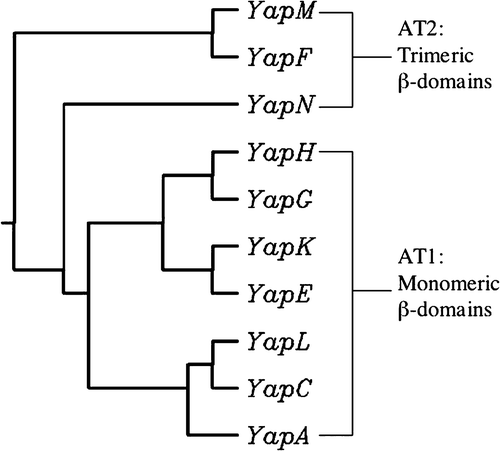
To evaluate the relationship of the 10 Yaps, a phylogenetic tree was constructed based on the alignment of their inferred β-domain sequences. Because these ATs show extensive size differences which would have greatly compromised the alignment quality, only the β-domain regions rather than the whole sequences were used for tree building. The constructed phylogenetic tree revealed closeness among YapA, YapC, and YapL; between YapH and YapG; and between YapE and YapK (). On the contrary, YapF, YapM and YapN appear to be less similar to the other seven ATs. Since these relationships were formed based on β-domains, we wanted to find out if groupings of these ATs could allow us to further classify them into AT-1 and AT-2 families (Jacob-Dubuisson et al. [Citation2004]). As described by Jacob-Dubuisson and colleagues, AT-1 is typified by AIDA-I, which forms a monomeric 48 kDa β-domain in the OM; AT-2 is exemplified by YadA, which forms a trimeric β structure in the OM (Jacob-Dubuisson et al. [Citation2004]). We aligned each AT β-domain sequence with AIDA-I or YadA. The alignment revealed that YapF, YapM, and YapN belong to the AT-2 family and are likely to form trimeric structures in the OM. Conversely, the other seven ATs are more similar to the AT-1 family and most likely form a monomeric β barrel in the OM.
Expression of the 10 yap genes in Y. pestis KIM
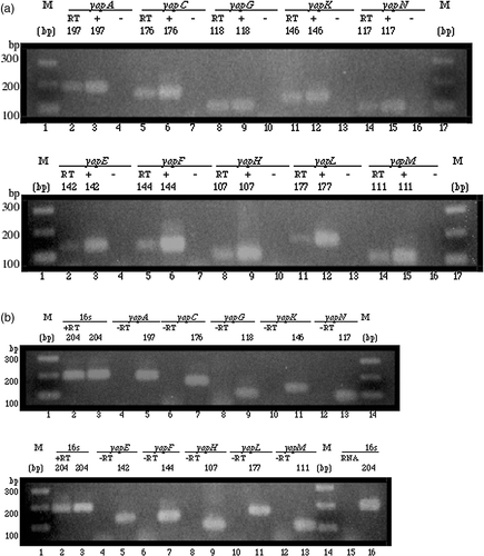
To assess the expression of the 10 yap genes in Y. pestis KIM, two-step RT-PCR was carried out. In the first step (RT reaction), total RNA extracted from Y. pestis was reverse transcribed using random hexamers as primers in order to generate an array of first-strand cDNA. In the subsequent amplification step, the expression of these 10 yap genes was assessed by PCR, which included cDNA as template and primers targeting specific genes. These primers were designed to generate final PCR products ranging from 107–197 bp: 197 bp (yapA amplicon), 176 bp (yapC amplicon), 118 bp (yapG amplicon), 146 bp (yapK amplicon), 117 bp (yapN amplicon), 142 bp (yapE amplicon), 144 bp (yapF amplicon), 107 bp (yapH amplicon), 177 bp (yapL amplicon), 111 bp (yapM amplicon). We were able to reverse transcribe all the 10 yaps from Y. pestis total RNA (a). To be certain that the RNA sample used in RT was free of DNA contamination, a PCR containing cleaned RNA (after DNaseI treatment and passing through the clean-up column) and primers targeting the 16s rRNA gene was run prior to performing the RT reaction (lane15, b). To assess DNA contamination in RT reagents, a RT reaction containing no template and random hexamers as primers was performed and the products subsequently PCR amplified using yap-gene-specific primers (“-RT”, b). To assess DNA contamination in PCR reagents, PCR reactions containing no template and primers targeting the yap genes were carried out (lanes 4, 7, 10, 13, and 16 in both panels of a). No gene products were detected from these negative control reactions, indicating that the RNA sample used in RT and all the reagents were free of DNA contamination. These results confirmed that the 10 yap genes are indeed expressed in Y. pestis KIM.
Figure 2. Expression of yap genes in Y. pestis KIM analysed by RT-PCR. Amplicon sizes are indicated above each panel in base pairs (bp). M: molecular markers. Each lane contained 5 µl of a 25 µl reaction resolved on a 2% agarose gel. (a) In both panels, RT-PCR products of yap genes (RT) are shown in lanes 2, 5, 8, 11, and 14; positive PCR control reactions (+) are shown in lanes 3, 6, 9, 12, and 15; and negative PCR control reactions (−) are shown in lanes 4, 7, 10, 13, and 16. The RT-PCR reactions included cDNA as template. The positive PCR reactions included genomic DNA as template. No template was used in the negative PCR reactions. (b) In both panels, positive RT control reactions (+RT) are shown in lane 2; and, negative RT control reactions (−RT) are shown in lanes 4, 6, 8, 10, and 12. Shown side-by-side in lanes 3, 5, 7, 9, 11, and 13 are positive PCR control reactions. The positive RT control reactions included cDNA as template and primers targeting the 16s rRNA gene. The negative RT control reactions contained the PCR amplified products of a negative control performed during reverse transcription. Shown in lane 15 is a control reaction that included 2.4 µg of cleaned RNA as template and primers targeting the 16s rRNA gene; shown side by side in lane 16 is a positive PCR control reaction.
Cloning of yapA, yapC, yapG, yapK, and yapN in E. coli K12
For secretion studies, we decided to focus our attention on five of the 10 identified Yaps: YapA, YapC, YapG, YapK, and YapN. These five Yaps are representative of the 10 KIM ATs: they include half of the previously identified ATs and half of the ATs identified by this study for the first time; they also have a wide range of sizes as well as diverse predicted functions. We cloned the five yap genes into E. coli K12. These yap genes were amplified from the genomic DNA of KIM by PCR. Each yap amplicon contained an open reading frame, extra nucleotides upstream of the initiation codon ATG (or GTG in yapN), and nucleotides downstream of the stop codon TAA. The region upstream of the initiation codon contained the putative −10 and −35 promoter sequences and also the putative Shine-Dalgarno ribosomal binding site. The sizes of the yap amplicons were 4622 bp (yapA), 2172 bp (yapC), 3361bp (yapG), and 4117 bp (yapK) and 2206 bp (yapN). Expression of the yap genes in E. coli relied on their native promoters without further induction.
DNA sequencing confirmed that the yapN insert had no mutation. One nucleotide mutation was found in yapC, but resulted in no mutation in amino acid. Three mutations were discovered in YapA: G586→Q, L1078→P, and D1294→G, two in YapG: V191→A and S486→F, and one in YapK: Q1012→L. These mutations occurred despite the use of a high fidelity DNA polymerase. Mutations such as these are expected to be found in large-size PCR-amplicons, as they have also been reported in the cloned gene of a Neisseria meningitidis AT (van Ulsen et al. [Citation2001]). Except for one mutation in YapA (D1294→G), the other mutations in YapA, YapG, and YapK were predicted to fall within the passenger domain, and therefore were not expected to interfere with protein secretion. Indeed, their secretion was confirmed later in this study.
Expression and localization of the YapA, YapC, YapG, YapK and YapN proteins in E. coli K12
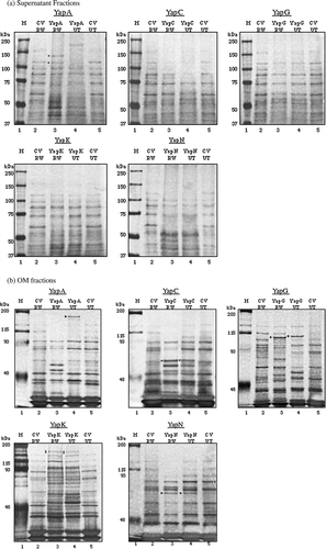
To determine cellular localization of the Yap proteins, yap-containing plasmids were transformed into the E. coli RW193 strain. Culture supernatant and OM fractions were subsequently isolated and analysed by SDS-PAGE. The protein expression profile of a yap-containing plasmid was then compared to that of the empty cloning vector so that unique protein bands close to predicted Mw representing putative AT proteins could be identified. Using these methods, we were able to find the YapA protein in the culture supernatant. When compared to the expression of the empty cloning vector, two unique protein bands of 130 and 115 kDa representing YapA were detected on the gel (a, YapA, lane 3). N-terminal sequencing further confirmed the identity of YapA and revealed that one form contains an N terminus of VXQIATT and the other VXXXATTDTP, both of which correspond to VSQIATTDTP, the N terminus of a mature YapA protein without a signal peptide. The predicted size of the YapA precursor and that of mature YapA are 150 and 146 kDa, respectively. It thus appears that the 130 kDa YapA protein corresponds to the entire passenger domain, while the 115 kDa YapA protein represents a form whose passenger domain has a deletion in the C terminus. Based on these results, we believe that after OM translocation YapA's passenger domain undergoes proteolytic processing from its β-domain and is subsequently released into the extracellular milieu. In contrast to the yapA clone, no unique bands could be detected in the supernatant fractions of clones expressing YapC, YapG, YapK, and YapN (a, panels 2–5, lane 3).
To ascertain OM localization of these other ATs in E. coli, OM isolation of the yap clones was carried out followed by SDS-PAGE analysis. Protein expression of these yaps was compared to that of E. coli RW193 containing the empty cloning vector. From the sample produced by the yapC RW193 clone, a unique 65 kDa band was identified on the gel (b, YapC, lane 3). The size of this putative YapC protein is comparable to the predicted size of mature YapC, 65 kDa. Mature YapG was predicted to be 101 kDa, and a unique band around 100 kDa could also be found (b, YapG, lane 3). Two close bands of ∼120 kDa representing putative YapK were detected on the gel as well (b, YapK, lane 3); these are comparable to the predicted mature YapK size of 126 kDa. Finally, a unique band corresponding to 60 kDa was detected from the sample produced by the yapN RW193 clone (b, YapN, lane 3); YapN was predicted to have a size of 62 kDa. All these data suggest that YapC, YapG, YapK, and YapN localize to the OM. Since their sizes determined by SDS-PAGE are comparable to their predicted sizes, we believe that the passenger domains of these Yaps are not cleaved from the β-domains after OM translocation. Furthermore, our SDS-PAGE analysis of the supernatant fractions gave no indications that YapC, YapG, YapK, or YapN was released from the cell surface (a, panels 2–5, lane 3). Such results support OM localization of YapC, YapG, YapK, and YapN.
Figure 3. Expression of YapA, YapC, YapG, YapK, and YapN in the RW193 (OmpT+ and OmpP+) and UT5600 (ΔOmpT and ΔOmpP) strains: (a) supernatant fractions (b) OM fractions. Both gels were silver stained. M: molecular markers. CV: cloning vector. RW: RW193. UT: UT5600. Dotted bands represent the identified Yap proteins.
To further verify OM localization of these proteins, OM samples containing YapC, YapG, YapK, and YapN were subjected to the NADH oxidase assay, which assessed IM contamination. The activity of NADH oxidase in each sample was reflected in the oxidation of NADH, which was monitored by measuring the decrease in absorbance at 340 nm. The positive control reaction containing 20 µg of total membranes had a change in the OD340 value of 0.521 in 5 min, whereas the negative control reaction containing 20 µg of total membranes and inhibited with 10 mM of KCN showed a value change of 0.015. The changes in OD340 of the OM samples ranged from 0.012–0.031. The amount of IM contamination in each OM sample was calculated to be less than 3%. The results suggest that these OM samples are virtually free of IM contaminants.
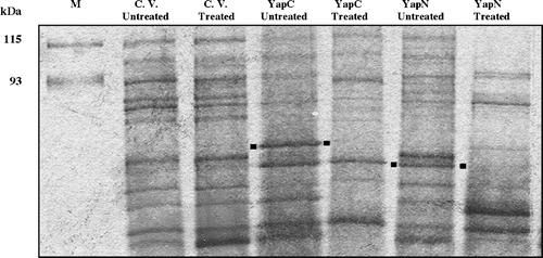
To determine whether these OM proteins are also surface exposed, protease accessibility experiments were carried out. Whole cells expressing the Yap proteins were treated with Proteinase K at a low concentration of 10 µg/ml. Surface localized proteins were indicated on the polyacrylamide gel by the disappearance of protein bands in the protease-treated samples as compared to the untreated control. The results verified that YapC and YapN were targeted to the OM surface and accessible for protease digestion (). Results of the protease assay for YapG and YapK were inconsistent (data not shown). Thus, cell fractionation and protease accessibility experiments allowed us to establish extracellular localization of YapA, OM localization of YapG and YapK, and cell surface localization of YapC and YapN.
Figure 4. Cell surface localization of YapC and YapN determined by protease accessibility experiments and SDS-PAGE analysis. The gel was silver stained. M: molecular markers. C. V: cloning vector. Treated samples came from whole RW193 cells incubated with 10 µg/ml of Proteinase K followed by OM isolation. Dotted positions are indicated to compare the presence and disappearance of YapC and YapN protein bands.
Role of OmpT/OmpP proteases in YapA processing
It is apparent that YapA undergoes processing during OM secretion because two sizes of this protein (130 and 115 kDa) could be detected in the supernatant fraction, and these sizes are smaller than what has been predicted. For some ATs, the OM protease OmpT or OmpP is responsible for cleaving between the C-terminal domain and the passenger domain, thereby releasing the passenger domain to the extracellular environment (Jacob-Dubuisson et al. [Citation2004]). In addition, several putative OmpT cleavage sites (Dekker et al. [Citation2001]) could be found in the YapA sequence (data not shown). To verify if YapA was indeed released from the cell by OmpT/OmpP, we expressed yap genes in the OmpT/OmpP mutant E. coli UT5600 strain and compared their expression to that in wild-type RW193. In SDS-PAGE analysis of the OM fractions, a unique band around 170 kDa was detected in the yapA UT5600 clone (b, YapA, lane 4). This 170-kDa OM protein represents the unprocessed form of YapA, as this protein is larger than the two processed forms of YapA expressed by RW193 (a, YapA, lane 3). The observation thus supports the hypothesis that OmpT/OmpP processing occurs during YapA secretion.
The finding of this overly large-size 170-kDa protein representing unprocessed YapA was puzzling since YapA was predicted to be only 150 kDa. We wondered if the increase in Mw could be due to glycosylation before OM translocation, as glycosylation has been observed in E. coli AT AIDA-I (Benz & Schmidt [Citation2001]) and in an Y. pestis OM hemagglutinin (Tikhomirova et al. [Citation2003]). To verify whether YapA is a glycoprotein, the supernatant fraction of E. coli RW193 expressing YapA was used in glycan detection assay. The assay revealed that YapA is not glycosylated (data not shown). Thus glycosylation is not the reason why YapA exhibits a higher Mw than what has been predicted.
Unlike YapA, other ATs (YapC, YapG, YapN, and a form of YapK) appear unprocessed during targeting to the OM and exhibit the same sizes in UT5600 as in RW193 (b, panels 2–5, lane 4). Interestingly, two forms of YapK (b, YapK, lanes 3 and 4) can also be found. The lower Mw band is likely a processed form of YapK. And yet this processed form appeared in the OM fractions of both the RW193 and UT5600 strains, suggesting that OM protease(s) other than OmpT/OmpP might play a role in its processing.
YapC, YapN, and YapK exhibit adherence activities
To infer the functions of the five Yap proteins, alignments of the passenger domains were carried out. The results revealed significant degrees of identity between Yaps and other known ATs: 22% between the passenger domain of YapA and that of Hia, an adhesin AT from H. influenzae; 27% between the passenger domain of YapC and that of pertactin, an adhesin AT of B. pertussis; 27% between the passenger domain of YapG and that of AIDA-I, an adhesin AT of E. coli; and 24% between the passenger domain of YapK and that of AIDA-I. Additionally, YapN was found to share 22% identity with ApeE, an esterase AT of S. enterica, but also possess the Hep-Hag hemagglutinin motif (Supplementary online). Most of these Yaps also harbor one or more RGD motifs (Supplementary online), further suggesting that they are likely adhesins.
To verify the predicted adherence ability of these Yaps, we performed hemagglutination () and autoaggregation () experiments. For the hemagglutination assay, E. coli UT5600 expressing the Yaps were serially diluted from 1:10–1:100 to test for Yaps’ ability to agglutinate sheep erythrocytes. Tsh of avian pathogenic E. coli, an established AT hemagglutinin (Stathopoulos et al. [Citation1999], Kostakioti & Stathopoulos [Citation2004]), was used as a positive control. Although Tsh was expressed from a different vector from the Yaps, it was included to validate this qualitative assay. Tsh showed a positive hemagglutination phenotype for all the dilutions tested. One of the Yap proteins, YapN, demonstrated a hemagglutination positive phenotype up to 1:80 dilution. Another Yap, YapK, moderately agglutinated the erythrocytes and positive agglutination activity was seen at 1:10 and 1:20 dilutions.
Table I. Hemagglutination activity of Yap proteins.
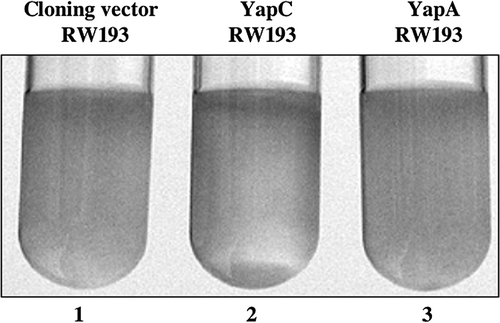
For the autoaggregation assay, E. coli RW193 cells expressing the Yaps were tested for their ability to adhere to each other and settle to the bottom of the test tubes. RW193 harboring the empty cloning vector and the one expressing YapA (a secreted protein in RW193) served as negative controls. Of all tested, only YapC exhibited a positive phenotype, as indicated by the formation of a pellet in the bottom of the test tube (). The results were consistent for overnight cultures and cultures grown to an OD600 of 1.5. Together with the results of the hemagglutination assay, these data support the computational predictions that YapC, YapK, and YapN are adhesins. The results suggest that YapC has the ability to cause bacterial aggregation, a characteristic exhibited by cells that form biofilms; and, YapN and YapK allow the bacteria to bind to eukaryotic cells, a property shown in invasive microbes.
Figure 5. Autoaggregation activity of YapC. E. coli RW193 expressing YapC exhibited a positive autoaggregation phenotype, as indicated by the presence of precipitated cells that form a pellet (lane 2). Strains harboring pCR2.1-TOPO (lane 1) and pYA12-YapA (lane 3) served as negative controls, in which a precipitated pellet is absent.
Discussion
Yersinia pestis secretes several known virulence factors that are responsible for the organism's pathogenicity (Perry & Fetherston [Citation1997]). It is likely that additional virulence factors secreted by Y. pestis remain to be identified. Although the type III secretion system of Yersinia has been well characterized (Cornelis [Citation2002]), not much is known about the type V (AT) system in this organism. Nearly all the identified ATs have been shown to play a role in bacterial virulence (Henderson & Nataro [Citation2001]). The IgA1 protease of N. gonorrhoeae has been shown to be capable of cleaving the hinge region of human secretory IgA1 (sIgA1) in vitro (Kilian et al. [Citation1996]) and is responsible for the bacterium's ability to reproduce intracellularly (Hauck & Meyer [Citation1997], Lin et al. [Citation1997]). Additionally, avian pathogenic E. coli Tsh has been shown to bind to extracellular matrix proteins (Kostakioti & Stathopoulos [Citation2004]). BrkA from B. pertussis has been implicated to contribute to serum resistance and is responsible for the resistance of Bordetellae to the classical pathway of complement-dependent killing by human serum (Fernandez & Weiss [Citation1994]). Others, like AIDA-I of E. coli, IcsA (VirG) of S. flexneri, Hap from H. influenzae, and Pet of E. coli (Dhermy [Citation1991]) have all been shown to play specific roles in the pathogenesis of their host organisms (Henderson & Nataro [Citation2001]).
All of these observations have prompted us to look for genes in Y. pestis that encode AT proteins, which may be important to the pathogenicity of this organism in plague infection. Our in silico analyses have resulted in the identification of 10 putative AT genes in the Y. pestis KIM genome. These candidates fulfill the criteria for ATs by the presence of an N-terminal signal sequence and a C-terminal β-domain containing the consensus residues. Possession of only a few cysteine residues is another characteristic shared by these identified ATs since formation of disulphide bonds can interfere with translocation of the passenger domains. Phylogenetic studies of the β-domains enabled us to classify the ATs into either AT-1 or AT-2 subfamilies, which also allowed us to infer the oligomerization states of the β-domains. While YapF, YapM and YapN belong to AT-2 subfamily and are likely to form trimers, the rest belong to AT-1, which according to most studies, form monomeric β barrels (Jacob-Dubuisson et al. [Citation2004]). Homologs of the 10 KIM ATs are present not only in CO92, but also in the more distantly related species Y. pseudotuberculosis, a pathogen that has a different route of infection and disease manifestation from Y. pestis (Salyers & Whitt [Citation1994]). While eight KIM ATs share high degrees of identity (>96%) with the homologs in Y. pseudotuberculosis, two ATs are significantly less similar: YapA (80.7%) and YapN (54.8%). It is possible that the sequence differences might contribute to the dissimilar modes of infection between these related pathogens.
Expression and secretion studies were subsequently performed to verify the results of computational predictions. The expression of the 10 yap genes in Y. pestis KIM was confirmed by RT-PCR. Five AT genes, yapA, yapC, yapG, yapK, and yapN, were PCR-amplified and cloned in E. coli K12. Included in the amplicons were the genes’ native promoter regions so as to express the yaps in E. coli without additional induction. Two forms of the YapA protein of approximately 130 and 115 kDa were found secreted into the culture medium. Our data confirmed the cleavage of its signal peptide and processing of the C-terminal domain by OmpT/OmpP during secretion. Expression of yapA in UT5600 (ΔOmpT/ΔOmpP) led to the production of a high Mw protein of ∼170 kDa localized to the OM, which most probably represents unprocessed YapA containing both its passenger domain and β-domain. Glycan detection assay of YapA revealed that this protein is not glycosylated. Thus the question as to why this protein produced by E. coli UT5600 has such a high Mw is unanswered. YapC, YapG, YapK, and YapN were not found in the culture medium but instead localized to the OM. In the SDS-PAGE analysis of OM fractions, YapC migrated to a distance corresponding to 65 kDa, YapG 100 kDa, YapK130 kD, and YapN 60 kDa. Expression of these four genes in the protease-deficient E. coli strain UT5600 verified that the proteins did not undergo post-translational processing. Cell surface exposure of YapC and YapN was further confirmed by the protease accessibility assay.
Our in silico analyses suggested the possibilities that YapA, YapC, YapG, and YapK might be adhesins, and YapN might be a hemagglutinin. The predictions imply that the passenger domains of Yaps possibly contribute to the virulence of Y. pestis by forming biofilms or adhering to the host cells. The results of the hemagglutination assay revealed a hemagglutination positive phenotype for YapN and YapK, confirming RBC-agglutination ability of these Y. pestis ATs. Additionally, YapC was found to contribute to bacterial autoaggregation. However, since the YapA, YapG, and YapK clones have mutations within their passenger (functional) domains, we cannot be certain whether these mutations have any effects on the negative adherence phenotypes exhibited by them, particularly YapA and YapG. Furthermore, Yaps may have other virulence functions in addition to cell adhesion, as multiple functions have been previously observed in several characterized ATs (Henderson & Nataro [Citation2001], Kostakioti & Stathopoulos [Citation2004]).
In conclusion, 10 Y. pestis ATs were identified in this study, five of them cloned, expressed, and characterized. One of these ATs (YapA) is a protein secreted in different forms and released into the extracellular milieu by OmpT/OmpP cleavage. Four other ATs (YapC, YapG, YapK, and YapN) are unprocessed OM proteins, with the possibility that two of them (YapC and YapN) are targeted to the cell surface. Three ATs (YapC, YapN, and YapK) also exhibit cell adherence activities. These Yap proteins represent the first ATs characterized in Y. pestis.
This paper was first published online on prEview on 29 September 2006.
We would like to thank Dr Viren Konde and Dr Laura Gutierrez at University of Houston for technical assistance in PCR and cloning; Dr James Graham and Dr Christopher Price at University of Kentucky, and Dr Theresa Koehler and Ms Maria Hadjifrangiskou at University of Texas, Houston, for technical assistance in RT-PCR; Dr David Thanassi, Dr James Bliska, and Ms Lisa Runo at Stony Brook University for providing Y. pestis bacterial strain, chromosomal DNA, RT-PCR protocols and technical assistance; and Dr Anne Delcour at University of Houston, Dr Shelley Payne at University of Texas-Austin, and Maria Kostakioti in our laboratory for critical review of the manuscript. This study is supported by a research grant (E-1548) from the Robert A. Welch Foundation.
References
- Achtman M, Zurth K, Morelli G, Torrea G, Guiyoule A, Carniel E. Yersinia pestis, the cause of plague, is a recently emerged clone of Yersinia pseudotuberculosis. Proc Natl Acad Sci USA 1999; 96: 14043–14048
- Benz I, Schmidt MA. Glycosylation with heptose residues mediated by the aah gene product is essential for adherence of the AIDA-I adhesin. Mol Microbiol 2001; 40: 1403–1413
- Cornelis GR. Yersinia type III secretion: send in the effectors. J Cell Biol 2002; 158: 401–408
- D'Souza SE, Ginsberg MH, Plow EF. Arginyl-glycyl-aspartic acid (RGD): a cell adhesion motif. Trends Biochem Sci 1991; 16: 246–250
- Dekker N, Cox RC, Kramer RA, Egmond MR. Substrate specificity of the integral membrane protease OmpT determined by spatially addressed peptide libraries. Biochemistry 2001; 40: 1694–1701
- Deng W, Burland V, Plunkett G, 3rd, Boutin A, Mayhew GF, Liss P, Perna NT, Rose DJ, Mau B, Zhou S, Schwartz DC, Fetherston JD, Lindler LE, Brubaker RR, Plano GV, Straley SC, McDonough KA, Nilles ML, Matson JS, Blattner FR, Perry RD. Genome sequence of Yersinia pestis KIM. J Bacteriol 2002; 184: 4601–4611
- Dhermy D. The spectrin super-family. Biol Cell 1991; 71: 249–254
- Drancourt M, Raoult D. Molecular insights into the history of plague. Microbes Infect 2002; 4: 105–109
- Egile C, d'Hauteville H, Parsot C, Sansonetti PJ. SopA, the outer membrane protease responsible for polar localization of IcsA in Shigella flexneri. Mol Microbiol 1997; 23: 1063–1073
- Elish ME, Pierce JR, Earhart CF. Biochemical analysis of spontaneous fepA mutants of Escherichia coli. J Gen Microbiol 1988; 134: 1355–1364
- Emsley P, Charles IG, Fairweather NF, Isaacs NW. Structure of Bordetella pertussis virulence factor P.69 pertactin. Nature 1996; 381: 90–92
- Fernandez RC, Weiss AA. Cloning and sequencing of a Bordetella pertussis serum resistance locus. Infect Immun 1994; 62: 4727–4738
- Filip C, Fletcher G, Wulff JL, Earhart CF. Solubilization of the cytoplasmic membrane of Escherichia coli by the ionic detergent sodium-lauryl sarcosinate. J Bacteriol 1973; 115: 717–722
- Galimand M, Guiyoule A, Gerbaud G, Rasoamanana B, Chanteau S, Carniel E, Courvalin P. Multidrug resistance in Yersinia pestis mediated by a transferable plasmid. N Engl J Med 1997; 337: 677–680
- Hauck CR, Meyer TF. The lysosomal/phagosomal membrane protein h-lamp-1 is a target of the IgA1 protease of Neisseria gonorrhoeae. FEBS Lett 1997; 405: 86–90
- Henderson IR, Navarro-Garcia F, Nataro JP. The great escape: structure and function of the autotransporter proteins. Trends Microbiol 1998; 6: 370–378
- Henderson IR, Nataro JP. Virulence functions of autotransporter proteins. Infect Immun 2001; 69: 1231–1243
- Henderson IR, Navarro-Garcia F, Desvaux M, Fernandez RC, Ala'Aldeen D. Type V protein secretion pathway: the autotransporter story. Microbiol Mol Biol Rev 2004; 68: 692–744
- Hendrixson DR, de la Morena ML, Stathopoulos C, St Geme JW, 3rd. Structural determinants of processing and secretion of the Haemophilus influenzae hap protein. Mol Microbiol 1997; 26: 505–518
- Jacob-Dubuisson F, Fernandez R, Coutte L. Protein secretion through autotransporter and two-partner pathways. Biochim Biophys Acta 2004; 1694: 235–257
- Kilian M, Reinholdt J, Lomholt H, Poulsen K, Frandsen EV. Biological significance of IgA1 proteases in bacterial colonization and pathogenesis: critical evaluation of experimental evidence. Apmis 1996; 104: 321–338
- Klietmann WF, Ruoff KL. Bioterrorism: implications for the clinical microbiologist. Clin Microbiol Rev 2001; 14: 364–381
- Kostakioti M, Stathopoulos C. Functional analysis of the Tsh autotransporter from an avian pathogenic Escherichia coli strain. Infect Immun 2004; 72: 5548–5554
- Kostakioti M, Newman CL, Thanassi DG, Stathopoulos C. Mechanisms of protein export across the bacterial outer membrane. J Bacteriol 2005; 187: 4306–4314
- Lin L, Ayala P, Larson J, Mulks M, Fukuda M, Carlsson SR, Enns C, So M. The Neisseria type 2 IgA1 protease cleaves LAMP1 and promotes survival of bacteria within epithelial cells. Mol Microbiol 1997; 24: 1083–1094
- Muller D, Benz I, Tapadar D, Buddenborg C, Greune L, Schmidt MA. Arrangement of the translocator of the autotransporter adhesin involved in diffuse adherence on the bacterial surface. Infect Immun 2005; 73: 3851–3859
- Newman CL, Stathopoulos C. Autotransporter and two-partner secretion: delivery of large-size virulence factors by gram-negative bacterial pathogens. Crit Rev Microbiol 2004; 30: 275–286
- Oliver DC, Huang G, Nodel E, Pleasance S, Fernandez RC. A conserved region within the Bordetella pertussis autotransporter BrkA is necessary for folding of its passenger domain. Mol Microbiol 2003; 47: 1367–1383
- Orent W. Plague. The mysterious past and terrifying future of the world's most dangerous disease. Free Press, New York 2004
- Otto BR, Sijbrandi R, Luirink J, Oudega B, Heddle JG, Mizutani K, Park SY, Tame JR. Crystal structure of hemoglobin protease, a heme binding autotransporter protein from pathogenic Escherichia coli. J Biol Chem 2005; 280: 17339–17345
- Parkhill J, Wren BW, Thomson NR, Titball RW, Holden MT, Prentice MB, Sebaihia M, James KD, Churcher C, Mungall KL, Baker S, Basham D, Bentley SD, Brooks K, Cerdeno-Tarraga AM, Chillingworth T, Cronin A, Davies RM, Davis P, Dougan G, Feltwell T, Hamlin N, Holroyd S, Jagels K, Karlyshev AV, Leather S, Moule S, Oyston PC, Quail M, Rutherford K, Simmonds M, Skelton J, Stevens K, Whitehead S, Barrell BG. Genome sequence of Yersinia pestis, the causative agent of plague. Nature 2001; 413: 523–527
- Perry RD, Pendrak ML, Schuetze P. Identification and cloning of a hemin storage locus involved in the pigmentation phenotype of Yersinia pestis. J Bacteriol 1990; 172: 5929–5937
- Perry RD, Fetherston JD. Yersinia pestis – etiologic agent of plague. Clin Microbiol Rev 1997; 10: 35–66
- Pohlner J, Halter R, Beyreuther K, Meyer TF. Gene structure and extracellular secretion of Neisseria gonorrhoeae IgA protease. Nature 1987; 325: 458–462
- Salyers A, Whitt D. Yersinia infections. Bacterial pathogenesis, a molecular approach. ASM Press, Washington, DC 1994; 213–228
- Stathopoulos C, Provence DL, Curtiss R, 3rd. Characterization of the avian pathogenic Escherichia coli hemagglutinin Tsh, a member of the immunoglobulin A protease-type family of autotransporters. Infect Immun 1999; 67: 772–781
- Stathopoulos C, Hendrixson DR, Thanassi DG, Hultgren SJ, St Geme JW, 3rd, Curtiss R, 3rd. Secretion of virulence determinants by the general secretory pathway in gram-negative pathogens: an evolving story. Microbes Infect 2000; 2: 1061–1072
- Thanassi DG, Stathopoulos C, Karkal A, Li H. Protein secretion in the absence of ATP: the autotransporter, two-partner secretion and chaperone/usher pathways of gram-negative bacteria (review). Mol Membr Biol 2005; 22: 63–72
- Tikhomirova, EI, Zadnova, SP, Volokh, OA, Karpunina, LV, Shcherbakov, AA. 2003. [Lectin of Yersinia pestis vaccine strain EV and its immunological properties]. Zh Mikrobiol Epidemiol Immunobiol :51–55.
- Titball RW, Hill J, Lawton DG, Brown KA. Yersinia pestis and plague. Biochem Soc Trans 2003; 31: 104–107
- van Ulsen P, van Alphen L, Hopman CT, van der Ende A, Tommassen J. In vivo expression of Neisseria meningitidis proteins homologous to the Haemophilus influenzae Hap and Hia autotransporters. FEMS Immunol Med Microbiol 2001; 32: 53–64
- Wang RF, Kushner SR. Construction of versatile low-copy-number vectors for cloning, sequencing and gene expression in Escherichia coli. Gene 1991; 100: 195–199
- Yen MR, Peabody CR, Partovi SM, Zhai Y, Tseng YH, Saier MH. Protein-translocating outer membrane porins of Gram-negative bacteria. Biochim Biophys Acta 2002; 1562: 6–31
- Yen, YT, Stathopoulos, C. 2006. Identification of autotransporter proteins secreted by type V secretion systems in gram-negative bacteria. In:. M van der Giezen, editor., Methods in molecular biology-protein targeting protocols. Totowa, New Jersey: Humana Press. Chapter 20, in press.
- Zhou S, Deng W, Anantharaman TS, Lim A, Dimalanta ET, Wang J, Wu T, Chunhong T, Creighton R, Kile A, Kvikstad E, Bechner M, Yen G, Garic-Stankovic A, Severin J, Forrest D, Runnheim R, Churas C, Lamers C, Perna NT, Burland V, Blattner FR, Mishra B, Schwartz DC. A whole-genome shotgun optical map of Yersinia pestis strain KIM. Appl Environ Microbiol 2002; 68: 6321–6331
