Abstract
Glycosyl-phosphatidylinositol (GPI)-anchored proteins are enriched in cholesterol- and sphingolipid-rich lipid rafts within the membrane. Rafts are known to have roles in cellular organization and function, but little is understood about the factors controlling the distribution of proteins in rafts. We have used atomic force microscopy to directly visualize proteins in supported lipid bilayers composed of equimolar sphingomyelin, dioleoyl-sn-glycero-3-phosphocholine and cholesterol. The transmembrane anchored angiotensin converting enzyme (TM-ACE) was excluded from the liquid ordered raft domains. Replacement of the transmembrane and cytoplasmic domains of TM-ACE with a GPI anchor (GPI-ACE) promoted the association of the protein with rafts in the bilayers formed with brain sphingomyelin (mainly C18:0). Association with the rafts did not occur if the shorter chain egg sphingomyelin (mainly C16:0) was used. The distribution of GPI-anchored proteins in supported lipid bilayers was investigated further using membrane dipeptidase (MDP) whose GPI anchor contains distearoyl phosphatidylinositol. MDP was also excluded from rafts when egg sphingomyelin was used but associated with raft domains formed using brain sphingomyelin. The effect of sphingomyelin chain length on the distribution of GPI-anchored proteins in rafts was verified using synthetic palmitoyl or stearoyl sphingomyelin. Both GPI-ACE and MDP only associated with the longer chain stearoyl sphingomyelin rafts. These data obtained using supported lipid bilayers provide the first direct evidence that the nature of the membrane-anchoring domain influences the association of a protein with lipid rafts and that acyl chain length hydrophobic mismatch influences the distribution of GPI-anchored proteins in rafts.
| Abbreviations | ||
| ACE | = | angiotensin converting enzyme |
| AFM | = | atomic force microscopy |
| CHO | = | Chinese hamster ovary |
| DOPC | = | dioleoyl-sn-glycero-3-phosphocholine |
| DRM | = | detergent resistant membrane |
| GPI | = | glycosyl-phosphatidylinositol |
| GPI-ACE | = | GPI anchored form of angiotensin converting enzyme |
| ld | = | liquid disordered |
| lo | = | liquid ordered |
| MDP | = | membrane dipeptidase |
| octyl glucoside | = | n-octyl β-D-glucopyranoside |
| TBS | = | Tris-buffered saline |
| TM-ACE | = | transmembrane form of angiotensin converting enzyme |
Introduction
The existence of distinct domains in biological membranes has prompted considerable interest in recent years Citation[1–3]. One such domain, enriched in sphingolipids and cholesterol, the so-called lipid raft, serves to cluster specific proteins and lipids and has been implicated in a variety of cellular functions including protein sorting, membrane trafficking and signal transduction Citation[4], Citation[5]. The specific lipid composition of rafts renders them resistant to solubilization by certain detergents and this has been exploited to isolate detergent resistant membrane (DRM) fractions from cells Citation[6–8]. Studies on model membrane systems have shown that lipid bilayers composed of sphingolipids, cholesterol and unsaturated phospholipids, phase separate to form liquid-ordered (lo) domains in a sea of liquid-disordered (ld) lipids Citation[9], Citation[10]. These lo domains, like rafts, are enriched in sphingolipids and cholesterol, and are resistant to detergent solubilization. Such model membrane systems provide a means of investigating the biophysical properties of rafts and can present valuable insights into lipid-lipid and lipid-protein interactions Citation[5], Citation[11].
One of the major classes of proteins associated with lipid rafts are the glycosyl-phosphatidylinositol (GPI)-anchored proteins Citation[6], Citation[12]. The distribution of GPI-anchored proteins in rafts is attributable predominantly to their lipid chains, which are typically saturated, and therefore preferentially pack with the more ordered, saturated chains of raft lipids Citation[13], Citation[14]. Several studies have shown that a GPI anchor will target proteins to rafts in both model membranes Citation[15–17] and cells Citation[18–20]. However, these investigations have relied upon detergent solubilization to determine the distribution of raft and non-raft proteins, a method which subsequent reports have suggested is susceptible to artefacts and may cause protein redistribution Citation[21–24].
In the present study the use of detergents was avoided by directly visualizing supported lipid bilayers using atomic force microscopy (AFM), a technique that has been successfully utilized to image lipid bilayers, lo raft domains and membrane proteins in recent years Citation[25–27]. Although it has been reported that the GPI-anchored placental alkaline phosphatase partitions into lo domains in model membranes Citation[28–30], no study has directly compared the distribution of a transmembrane polypeptide anchored protein with that of a GPI-anchored protein in model membranes. Here we present data on the distribution of the transmembrane protein, angiotensin converting enzyme (TM-ACE) within supported lipid bilayers composed of equimolar sphingomyelin, dioleoyl-sn-glycero-3-phosphocholine (DOPC) and cholesterol, and compare this distribution with that obtained using a GPI-anchored form of ACE (GPI-ACE) and the GPI-anchored membrane dipeptidase (MDP). In addition we study the effect of sphingomyelin chain length on the raft localisation of GPI-ACE and MDP using natural sphingomyelin mixtures from egg and brain which are comprised of mainly C16:0 and C18:0 chains, respectively. Synthetic palmitoyl and stearoyl sphingomyelin were used to verify that the acyl chain length was responsible for the distribution of GPI-anchored proteins in egg and brain sphingomyelin rafts.
Materials and methods
Purification of TM-ACE, GPI-ACE and MDP
TM-ACE was purified from porcine kidney cortex, following solubilization of the membranes with Triton X-100, by lisinopril-Sepharose affinity chromatography as described previously Citation[31]. The protocol was adapted to replace the Triton X-100 with 2 mM n-octyl -D-glucopyranoside (octyl glucoside) in the final isolation buffer. Chinese hamster ovary (CHO) cells stably expressing GPI-ACE Citation[18], Citation[32] were cultured in Ham's F12 medium (Cambrex Bio Science, Berkshire, UK) supplemented with 10% GIBCO foetal bovine serum (Invitrogen, Paisley, UK), penicillin (50 units/ml) and streptomycin (50 units/ml) (both from Cambrex Bio Science, Berkshire, UK). Confluent cells were washed and scraped into phosphate buffered saline (20 mM Na2HPO4, 2 mM NaH2PO4, 150 mM NaCl, pH 7.4). Cells were pelleted at 500 g for 5 min, sonicated, centrifuged at 100,000 g for 1 h and then solubilized using octyl glucoside (60 mM). Following centrifugation at 100,000 g for 1 h, GPI-ACE was purified from the supernatant fraction by lisinopril-Sepharose affinity chromatography Citation[31] with the final isolation buffer containing 2 mM octyl glucoside. MDP was purified from porcine kidney cortex, following solubilization from the membranes with octyl glucoside, by cilastatin-Sepharose affinity chromatography as described previously Citation[33] with 2 mM octyl glucoside in the final protein isolation buffer. Enzyme activity was determined using Hippuryl-His-Leu as substrate for TM-ACE and GPI-ACE Citation[31] and Gly-D-Phe as substrate for MDP Citation[33]. Protein concentration was determined using the bicinchoninic acid assay with bovine serum albumin as standard Citation[34]. For SDS polyacrylamide gel electrophoresis, proteins were separated on 7–17% (w/v) acrylamide gradient gels as described previously Citation[31] with Precision protein prestained standards (Bio-Rad, Hertfordshire, UK).
Formation of supported lipid bilayers
Egg sphingomyelin, brain sphingomyelin, N-palmitoyl-D-erythro-sphingosylphosphoryl-choline (palmitoyl sphingomyelin), and N-stearoyl-D-erythro-sphingosylphosphoryl-choline (stearoyl sphingomyelin) were purchased from Avanti Polar Lipids (Alabaster, USA). DOPC and cholesterol were purchased from Sigma-Aldrich (Dorset, UK). Equimolar mixtures of sphingomyelin, DOPC and cholesterol were prepared in chloroform:methanol (3:1 volume ratio) and dried under argon for 2 h. The dried lipid mixtures were rehydrated in Tris-buffered saline (TBS; 5 mM Tris/HCl, 100 mM NaCl, pH 7.6) to a concentration of 2 mg/ml and the purified protein (1–2 µg per ml hydrated lipid) was added. For control samples, the final protein isolation buffer containing 2 mM octyl-glucoside was used in the absence of purified protein. Vesicles were formed by a process of vortexing, dialyzing in TBS overnight and then sonicating in a bath sonicator (Ultrawave Ltd, Cardiff, UK) for 30 min. Supported lipid bilayers were prepared by transferring 10 µl of sample on to freshly cleaved mica followed by 80 µl TBS containing 2 mM CaCl2. After 3 min the bilayer was washed twice with TBS before imaging by AFM.
Atomic force microscopy
A multimode atomic force microscope with a Nanoscope IIIa controller (Digital Instruments, Santa Barbara, USA) and an E-scanner was used to image samples in TBS with a fluid cell (Digital Instruments). Images were recorded in tapping mode using oxide-sharpened, silicon nitride tips mounted on cantilevers with nominal spring constants of 0.32 Newton/m, oscillating to a frequency between 7 and 9 KHz. The set point was adjusted during imaging to minimize the force whilst scanning at a rate of 1–2 Hz. Nanoscope software was used to flatten the images.
Statistical analysis of protein distribution
A minimum of three separate experiments were performed for each of the proteins in each of the lipid mixtures. Multiple scans of 5–10 µm square were recorded for each experiment and 9 representative images were used for analysis. The total number of proteins in raft and non-raft regions was analyzed using Statistica (Statsoft, UK). Analysis of the AFM data showed that the number of proteins which were incorporated into lo rafts was a continuous random variable with a probability density function which approximated to a normal distribution. Significance testing was carried out to determine whether there was a difference between the mean protein incorporation into lo and a value for Z determined. A one-tailed test was performed, with the null hypothesis being rejected if -3.291 < Z<3.291 inferring significance at the 0.5% level.
Results
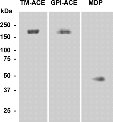
To investigate the effect of the type of anchor on the distribution of a protein in the membrane, two forms of ACE were purified. The endogenous transmembrane form of ACE (TM-ACE) was purified from porcine kidney, while a GPI-anchored form of ACE (GPI-ACE) was purified from CHO cells that had been transfected with the cDNA in which the sequence encoding the transmembrane and cytosolic domains of human ACE had been replaced with the sequence encoding a C-terminal GPI anchor attachment signal Citation[18], Citation[32]. Both proteins migrated as a single band of 180 kDa on SDS polyacrylamide gel electrophoresis (). TM-ACE and GPI-ACE had similar specific activities of 1.98 µmol/min/mg and 1.91 µmol/min/mg, respectively, with Hippuryl-His-Leu as substrate, similar to that reported previously Citation[31].
Figure 1. Purification of TM-ACE, GPI-ACE and MDP. TM-ACE, GPI-ACE and MDP were purified as described in the Experimental section, analysed on a 7–17% polyacrylamide SDS gel and stained with Coomassie Brilliant Blue. The positions of the molecular weight markers (kDa) are shown.
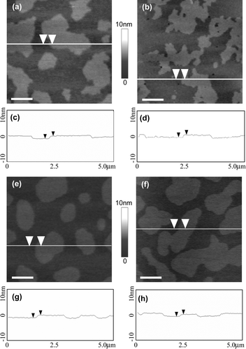
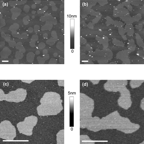
Supported lipid bilayers composed of equimolar sphingomyelin, DOPC and cholesterol using either egg (mainly C16:0; Avanti Polar Lipids) or brain (mainly C18:0; Avanti Polar Lipids) sphingomyelin were formed and imaged by AFM (a–2d). Both the lipid mixtures exhibited phase separation and formed lo ‘raft’ domains which were approx. 0.7 nm higher than the surrounding ld ‘non-raft’ lipids, as described previously Citation[25], Citation[29]. In the bilayers containing egg sphingomyelin 35.3% of the surface area was in the higher lo raft domains, while in the brain sphingomyelin bilayers 34.0% was in the lo domain.
Figure 2. AFM images of supported lipid bilayers containing either egg, brain, palmitoyl or stearoyl sphingomyelin. Supported lipid bilayers composed of equimolar sphingomyelin, DOPC and cholesterol were imaged in fluid using tapping mode AFM. Surface images of bilayers containing (a) egg sphingomyelin, (b) brain sphingomyelin, (e) palmitoyl sphingomyelin or (f) stearoyl sphingomyelin. (c), (d), (g) and (h) cross-sections of images in (a), (b), (e) and (f), respectively, at the lines indicated. The arrows indicate a height difference of ∼0.7 nm between the phases in all the lipid bilayers. All images are 5 µm scans with 10 nm height scale. Bar = 1 µm.
The addition of TM-ACE and GPI-ACE to the lipid vesicles during preparation enabled these two proteins to be incorporated into the lipid bilayer and their distribution between the phases to be investigated. AFM images (n=9) were analyzed from repeated experiments for both TM-ACE and GPI-ACE in lipid mixtures containing either egg or brain sphingomyelin (). Both proteins were visualized as small protruding particles in the AFM image. The total number of protein molecules in the lo and ld regions was counted in order to determine the percentage of each protein associated with the lo rafts (). Surprisingly, in the supported lipid bilayers containing egg sphingomyelin both the TM-ACE and GPI-ACE were located almost exclusively in the ld non-raft phase with only 2.4% and 1.5%, respectively, present in the lo rafts (a, 3c). However, in bilayers formed from the lipid mixture containing brain sphingomyelin, although the TM-ACE was still found almost exclusively (96.8%) in the ld phase, a significant proportion (41.3%) of the GPI-ACE was associated with the lo raft domains (b, 3d). These results indicate directly that the type of membrane anchor on ACE determines its distribution between raft and non-raft domains of the membrane, although this differential distribution is only seen when bilayers contain brain sphingomyelin.
Figure 3. Distribution of TM-ACE and GPI-ACE in supported lipid bilayers. Supported lipid bilayers of equimolar sphingomyelin, DOPC and cholesterol containing either TM-ACE or GPI-ACE were imaged in fluid using tapping mode AFM. (a) TM-ACE in bilayers containing egg sphingomyelin; the protein is almost exclusively located in ld non-raft regions. (b) TM-ACE in bilayers containing brain sphingomyelin; TM-ACE is excluded from the lo raft domains. (c) GPI-ACE in bilayers containing egg sphingomyelin; the protein is confined to ld non-raft regions. (d) GPI-ACE in bilayers containing brain sphingomyelin; 38% of the protein is located in lo rafts. All images are 10 µm scans with 10 nm height scale. Bar = 1 µm.
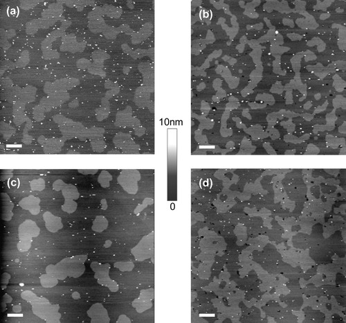
Table I. Statistical analysis of the distribution of proteins located in lo rafts.
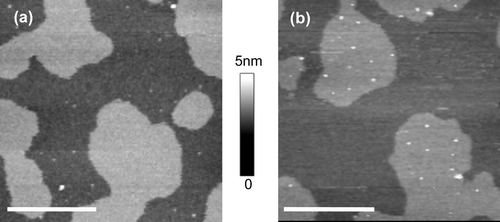
In order to determine whether the sphingomyelin species affected the distribution of another GPI-anchored protein between rafts and non-raft domains, we investigated the distribution of MDP, a well-characterized GPI-anchored protein whose complete anchor structure has been determined Citation[35]. MDP purified from porcine kidney migrated as a single band of 45 kDa on SDS polyacrylamide gel electrophoresis () and had a specific activity of 42.3 µmol/min/mg with Gly-D-Phe as substrate, similar to that reported previously Citation[33]. MDP was incorporated into lipid vesicles of equimolar sphingomyelin, DOPC and cholesterol using either egg or brain sphingomyelin (). Images (n=9) from repeated experiments were analyzed in order to determine the percentage of the protein in lo rafts with each sphingomyelin species (). As with GPI-ACE, MDP was also essentially excluded from lo rafts when egg sphingomyelin was used in the lipid mixture (a). However, when brain sphingomyelin was used, the majority (92.8%) of the MDP was localized in the lo raft domains (b).
Figure 4. Distribution of MDP in supported lipid bilayers. Supported lipid bilayers of equimolar sphingomyelin, DOPC and cholesterol containing MDP were imaged in fluid using tapping mode AFM. (a) MDP in bilayers containing egg sphingomyelin; the protein is almost exclusively located in ld non-raft regions. (b) MDP in bilayers containing brain sphingomyelin; the protein is almost exclusively located in lo raft domains. All images are 2.5µm scans with 5nm height scale. Bar = 1 µm.
To verify that the association of the GPI-anchored proteins with brain sphingomyelin rafts was attributable to the length of the sphingomyelin acyl chains, bilayers were formed from lipid mixtures containing either synthetic palmitoyl or stearoyl sphingomyelin (Avanti Polar Lipids) (e–2h). Both the lipid mixtures exhibited phase separation and formed lo domains which were approx. 0.7 nm higher than the surrounding ld lipids. In the bilayers made from palmitoyl sphingomyelin 33.9% of the surface area was in the lo raft domains, while in the stearoyl sphingomyelin bilayers 36.5% was in the lo domains. Both GPI-ACE and MDP exhibited a similar distribution in the synthetic palmitoyl and stearoyl sphingomyelin rafts as they did in egg and brain sphingomyelin rafts (); 42.2% of GPI-ACE and 93.9% of MDP were localized to lo raft domains when stearoyl sphingomyelin was used in the lipid bilayer mixture (). Conversely, only 0.8% of GPI-ACE and 0.9% of MDP associated with the palmitoyl sphingomyelin rafts ().
Figure 5. Distribution of GPI-ACE and MDP in supported lipid bilayers containing either palmitoyl or stearoyl sphingomyelin. Supported lipid bilayers composed of equimolar sphingomyelin, DOPC and cholesterol were imaged in fluid using tapping mode AFM. (a) GPI-ACE in bilayers containing palmitoyl sphingomyelin; the protein is confined to ld non-raft regions. (b) GPI-ACE in bilayers containing stearoyl sphingomyelin; 46.3% of the protein is located in lo rafts. (c) MDP in bilayers containing palmitoyl sphingomyelin; the protein is almost exclusively located in ld non-raft regions. (d) MDP in bilayers containing stearoyl sphingomyelin; the protein is predominantly (91.2%) located in lo raft domains. Images (a) and (b) are 10 µm scans with 10 nm height scale. (c) and (d) are 2.5 µm scans with 5 nm height scale. ?Bar = 1 µm.
Discussion
The distribution of membrane proteins in a lipid bilayer can be influenced by a variety of factors, including the length of the transmembrane domain, oligomerization with other proteins, and the nature of its acylation Citation[36]. By incorporating proteins into supported lipid bilayers, we have for the first time directly visualized by AFM the effect of (i) a GPI anchor versus a transmembrane anchor on the partitioning of a membrane protein in rafts, and (ii) sphingomyelin chain length on the distribution of GPI-anchored proteins in rafts. TM-ACE, a transmembrane protein which is not located in lipid rafts in the plasma membrane of cells Citation[37], Citation[38], was essentially excluded from the lo raft domains in the supported lipid bilayer. However, when the transmembrane and cytosolic domains in TM-ACE were exchanged for a GPI anchor and the resulting GPI-ACE was incorporated into a bilayer, 41% of the protein entered the lo rafts when the bilayer contained brain sphingomyelin. This result directly demonstrates that it is the GPI anchor and not the ectodomain that is responsible for targeting the protein to lo rafts in this system. Although one might have expected the orientation of reconstituted proteins to be randomized, with half the molecules facing inwards, experimental data show that the asymmetric orientation of ectoenzymes like ACE and MDP is maintained upon reconstitution into artificial lipid bilayers Citation[39], Citation[40].
AFM revealed that placental alkaline phosphatase was located almost exclusively (92%) in lo rafts in supported lipid bilayers containing brain sphingomyelin Citation[29]. Whether our observation that only 41% of GPI-ACE was associated with lo rafts suggests that another factor could be limiting the inclusion of this protein in the raft domains is not clear. Interestingly though, analysis of the transfected CHO cells that express GPI-ACE showed that upon treatment with Triton X-100 and separation on a sucrose gradient, only 52.8% of the GPI-ACE was located in DRMs Citation[18], suggesting that its partial localization to rafts in both cells and model membranes may be an intrinsic feature of this protein.
Accurate measurement of protein dimensions by AFM is extremely challenging. The triangular shape of the AFM tip can impede accurate width measurements due to the side of the tip making contact with the particle before the apex – referred to as ‘tip convolution’. As a result width measurements can be greatly amplified and, for irregular shaped particles such as proteins, these effects may be further exaggerated, especially when the particles are not directly fixed to the mica surface as in a lipid bilayer. Height measurements, although more accurate than width, are also susceptible to inaccuracies due to lack of control of electrostatic forces between the tip and the protein Citation[41] as well as the physical forces of tapping. Therefore, in order to obtain accurate measurements of protein dimensions by AFM, conditions have to be specifically tailored to the individual protein. This was not possible within this study which required consistent conditions for direct comparison between different proteins. So it is not possible for us to obtain accurate measurements for the dimensions of the proteins in our AFM images and therefore it is not possible for us to determine if the protein particles within our images correspond to individual proteins.
However, we have compared the relative dimensions of the protein particles within the same images, between replica samples and in the various lipid mixtures and have concluded: (i) For all 3 proteins used, in all the replica images, the largest particle does not exceed 4× the smallest particle. Therefore the largest possible aggregate present contains 4 of the smallest particles. For all images, this 4× aggregate represents less than 8% of the total number of particles in a single image; (ii) for all 3 proteins used, in all the replica images, the smallest particle represents at least 60% of the total number of particles in a single image; (iii) for images where the protein is distributed between the lo and ld phase, there is no significant difference between the particle sizes in each phase. Therefore, inclusion or exclusion into the lo phase does not appear to be dependent on particle size or aggregation; and (iv) for all 3 proteins used, comparing the average particle size in each of the lipid mixtures shows no significant difference in particle size when different sphingomyelin species are used in the bilayer. Therefore, the lipid mixture does not appear to affect protein size or aggregation. In conclusion, although we cannot rule out some degree of protein aggregation, we consider the observations and conclusions made in our study are independent of aggregation. It should also be noted that other AFM studies show a similar distribution of particle sizes when the protein is reconstituted in bilayers Citation[29], Citation[42].
Although the nature of the lipids which form the bilayer has been shown to affect the distribution of some transmembrane proteins Citation[42–44], this has not been explored before for GPI-anchored proteins. Exchanging brain sphingomyelin for egg sphingomyelin caused GPI-ACE to be excluded from the lo raft domains. To further investigate the significance of the sphingomyelin species on the distribution of another GPI-anchored protein in lo rafts, we utilized MDP, an endogenous GPI-anchored protein that, following Triton X-100 extraction, is found exclusively in DRMs Citation[45]. When incorporated into supported lipid bilayers containing egg sphingomyelin, MDP was excluded from lo rafts but the exchange of egg sphingomyelin for brain sphingomyelin resulted in 93% of MDP being located in the lo raft domains. The predominant difference between egg and brain sphingomyelin is the acyl chain length. Egg sphingomyelin is primarily (84%) composed of palmitoyl (C16:0) acyl chains and brain sphingomyelin mainly (88%) consists of lipids with chain lengths of C18 or longer. The effect of sphingomyelin acyl chain length on the raft association of GPI-anchored proteins was confirmed using synthetic palmitoyl and stearoyl sphingomyelin lipids. Both GPI-ACE (42%) and MDP (94%) localized to the stearoyl sphingomyelin raft domains but were excluded from lo rafts when palmitoyl sphingomyelin was used.
Despite brain sphingomyelin containing approx. 20% unsaturated acyl chains the surface area covered by the lo phase was very similar to that seen with stearoyl sphingomyelin. We assume that the unsaturated sphingomyelin molecules (all of which are monounsaturated) have been incorporated into the lo phase. Monounsaturated sphingolipids can form the lo phase as long as there is sufficient cholesterol. Previous studies have shown that the phase transition of brain sphingomyelin is eliminated by the addition of equimolar cholesterol, indicative of the formation of a single lo phase Citation[46]. In addition, the unsaturated chains are all C24:1Δ15 (Avanti) and there is evidence to suggest that double bonds which occur beyond CΔ13 are too far down the acyl chain to interfere with the interaction between the cholesterol and sphingomyelin Citation[47].
The GPI anchor of MDP consists almost exclusively of distearoyl (C18:0) acyl chains Citation[35]. The incorporation of MDP into egg or palmitoyl sphingomyelin rafts would require the slightly longer acyl chains in the GPI anchor of MDP to associate with the tightly packed, shorter acyl chains of the sphingomyelin. Such an interaction has been shown to occur in lipid bilayers where the longer fatty acid interdigitates into the lower lipid leaflet Citation[48]. The findings of the present study suggest that such an interaction is unfavourable in the ordered lo domains, at least in supported lipid bilayers, and that the saturated acyl chains of the GPI anchor associate with the unsaturated ld lipids in preference to interdigitation. In contrast, brain and stearoyl sphingomyelin provide slightly longer acyl chains, of equivalent length to the acyl chains in the GPI anchor of MDP, and the MDP GPI anchor is able to insert into the outer leaflet of the lo raft regions formed from the sphingomyelin without causing interdigitation. In model bilayers prepared from mixtures containing sphingomyelin, unsaturated phosphatidylcholine and cholesterol, the outer and inner leaflets in lo domains are coupled Citation[5]. However, in cell membranes, the inner leaflet has a different lipid composition to that of the outer leaflet Citation[49]. Whether the transmembrane asymmetry found in biological membranes would allow the interdigitation of the long acyl chains in a GPI anchor with the inner bilayer remains to be seen.
The mismatch of the length of the hydrophobic portion of the polypeptide chain in transmembrane proteins with that of the surrounding lipids has been reported as a method of sorting such proteins into lipid rafts Citation[50–53]. The results of the present study suggest that hydrophobic mismatch also influences the distribution of GPI-anchored proteins in rafts. The studies which reported the association of placental alkaline phosphatase, which, like MDP, consists of a distearoyl GPI anchor Citation[54], with lipid rafts also used brain sphingomyelin or a synthetic stearoyl sphingolipid Citation[29], Citation[30]. Another study found that the variant surface glycoprotein from Trypanosoma brucei which contains C14 acyl and alkyl chains did not readily reconstitute into model membranes containing brain sphingomyelin Citation[17]. Together these studies give further support to our hypothesis that GPI-anchored proteins are targeted to rafts when the sphingomyelin species has an equivalent acyl chain length as the GPI anchor. The analysis of the acyl chain lengths of the sphingomyelin species in DRMs extracted from mast cells revealed a similar composition of both C16:0 and C18:0 chains Citation[55], while DRMs from rat brain membranes contained predominantly C18:0 sphingomyelin Citation[56]. What is not known from these studies is whether some individual rafts in the membrane consist predominantly of C16:0 sphingomyelin, while others consist primarily of C18:0 sphingomyelin, but our data would suggest that differences in sphingomyelin chain length composition may determine the segregation of particular GPI-anchored proteins into particular rafts.
This paper was first published online on iFirst on 03 May 2007.
Acknowledgements
A. E. Garner is in receipt of a studentship from the Biotechnology and Biological Sciences Research Council (BBSRC) of Great Britain. The financial support of the BBSRC and the Medical Research Council of Great Britain is gratefully acknowledged. We thank Dr S. Connell for assistance with the AFM, M. Nimick for assistance with the purification of ACE and MDP, Dr N. T. Watt for assistance with the statistical analysis and Dr R. A. Skidgel (University of Illinois at Chicago, USA) for the cDNA encoding GPI-ACE.
References
- Lagerholm BC, Weinreb GE, Jacobson K, Thompson NL. Detecting microdomains in intact cell membranes. Annu Rev Phys Chem 2005; 56: 309–336
- Morris R, Cox H, Mombelli E, Quinn PJ. Rafts, little caves and large potholes: How lipid structure interacts with membrane proteins to create functionally diverse membrane environments. Subcell Biochem 2004; 37: 35–118
- Mayor S, Rao M. Rafts: Scale-dependent, active lipid organization at the cell surface. Traffic 2004; 5: 231–240
- Simons K, Toomre D. Lipid rafts and signal transduction. Nat Rev Mol Cell Biol 2000; 1: 31–39
- Simons K, Vaz WL. Model systems, lipid rafts, and cell membranes. Annu Rev Biophys Biomol Struct 2004; 33: 269–295
- Brown DA, Rose JK. Sorting of GPI-anchored proteins to glycolipid-enriched membrane subdomains during transport to the apical cell surface. Cell 1992; 68: 533–544
- Hooper NM. Detergent-insoluble glycosphingolipid/cholesterol-rich membrane domains, lipid rafts and caveolae. Mol Membr Biol 1999; 16: 145–156
- Brown DA, London E. Structure and function of sphingolipid- and cholesterol-rich membrane rafts. J Biol Chem 2000; 275: 17221–17224
- Ipsen JH, Karlstrom G, Mouritsen OG, Wennerstrom H, Zuckermann MJ. Phase equilibria in the phosphatidylcholine-cholesterol system. Biochim Biophys Acta 1987; 905: 162–172
- Brown DA, London E. Structure and origin of ordered lipid domains in biological membranes. J Membr Biol 1998; 164: 103–114
- Brown DA. Seeing is believing: Visualization of rafts in model membranes. Proc Natl Acad Sci USA 2001; 98: 10517–10518
- Hooper NM, Turner AJ. Ectoenzymes of the kidney microvillar membrane. Differential solubilization by detergents can predict a glycosyl-phosphatidylinositol membrane anchor. Biochem J 1988; 250: 865–869
- Brown DA, London E. Functions of lipid rafts in biological membranes. Ann Rev Cell Develop Biol 1998; 14: 111–136
- Ferguson MAJ. The structure, biosynthesis and functions of glycosylphosphatidylinositol anchors, and the contributions of trypanosome research. J Cell Sci 1999; 112: 2799–2809
- Schroeder R, London E, Brown D. Interactions between saturated acyl chains confer detergent resistance on lipids and glycosylphosphatidylinositol (GPI)-anchored proteins: GPI-anchored proteins in liposomes and cells show similar behaviour. Proc Natl Acad Sci USA 1994; 91: 12130–12134
- Schroeder RJ, Ahmed SM, Zhu Y, London E, Brown DA. Cholesterol and sphingolipid enhance the Triton X-100 insolubility of glycosylphosphatidylinositol-anchored proteins by promoting the formation of detergent-insoluble ordered membrane domains. J Biol Chem 1998; 273: 1150–1157
- Benting J, Rietveld A, Ansorge I, Simons K. Acyl and alkyl chain length of GPI-anchors is critical for raft association in vitro. FEBS Lett 1999; 462: 47–50
- Parkin ET, Tan F, Skidgel RA, Turner AJ, Hooper NM. The ectodomain shedding of angiotensin-converting enzyme is independent of its localisation in lipid rafts. J Cell Sci 2003; 116: 3079–3087
- Cordy JM, Hussain I, Dingwall C, Hooper NM, Turner AJ. Exclusively targeting beta-secretase to lipid rafts by GPI-anchor addition up-regulates beta-site processing of the amyloid precursor protein. Proc Natl Acad Sci USA 2003; 100: 11735–11740
- Legler DF, Doucey M-A, Schneider P, Chapatte L, Bender FC, Bron C. Differential insertion of GPI-anchored GFPs into lipid rafts of live cells. FASEB J 2005; 19: 73–75
- Heerklotz H. Triton promotes domain formation in lipid raft mixtures. Biophys J 2002; 83: 2693–2701
- Heerklotz H, Szadkowska H, Anderson T, Seelig J. The sensitivity of lipid domains to small perturbations demonstrated by the effect of Triton. J Mol Biol 2003; 329: 793–799
- Heffer-Lauc M, Lauc G, Nimrichter L, Fromholt SE, Schnaar RL. Membrane redistribution of gangliosides and glycosylphosphatidylinositol-anchored proteins in brain tissue sections under conditions of lipid raft isolation. Biochim Biophys Acta 3 2005; 1686: 200–208
- Lichtenberg D, Goni FM, Heerklotz H. Detergent-resistant membranes should not be identified with membrane rafts. Trends Biochem Sci 2005; 30: 430–436
- Rinia HA, de Kruijff B. Imaging domains in model membranes with atomic force microscopy. FEBS Lett 2001; 504: 194–199
- Lawrence JC, Saslowsky DE, Edwardson JM, Henderson RM. Real-time analysis of the effects of cholesterol on lipid raft behavior using atomic force microscopy. Biophys J 2003; 84: 1827–1832
- Hussain MA, Agnihotri A, Siedlecki CA. AFM imaging of ligand binding to platelet integrin alphaIIbbeta3 receptors reconstituted into planar lipid bilayers. Langmuir 2005; 21: 6979–6986
- Milhiet PE, Giocondi MC, Baghdadi O, Ronzon F, Roux B, Le Grimellec C. Spontaneous insertion and partitioning of alkaline phosphatase into model lipid rafts. EMBO Rep 2002; 3: 485–490
- Saslowsky DE, Lawrence J, Ren X, Brown DA, Henderson RM, Edwardson JM. Placental alkaline phosphatase is efficiently targeted to rafts in supported lipid bilayers. J Biol Chem 2002; 277: 26966–26970
- Kahya N, Brown DA, Schwille P. Raft partitioning and dynamic behavior of human placental alkaline phosphatase in giant unilamellar vesicles. Biochemistry 2005; 44: 7479–7489
- Hooper NM, Turner AJ. Isolation of two differentially glycosylated forms of peptidyl-dipeptidase A (angiotensin converting enzyme) from pig brain: a re-evaluation of their role in neuropeptide metabolism. Biochem J 1987; 241: 625–633
- Marcic B, Deddish PA, Skidgel RA, Erdos EG, Minshall RD, Tan F. Replacement of the transmembrane anchor in angiotensin I-converting enzyme (ACE) with a glycosylphosphatidylinositol tail affects activation of the B2 bradykinin receptor by ACE inhibitors. J Biol Chem 2000; 275: 16110–16118
- Littlewood GM, Hooper NM, Turner AJ. Ectoenzymes of the kidney microvillar membrane. Affinity purification, characterization and localization of the phospholipase C-solubilized form of renal dipeptidase. Biochem J 1989; 257: 361–367
- Smith PK, Krohn RI, Hermanson GT, Mallia AK, Gartner FH, Provenzano MD, Fujimoto EK, Goeke BJ, Olson BJ, Klenk DC. Measurement of protein using bicinchoninic acid. Anal Biochem 1985; 150: 76–85
- Brewis IA, Ferguson MAJ, Mehlert A, Turner AJ, Hooper NM. Structures of the glycosyl-phosphatidylinositol anchors of porcine and human membrane dipeptidase. Interspecies comparison of the glycan core structures and further structural studies on the porcine anchor. J Biol Chem 1995; 270: 22946–22956
- Sprong H, van der Sluijs P, van Meer G. How proteins move lipids and lipids move proteins. Nat Rev Mol Cell Biol 2001; 2: 504–513
- Schnitzer JE, Oh P, Jacobson BS, Dvorak AM. Caveolae from luminal plasmalemma of rat lung endothelium: microdomains enriched in caveolin, Ca2 + -ATPase, and inositol trisphosphate receptor. Proc Natl Acad Sci USA 1995; 92: 1759–1763
- Parkin ET, Turner AJ, Hooper NM. Isolation and characterization of two distinct low-density, Triton-insoluble complexes from porcine lung membranes. Biochem J 1996; 319: 887–896
- Kenny AJ, Fulcher IS, McGill KA, Kershaw D. Proteins of the kidney microvillar membrane. Reconstitution of endopeptidase in liposomes shows that it is a short stalked protein. Biochem J 1983; 211: 755–762
- Gee NS, Kenny AJ. Proteins of the kidney microvillar membrane. The 130kDa protein in pig kidney recognised by monoclonal antibody GK5C1 is an ectoenzyme with aminopeptidase activity. Biochem J 1985; 230: 753–764
- Muller DJ, Engel A. The height of biomolecules measured with the atomic force microscope depends on electrostatic interactions. Biophys J 1997; 73: 1633–1644
- Geisse NA, Cover TL, Henderson RM, Edwardson JM. Targeting of Helicobacter pylori vacuolating toxin to lipid raft membrane domains analysed by atomic force microscopy. Biochem J 2004; 381: 911–917
- McIntosh TJ, Vidal A, Simon SA. Sorting of lipids and transmembrane peptides between detergent-soluble bilayers and detergent-resistant rafts. Biophys J 2003; 85: 1656–1666
- Ridder AN, van de Hoef W, Stam J, Kuhn A, de Kruijff B, Killian JA. Importance of hydrophobic matching for spontaneous insertion of a single-spanning membrane protein. Biochemistry 2002; 41: 4946–4952
- Parkin ET, Turner AJ, Hooper NM. Differential effects of glycosphingolipids on the detergent-insolubility of the glycosylphosphatidylinositol-anchored membrane dipeptidase. Biochem J 2001; 358: 209–216
- McIntosh TJ, Simon SA, Needham D, Huang CH. Structure and cohesive properties of sphingomyelin/cholesterol bilayers. Biochemistry 1992; 31: 2012–2020
- Ramstedt B, Slotte JP. Interaction of cholesterol with sphingomyelins and acyl-chain-matched phosphatidylcholines: a comparative study of the effect of the chain length. Biophys J 1999; 76: 908–915
- Mehlhorn IE, Florio E, Barber KR, Lordo C, Grant CW. Evidence that trans-bilayer interdigitation of glycosphingolipid long chain fatty acids may be a general phenomenon. Biochim Biophys Acta 1988; 939: 151–159
- Devaux PF, Morris R. Transmembrane asymmetry and lateral domains in biological membranes. Traffic 2004; 5: 241–246
- Dumas F, Lebrun MC, Tocanne JF. Is the protein/lipid hydrophobic matching principle relevant to membrane organization and functions?. FEBS Lett 1999; 458: 271–277
- de Planque MR, Killian JA. Protein-lipid interactions studied with designed transmembrane peptides: role of hydrophobic matching and interfacial anchoring. Mol Membr Biol 2003; 20: 271–284
- Lee AG. How lipids affect the activities of integral membrane proteins. Biochim Biophys Acta 2004; 1666: 62–87
- Vidal A, McIntosh TJ. Transbilayer peptide sorting between raft and nonraft bilayers: comparisons of detergent extraction and confocal microscopy. Biophys J 2005; 89: 1102–1108
- Redman CA, Thomas-Oates JE, Ogata S, Ikehara Y, Ferguson MAJ. Structure of the glycosyl-phosphatidylinositol membrane anchor of human placental alkaline phosphatase. Biochem J 1994; 302: 861–865
- Fridriksson EK, Shipkova PA, Sheets ED, Holowka D, Baird B, McLafferty FW. Quantitative analysis of phospholipids in functionally important membrane domains from RBL-2H3 mast cells using tandem high-resolution mass spectrometry. Biochemistry 1999; 38: 8056–8063
- Brugger B, Graham C, Leibrecht I, Mombelli E, Jen A, Wieland F, Morris R. The membrane domains occupied by glycosylphosphatidylinositol-anchored prion protein and Thy-1 differ in lipid composition. J Biol Chem 2004; 279: 7530–7536