
Abstract
Maternal stress, especially during early pregnancy, predisposes offspring to neuropsychiatric disorders. We hypothesized that maternal psychosocial stress (MPS) during pregnancy affects fetal structural neurodevelopment depending on the gestational age of exposure. Fetal sheep brains were harvested at 130 days gestation (dG, term 150 dG) from ewes frequently isolated from flock-mates during early gestation (first and second trimester; n = 10) or late gestation (third trimester; n = 10), or from control flock-mates (n = 8). Immunohistochemistry for formation of neuronal processes, myelination, synaptic density, cell proliferation and programed cell death was performed on brain tissue sections. Sections of the cortical gray matter, the hippocampal CA3 region and the superficial, subcortical and deep white matter were examined morphometrically. Stress effects depended on the brain region and time of exposure. Stress during early gestation but not during late gestation reduced the amount of neuronal processes in the cerebral cortex and hippocampus by 36.9 ± 10.1% (p < 0.05, mean ± SEM) and 36.9 ± 15.8% (p < 0.05), respectively, accompanied by a decrease in synaptic density in the cerebral cortex and hippocampus by 39.8 ± 23.1% (p < 0.05) and 32.9 ± 13.4% (p < 0.01). Myelination was decreased in white matter layers on average by 44.8 ± 11.7% (p < 0.05) accompanied by reduced (glial) cell proliferation in the deep white matter by 83.6 ± 12.4% (p < 0.05). In contrast, stress during the third trimester had no effect in any brain region. Chronic MPS during the first and second trimester induced prolonged effects on neuronal network and myelin formation which might contribute to disturbed neurobehavioral, cognitive and motor development in offspring of stressed mothers.
Many women are exposed to stressful events during pregnancy. Maternal stress especially during early pregnancy predisposes for offspring’s neuropsychiatric disorders. In our sheep study, we show that disturbance of fetal brain development is a potential mechanism and is worst during 1st and 2nd trimester.
Lay summary
Introduction
Fetal exposure to environmental factors in utero such as increased stress hormones may not only disturb fetal development but also increase the risk for acquiring health problems later in life (Charil et al., Citation2010; Van den Bergh et al., Citation2017). Maternal psychosocial stress (MPS) is one of the most common causes of increased fetal stress hormone levels. Between 30% and 41% of pregnant women experience psychosocial stress during pregnancy (Fontein-Kuipers et al., Citation2014; Van den Bergh et al., Citation2017; WHO, Citation2008).
Previous studies have shown that chronic MPS is detrimental to fetal brain development (Charil et al., Citation2010; Van den Bergh et al., Citation2017). In humans, MPS disturbs motor development (Grace, Bulsara, Robinson, & Hands, Citation2016) and increases the risk for cognitive, behavioral, and emotional problems such as autism, attention-deficit hyperactivity disorder (ADHD), depression and schizophrenia in later life (Charil et al., Citation2010; Raikkonen, Seckl, Pesonen, Simons, & Van den Bergh, Citation2011; Van den Bergh et al., Citation2017). Similarly, MPS is associated with behavioral disorders or motor deficits in the offspring of rodents (Patin, Vincent, Lordi, & Caston, Citation2004; Weinstock, Citation2008), sheep (Coulon et al., Citation2015) and nonhuman primates (Schneider, Moore, Kraemer, Roberts, & DeJesus, Citation2002). These effects may be the result of both programing of functional systems like the hypothalamic–pituitary–adrenal (HPA) axis and maturational effects on structural brain development. In addition, recent studies have proposed the significance of microglial programing and neuroinflammation as a result of MPS (Desplats, Gutierrez, Antonelli, & Frasch, Citation2019).
While much data exist on the glucocorticoid (GC)-induced programing of the activity of the HPA axis by epigenetic mechanisms (Glover, O'Connor, & O'Donnell, Citation2010; Moisiadis & Matthews, Citation2014b), not much is known on the influence of MPS on structural brain development. Human MRI studies have shown abnormalities in offspring brain development in neonates, adolescents and adults due to MPS or increased anxiety during pregnancy (Buss, Davis, Muftuler, Head, & Sandman, Citation2010; Dufoix et al., Citation2015; Favaro, Tenconi, Degortes, Manara, & Santonastaso, Citation2015; Qiu et al., Citation2013). Studies in rodents have shown that MPS reduces neurogenesis, neurite formation, synaptic density, as well as myelination of the hippocampus and frontal cortex in the offspring (Boersma et al., Citation2014; Braun et al., Citation2017; Charil et al., Citation2010). However, little is known about stress sensitive periods of fetal brain development. Data on rodents are difficult to translate to humans, since rodents are postnatal brain developers compared to humans (Darlington, Dunlop, & Finlay, Citation1999; Dobbing & Sands, Citation1979).
We hypothesized that MPS during pregnancy affects fetal structural neurodevelopment in relation to the gestational age of exposure since previous studies have shown that effects of MPS on functional, behavioral and neuropsychiatric outcome are dependent on the gestational age of the fetus (Charil et al., Citation2010; Van den Bergh et al., Citation2017). We examined the effects of MPS on neurodevelopment in fetal sheep, an animal model that is closely comparable to human physiology in multiple aspects (Morrison et al., Citation2018). Sheep are prenatal brain developer similar to humans (Dobbing & Sands, Citation1979). Like humans, they have a prolonged gestational period of 150 days compared to 20–24 days in rodents. Unlike rodents, sheep give birth only to one or two offspring. The stress model we used involved repetitive isolation of pregnant ewes from the rest of the flock which is widely regarded as a model of human psychosocial stress (Rakers et al., Citation2013). We examined the effects of MPS on the formation of neuronal processes and synapses as well as on myelination, cell proliferation and cell death since these developmental processes are affected by GC (Antonow-Schlorke et al., Citation2009; Brodhun et al., Citation2004).
Materials and methods
Animals
All experiments were approved by the Animal Welfare Commission of the Thuringian government. Long-Wool Merino Sheep were crossbred on a single occasion. After verification of pregnancy using ultrasound at 30 ± 2 days of gestation (dG, term 150 dG), ewes were randomly divided into three experimental groups that were exposed to stress either during the first and second trimester or during the third trimester of pregnancy or served as respective controls. Control and stressed ewes were held in different paddocks at the same facility. Ewes were fed 100% of appropriate nutritional requirements during pregnancy, and water was provided ad libitum. Temperature and the light-dark-rhythm resembled that of the natural environment. The study was conducted during springtime.
Maternal stress protocol
We used isolation as a well-described animal model of MPS in sheep (for details see (Rakers et al., Citation2013)). Briefly, ewes were repeatedly separated in a well-lit box (3.0 m × 3.0 m × 1.4 m) with no visual, tactile, or auditory contact with their flock-mates and no access to food or water twice per week for a duration of three hours between 30 and 100 ± 2 dG (equals 0.2–0.67 gestation, stress during the first and second trimester, n = 10) or between 100 and 120 ± 2 dG (equals 0.67–0.8 gestation, stress during the third trimester, n = 10). The animals could move freely in the confined space. Control ewes did not undergo isolation stress (controls, n = 8). Repetitive isolation provokes a physiological and hormonal stress response with only minor habituation (Rakers et al., Citation2013).
Fetal instrumentation and brain collection
After the end of the control or stress periods, ewes were transported to the surgical facility three days before chronic fetal instrumentation at 104 ± 1 dG (controls, stress during the first and second trimester) or 124 ± 1 dG (stress during the third trimester). Fetuses were chronically instrumented with catheters inserted into the carotid artery and the jugular vein and ECG electrodes to measure stress responsiveness of the HPA axis using a single hypotensive challenge (Rakers et al., Citation2013). Maternal and fetal arterial blood samples were taken before anesthesia to analyze blood gases using a blood gas analyzer (ABL600, Radiometer, Copenhagen, Denmark; measurements corrected to 39 °C). At 130 dG, ewes were anesthetized with 4% halothane and fetuses were delivered by Cesarean section. Fetuses were euthanized by rapid exsanguination while still under general anesthesia. Thereafter, ewes were euthanized by i.v. injection of a 16% solution of pentobarbital sodium (Fatal-Plus, Vortech Pharmaceuticals, Dearborn, MI, USA). Brains were harvested, and one hemisphere was cut in tissue blocks of 7 mm thickness that were immediately transferred into 4% neutrally buffered paraformaldehyde solution.
Histological processing
After immersion fixation of 7 to 9 days, blocks were embedded in paraffin. Paraffin slices (6 µm) were cut according to a fetal sheep brain atlas (Johnson, Sudheimer, Davis, & Winn, Citation2016) at the level of the frontal cortex (sections 0481-0599) and at the level of the hippocampal formation (sections 1121–1241). For anatomical orientation, slices were treated with hematoxylin–eosin (HE). In order to evaluate myelin formation, myelin basic protein immunohistochemistry was performed using the ABC technique as described in detail earlier (Antonow-Schlorke et al., Citation2009). Briefly, after antigen-retrieval (microwave, 750 W, 10 min, 0.01 M citrate buffer), immunohistochemistry was performed on tissue slices with a monoclonal rat anti-MBP antibody (MAB386, 1:500, Chemicon, Temecola, CA, USA) using the avidin–biotin–peroxidase complex method (Vectastain Elite ABC staining kit, Vector Labs, Burlingame, USA). MBP is selectively expressed by mature cells of the oligodendroglial lineage (Sternberger, Itoyama, Kies, & Webster, Citation1978). MBP IR marks oligodendrocyte somata and the myelin sheath that contains MBP as an integral and essential component of brain myelin (Sternberger et al., Citation1978). Adjacent tissue sections were used for silver staining (Sevier & Munger, Citation1965) to estimate neurite density, and to relate possible changes of myelination to neural process formation. In order to estimate the synaptic density of the cerebral cortex, slices were treated with anti-synaptophysin antibody (mouse anti-synaptophysin; S5768, Sigma-Aldrich, St. Louis, MO, U.S.A.; 1:800) using the ABC technique. The immunohistochemical distribution of synaptophysin shows 95% of all neocortical synapses as an integral Ca2+-binding membrane protein of their presynaptic vesicles (Navone et al., Citation1986). Cell proliferation was assessed using the ABC method for immunohistochemistry against ki67 (mouse anti-NCL-ki67-MM1; #600185, Novocastra, Newcastle, UK; 1:100), a marker protein in proliferative-active cells that was shown to be expressed during all phases of the cell cycle with exception of the resting phase (Scholzen & Gerdes, Citation2000). Developmental cell death was visualized by the TUNEL in situ method (TUNEL kit, Roche, Indianapolis, IN, U.S.A.).
Histological examination
Histological examination was performed as described previously (Antonow-Schlorke et al., Citation2009; Colberg et al., Citation2004) using light microscopy within the frontal lobe – the cerebral cortex, superficial white matter, subcortical white matter, deep white matter – and the CA3 region of the hippocampal formation. Light-microscopic images (Axioplan2, Zeiss, Oberkochen, Germany; x 63) were digitized using a three CCD color video camera (Axiocam HR, Zeiss) under standardized conditions. All images were analyzed using an image analysis program (Image J 1.48v, NIH, USA). Scale was calibrated using a microscope stage micrometer. The number of ki67+ cells and TUNEL + cells were counted manually in all regions within an area of 1.50 mm2 in a standardized field. Microscopic pictures of MBP IR, SYN IR and silver staining were transformed into binary images. The area of marked structures was measured after applying Otsu’s threshold clustering algorithm (Otsu, Citation1979) to select the region of interest. Otsu’s algorithm was chosen because it performed best for our samples compared to other algorithms available in Image J. Large blood vessels were excluded manually to prevent false-positive measurement. The area of MBP IR and silver staining was quantified in all brain regions, whereas the area of SYN IR was only assessed in selected regions of gray matter, here the cerebral cortex and the CA3 region of the hippocampus.
Statistics
Differences in the topographical distribution of silver staining, MBP IR, SYN IR and in the number of ki-67+ cells and TUNEL + cells between brain regions within the control group and differences in the area of silver staining, MBP IR, SYN IR as well as in the number of ki-67+ cells and TUNEL + cells between the experimental groups were tested with Generalized Estimating Equations (GEE) (Hardin & Hilbe, Citation2014) and the Sidak test was used as post hoc test to correct for multiple testing. We used sheep as subject variables, the immunohistochemical method as dependent variable and both experimental group and brain region as factors. Differences in physiological parameters of maternal and fetal blood gas analysis between the experimental groups were tested using one-way ANOVA, and the Sidak test was used as post hoc test to correct for multiple testing. Significance was chosen at p level of <0.05. Results are presented as mean ± SEM.
Results
Blood gas analysis
There were no differences in the pH values and concentration of maternal or fetal arterial blood gases between the first and second trimester or third trimester stress groups and controls. All parameters were within the physiological range ().
Table 1. Maternal and fetal blood gases before brain collection.
Formation of neuronal processes
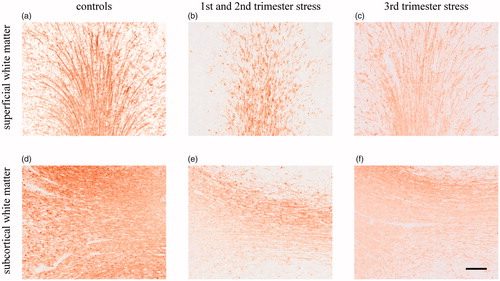
Neurons and their processes were visualized in all cortical layers using silver staining. The topographical distribution of the area of silver staining in control fetuses increased in direction from the superficial to the deep white matter reflecting increasing density of axonal processes (p < 0.01, , ). The area of silver staining in the hippocampus was similar to that seen in superficial white matter (, ). MPS during the first and second trimester reduced the area of silver staining in the cerebral cortex by 36.9 ± 10.1% (p < 0.05) and in the hippocampus by 36.9 ± 15.8% (p < 0.05) but it did not change the area of silver staining in the superficial, subcortical and deep white matter (, and ). In contrast to the effects of stress during the first and second trimester, stress during the third trimester did not change the area of silver staining in any brain region (, and ).
Figure 1. Effects of chronic maternal stress on neuronal network formation in fetal sheep brain at 0.87 gestation. Silver staining of the cerebral cortex (cor), superficial white matter (sfwm), subcortical white matter (swm), deep white matter (dwm) and the CA3 region of the hippocampus (hc). *p<.05, compared to controls; $$p<.01 compared to the next more superficial brain region; §§p<.01 compared to the hippocampus. Controls: n = 8, 1st/2nd trimester stress: n = 10, 3rd trimester stress: n = 10.
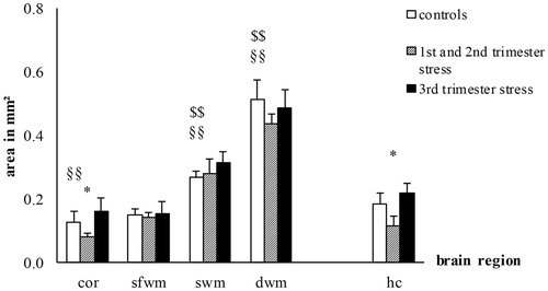
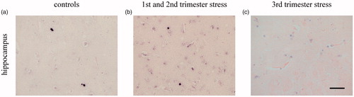
Figure 2. Effects of chronic maternal stress on neuronal network formation in frontal cortex and hippocampus of the fetal sheep brain at 0.87 gestation. Representative photomicrographs of silver staining of the cerebral cortex (a–c) and the CA3 region of the hippocampus (d–f). Stress during the first and second trimester but not stress during the third trimester reduced the area of silver staining. Scale bar 50 µm.
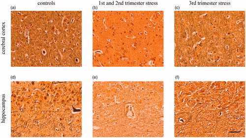
Table 2. Chronic effects of maternal psychosocial stress on structural brain development at 0.87 gestation.
Synaptic density
Synaptophysin immunoreactivity (SYN IR) was widely distributed in the cerebral cortex and hippocampus. Here, SYN IR appeared as a granular pattern in cell bodies and in the neuropil. In controls, the area of SYN IR in the cerebral cortex was higher than in the hippocampus (p < 0.01, , ). MPS during the first and second trimester reduced SYN IR in the cerebral cortex by 39.8 ± 23.1% (p < 0.05) and in the hippocampus by 32.9 ± 13.4% (p < 0.01, , and ). In contrast to the effects of stress during the first and second trimester, stress during the third trimester did not change the area of SYN IR in both brain regions (, and ).
Figure 3. Effects of chronic maternal stress on synaptic density in fetal sheep brain at 0.87 gestation. Anti-synaptophysin-immunohistochemistry in the cerebral cortex and the CA3 region of the hippocampus. *p<.05, **p<.01 compared to controls; §§p<.01 compared to the hippocampus. Controls: n = 8, 1st/2nd trimester stress: n = 10, 3rd trimester stress: n = 10.
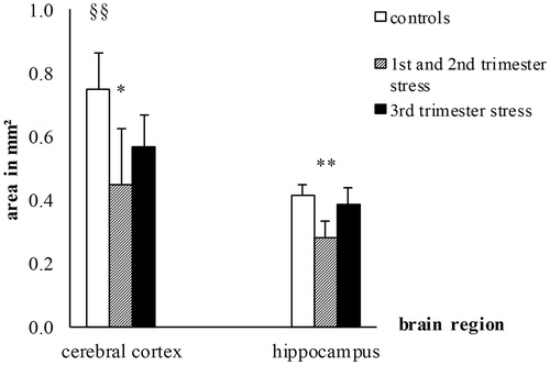
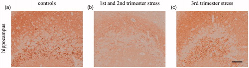
Figure 4. Effects of chronic maternal stress on synaptic density in frontal cortex of the fetal sheep brain at 0.87 gestation. Representative photomicrographs of synaptophysin immunohistochemistry (brown precipitation) of the CA3 region of the hippocampus (a–c). Stress during the first and second trimester but not stress during the third trimester reduced synaptophysin IR. Scale bar 100 µm.
Formation of myelin
Myelin basic protein immunoreactivity (MBP IR) clearly revealed single MBP + oligodendrocytes and myelinated fibers in the cerebral cortex and in the superficial, subcortical and deep white matter. In parallel to the increase in the density of axonal processes, the amount of MBP IR increased from the cerebral cortex towards the subcortical and deep white matter in control fetuses (p < 0.01, , ). MPS during the first and second trimester reduced MBP IR in the superficial, subcortical and deep white matter by 49.9 ± 18.0% (p < 0.01), 40.3 ± 5.0% (p < 0.05) and 47.1 ± 15.9% (p < 0.05) respectively (, and ) reflecting lower myelination. Stress during the first and second trimester did not change MBP IR in both the cerebral cortex and in the hippocampus (, ). In contrast to the effects of MPS during the first and second trimester, stress during the third trimester did not change MBP IR in any brain region (, and ).
Figure 5. Effects of chronic maternal stress on myelination in fetal sheep brain at 0.87 gestation. Anti-MBP-immunohistochemistry of the cerebral cortex (cor), superficial white matter (sfwm), subcortical white matter (swm), deep white matter (dwm), and the CA3 region of the hippocampus (hc). *p<.05, **p<.01 compared to controls; $$p<.01 compared to the next more superficial brain region; §§p<.01 compared to the hippocampus. Controls: n = 8, 1st/2nd trimester stress: n = 10, 3rd trimester stress: n = 10.

Figure 6. Effects of chronic maternal stress on myelination in white matter of fetal sheep brain at 0.87 gestation. Representative photomicrographs of MBP immunohistochemistry (brown precipitation) of the superficial white matter (a–c) and the subcortical white matter (d–f). Stress during the first and second trimester but not stress during the third trimester reduced MBP IR. Scale bar 200 µm.
Cell proliferation
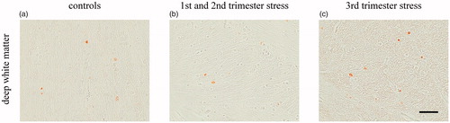
Ki-67 IR selectively revealed proliferative-active cells. In control fetuses, the number of ki67+ cells in subcortical white matter was higher than in superficial white matter (p < 0.05, , ). MPS during the first and second trimester diminished the number of ki-67+ cells in the deep white matter by 83.6 ± 12.4% (p < 0.05, , and ) but did not change the number of ki-67+ cells in the cortical gray matter, in the superficial and subcortical white matter or in the hippocampus (, ). In contrast to the effects of MPS during the first and second trimester, stress during the third trimester did not change the number of ki67+ cells in any region (, and ).
Figure 7. Effects of chronic maternal stress on cell proliferation in fetal sheep brain at 0.87 gestation. Anti-ki-67-immunohistochemistry of the cerebral cortex (cor), superficial white matter (sfwm), subcortical white matter (swm), deep white matter (dwm) and the CA3 region of the hippocampus (hc). *p<.05 compared to controls; $p<.05 compared to the next more superficial brain region. Controls: n = 8, 1st/2nd trimester stress: n = 10, 3rd trimester stress: n = 10.
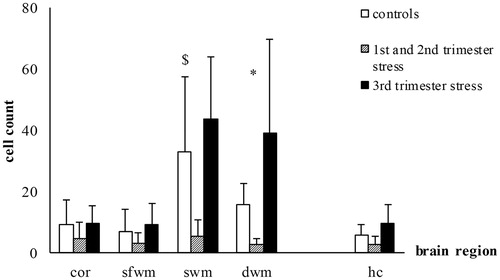
Figure 8. Effects of chronic maternal stress on cell proliferation in white matter of fetal sheep brain at 0.87 gestation. Representative photomicrographs of cell proliferation (ki-67 immunohistochemistry, brown precipitation) of the deep white matter (a–c). Stress during the first and second trimester but not stress during the third trimester decreased cell proliferation seen by the lower number of ki-67 marked cells. Scale bar 50 µm.
Developmental cell death
Individual TUNEL + cells appeared in all the regions examined. In control fetuses, the number of TUNEL + cells was similarly low in all brain regions (, ). Both MPS during the first and second trimester or during the third trimester did not change the number of TUNEL + cells in any brain region (, and ).
Figure 9. Effects of chronic maternal stress on developmental cell death in fetal sheep brain at 0.87 gestation. TUNEL-method of the cerebral cortex (cor), superficial white matter (sfwm), subcortical white matter (swm), deep white matter (dwm) and the CA3 region of the hippocampus (hc). Controls: n = 8, 1st/2nd trimester stress: n = 10, 3rd trimester stress: n = 10.
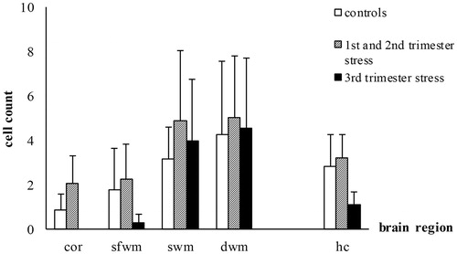
Figure 10. Effects of chronic maternal stress on developmental cell death in white matter of fetal sheep brain at 0.87 gestation. Representative photomicrographs of developmental cell death (TUNEL, black color, counterstaining with Nuclear Fast Red) of the deep white matter (a–c). Partly due to the low number of TUNEL marked cells and the high variance there were no effects on developmental cell death. Scale bar 50 µm.
Discussion
Most animal studies investigating the effects of prenatal stress on structural brain development used rodents and focused on a single time of stress exposure or a specific aspect of structural brain development such as myelination or neurogenesis (Boersma et al., Citation2014; Braun et al., Citation2017; Charil et al., Citation2010). We performed a comprehensive analysis of stress effects on brain development in multiple brain regions at two different fetal developmental periods in sheep, an animal model that is close to the time-course of brain development in humans. Our study provides evidence that prolonged MPS changes structural brain development of the fetus. Stress effects were dependent on the cerebral region and time period of exposure to maternal stress. Stress during the first and second trimester but not during the third trimester induced an alteration of the neuronal network formation shown by a reduced number of neuronal processes and synaptic density in the cerebral cortex and hippocampus, as well as decreased proliferative activity and reduced myelination in the white matter.
Neuronal network formation
The reduced neuronal network formation in the fetal brain highly likely reflects aberrant network maturation. It has previously been shown that MPS in the form of aversive handling of ewes during the second half of gestation delayed spine maturation in the hippocampus and the prefrontal cortex (Petit et al., Citation2015). On the mechanistic level, stress-associated exposure to increased cortisol concentrations seems to play an important role. In our stress model, MPS induces increased fetal cortisol levels (Dreiling et al., Citation2016). Corticosterone, the principal murine GC is a critical regulator of dendritic spine development (Liston & Gan, Citation2011). In previous studies in sheep and baboons, the cytoskeletal microtubule-associated proteins MAP1B and MAP2 that are critically involved in neuronal development were reduced transiently after administration of betamethasone during the last trimester of pregnancy (Antonow-Schlorke et al., Citation2003, Citation2007). Causally, the authors assumed catabolic GC effects, since GC suppress activity of mitochondrial respiratory chain enzymes (Schwab et al., Citation2006).
A lower energy supply could have reduced synaptic protein synthesis in our study. The comparable decrease in synaptic density and axonal density in the cerebral cortex and the hippocampus suggests that the reduction in synaptic density could be an epiphenomenon of the reduced axonal density. Our result on the decrease in synaptic density is in agreement with adverse effects of MPS on fetal hippocampal synaptic density in rats and non-human primates (Charil et al., Citation2010; Xu et al., Citation2013).
Myelin formation
Parallel to the altered network formation after MPS during the first and second trimester, we demonstrated decreased myelination and proliferative cell activity specifically in the subcortical and deep white matter where myelination was most advanced. Because the stress-related decrease of MBP IR was not accompanied by a loss of axonal processes, our results point to a specific stress effect on myelination. This effect may be mediated by decreased oligodendroglial proliferation. Although we did not differentiate between neuronal and glial proliferative activity, it is highly likely that the decrease in proliferative-active cells mainly involves glial cells since glial but not neuronal proliferation peaks during the second half of gestation (Barlow, Citation1969; McIntosh, Baghurst, Potter, & Hetzel, Citation1979). The low susceptibility of the cerebral cortex to stress may be due to the later onset of cortical myelination at the end of third trimester of pregnancy (Barlow, Citation1969). Indeed, myelination was still very low in the cerebral cortex during the third trimester. The effects of MPS on myelination in the white matter during the first and second trimester are in general agreement with findings in rodents (Suzuki et al., Citation2016; Xu et al., Citation2013). Although rodents were exposed to mid-to-late gestational stress, the stress period resembles that in our study since mice are born very prematurely with brains at a developmental stage of 0.25 to 0.5 gestation compared to sheep and humans (Bayer, Altman, Russo, & Zhang, Citation1993; Darlington et al., Citation1999).
The absence of the effects of MPS on brain development during the third trimester in the present study is in contrast to the effects of synthetic GC in the third trimester (Antonow-Schlorke et al., Citation2009; Huang, Harper, Evans, Newnham, & Dunlop, Citation2001) and could be explained by the ability of synthetic GC to cross the placental blood barrier unaffected by the placental 11ß-HSD 2 (Benediktsson, Calder, Edwards, & Seckl, Citation1997) which inactivates 80–90% of maternal cortisol to cortisone (Rakers et al., Citation2017). Moreover synthetic GC do not bind to mineralocorticoid receptors which are known to produce neuroprotective effects (Hassan, von Rosenstiel, Patchev, Holsboer, & Almeida, Citation1996).
Developmental cell death
Although previous studies have shown an increase of developmental cell death in nonhuman primates (Uno et al., Citation1994) and humans (Tijsseling et al., Citation2012) following antenatal GC treatment during the third trimester, we did not find any effect of MPS on TUNEL + cells. This may be in part due to low number and high variance of TUNEL + cells in our study. Again, the different binding characteristics of synthetic GC and cortisol to GR and mineralocorticoid receptors may also explain the absent effect of MPS in our study (Hassan et al., Citation1996).
Vulnerable periods and long-term implications
There was no effect of MPS during the third trimester on markers of structural brain development. The higher vulnerability of brain maturation to MPS during the first and second trimester is most likely due to the ongoing processes of neuronal migration, network formation and myelination at this earlier stage of development (Rakic, Citation1988). In agreement with our results, Xu et al. (Citation2013) showed that restraint stress in rats during mid-to-late gestation has more pronounced effects on myelination and synaptophysin expression in the fetal brain than restraint stress in late gestation.
The high sensitivity of structural brain development to stress during the first and second trimester parallels the vulnerability of the fetus at this gestational age to programing effects of the activity of the HPA axis (Rakers et al., Citation2013). Thus, the effects of MPS on structural brain development may be mediated in part by programing of the hyperactivity of the HPA axis. On the other hand, GC receptor mRNA was not detected before 60 dG in fetal sheep (Andrews & Matthews, Citation2000). However, it is not clear whether GC receptors are indispensable for mediating stress effects on brain development. For example, oxidative stress, immunomodulatory cytokines and catecholamines (via an acute reduction of the utero-placental perfusion) can also transfer maternal stress to the fetus (Rakers et al., Citation2017).
It is unlikely that chronic fetal instrumentation for a three week period following stress during the first and second trimester was responsible for the adverse effects on structural brain development, since maternal and fetal blood gas analysis showed normal physiological values throughout the period of instrumentation. Moreover, the period of fetal instrumentation in this group was during the third trimester when stress did not show any effect on structural brain development. Thus, the changes in neuronal network formation and myelination even 30 days after the last stress exposure indicate lasting structural effects of MPS in the fetal brain. Indeed, adverse effects of MPS on spine density of hippocampal and prefrontal cortical neurons could be still shown in adult rats (Radley et al., Citation2006). In rhesus monkeys, in whom a loss of cortical and hippocampal pyramidal neurons was seen at term after antenatal GC treatment in the early third trimester, a reduced hippocampal volume was still detectable by MRI at 19 to 20 months of age (Uno et al., Citation1994). In humans, delayed hippocampal growth was shown in children of mothers who reported either increased anxiety during the first or second trimester (Qiu et al., Citation2013) or experienced stress due to a natural disaster while pregnant (Dufoix et al., Citation2015). Similarly, gray matter volume was decreased in children of mothers who reported high anxiety during mid gestation (Buss et al., Citation2010) or had experienced highly stressful events during pregnancy (Favaro et al., Citation2015). The higher sensitivity of structural brain development to MPS during the first and second than during the third trimester is also associated with more pronounced long-term behavioral and neuropsychiatric consequences in rats (Patin et al., Citation2004), non-human primates (Schneider, Roughton, Koehler, & Lubach, Citation1999) and humans (Van den Bergh et al., Citation2017) although some studies in humans found more pronounced effects of late gestation stress on motor development (Grace et al., Citation2016; Simcock et al., Citation2016).
Limitations
Since we only distinguished between MPS during the first and second trimester or the third trimester, we cannot determine the exact time period during the first or second trimester that is particularly vulnerable. We also cannot exclude that the longer duration of fetal exposure to stress over the first and second trimester compared to the shorter exposure over the third trimester contributed to the more pronounced effects of MPS. It is important to note that we did not examine sex effects in this exploratory study. A few previous studies suggest greater vulnerability to MPS in male rodent offspring, possibly because of lower expression and activity of 11ß-HSD 2 and therefore higher maternal-fetal cortisol transfer in males (Charil et al., Citation2010). Fetal neuroinflammation and microglial programing which may affect brain development also seem to be sex-specific and to be more pronounced in males (Desplats et al., Citation2019; Hanamsagar & Bilbo, Citation2016). However, studies which examined sex-specific effects of MPS on brain development in humans showed inconsistent results, depending on the kind of stressor used and the structural or functional system that was examined (Van den Bergh et al., Citation2017).
Conclusions
In agreement with clinical studies showing more pronounced effects of MPS exposure on behavioral and neuropsychiatric outcome during early rather than late pregnancy (Van den Bergh et al., Citation2017), we could demonstrate that prolonged MPS exposure during the first and second but not during the third trimester has complex and multifactorial effects on neuronal network and myelin formation in the fetal sheep brain. Neuronal network and myelin formation represent hallmarks of brain development which determine proper brain function. These stress effects on structural brain development are complementary to the higher sensitivity of the HPA axis to the programing effects of MPS during the first and second rather than during the third trimester (Rakers et al., Citation2013). The stress effects on structural brain development seem to be longer lasting, since they were still present after a recovery period of 30 days. Future studies are needed to determine the long-term functional relevance of the aberrations in neuronal network and myelin formation. Furthermore, region-specific differences in neuroinflammation and microglial activity would be of interest to extend the findings in our study.
Acknowledgement
The authors thank Ina Ingrisch, Claudia Sommer and Thi Kim Chi Le for excellent technical assistance and are grateful to Nasim Kroegel for language editing.
Disclosure statement
All authors report no conflicts of interest.
Additional information
Funding
References
- Andrews, M.H., & Matthews, S.G. (2000). Regulation of glucocorticoid receptor mRNA and heat shock protein 70 mRNA in the developing sheep brain. Brain Research, 878, 174–182. doi:10.1016/S0006-8993(00)02735-9
- Antonow-Schlorke, I., Helgert, A., Gey, C., Coksaygan, T., Schubert, H., Nathanielsz, P.W., … Schwab, M. (2009). Adverse effects of antenatal glucocorticoids on cerebral myelination in sheep. Obstetrics & Gynecology, 113, 142–151. doi:10.1097/AOG.0b013e3181924d3b
- Antonow-Schlorke, I., Müller, T., Brodhun, M., Wicher, C., Schubert, H., Nathanielsz, P.W., … Schwab, M. (2007). Betamethasone-related acute alterations of microtubule-associated proteins in the fetal sheep brain are reversible and independent of age during the last one-third of gestation. American Journal of Obstetrics and Gynecology, 196, 553.e1–556. doi:10.1016/j.ajog.2006.10.898
- Antonow-Schlorke, I., Schwab, M., Li, C., & Nathanielsz, P.W. (2003). Glucocorticoid exposure at the dose used clinically alters cytoskeletal proteins and presynaptic terminals in the fetal baboon brain. The Journal of Physiology, 547, 117–123. doi:10.1111/j.1469-7793.2003.00117.x
- Barlow, R.M. (1969). Foetal sheep – morphogenesis of nervous system and histochemical aspects of myelination. The Journal of Comparative Neurology, 135, 249–262. doi:10.1002/cne.901350302
- Bayer, S.A., Altman, J., Russo, R.J., & Zhang, X. (1993). Timetables of neurogenesis in the human brain based on experimentally determined patterns in the rat. Neurotoxicology, 14, 83–144.
- Benediktsson, R., Calder, A.A., Edwards, C.R.W., & Seckl, J.R. (1997). Placental 11 beta-hydroxysteroid dehydrogenase: A key regulator of fetal glucocorticoid exposure. Clinical Endocrinology, 46, 161–166. doi:10.1046/j.1365-2265.1997.1230939.x
- Boersma, G.J., Bale, T.L., Casanello, P., Lara, H.E., Lucion, A.B., Suchecki, D., & Tamashiro, K.L. (2014). Long-term impact of early life events on physiology and behaviour. Journal of Neuroendocrinology, 26, 587–602. doi:10.1111/jne.12153
- Braun, K., Bock, J., Wainstock, T., Matas, E., Gaisler-Salomon, I., Fegert, J., … Segal, M. (2017). Experience-induced transgenerational (re-)programming of neuronal structure and functions: Impact of stress prior and during pregnancy. Neuroscience & Biobehavioral Reviews, pii: S0149-7634(16)30731-X.
- Brodhun, M., Mueller, T., Coksaygan, T., Antonow-Schlorke, I., Schubert, H., Patt, S., … Schwab, M. (2004). Neurogenesis and programmed cell death (PCD) during development of the fetal sheep brain. Journal of the Society for Gynecologic Investigation, 11, 253a–253a.
- Buss, C., Davis, E.P., Muftuler, L.T., Head, K., & Sandman, C.A. (2010). High pregnancy anxiety during mid-gestation is associated with decreased gray matter density in 6–9-year-old children. Psychoneuroendocrinology, 35, 141–153. doi:10.1016/j.psyneuen.2009.07.010
- Charil, A., Laplante, D.P., Vaillancourt, C., & King, S. (2010). Prenatal stress and brain development. Brain Research Reviews, 65, 56–79. doi:10.1016/j.brainresrev.2010.06.002
- Colberg, C., Antonow-Schlorke, I., Muller, T., Schubert, H., Witte, O.W., & Schwab, M. (2004). Recovery of glucocorticoid-related loss of synaptic density in the fetal sheep brain at 0.75 of gestation. Neuroscience Letters, 364, 130–134. doi:10.1016/j.neulet.2004.04.052
- Coulon, M., Nowak, R., Andanson, S., Petit, B., Levy, F., & Boissy, A. (2015). Effects of prenatal stress and emotional reactivity of the mother on emotional and cognitive abilities in lambs. Developmental Psychobiology, 57, 626–636. doi:10.1002/dev.21320
- Darlington, R.B., Dunlop, S.A., & Finlay, B.L. (1999). Neural development in metatherian and eutherian mammals: Variation and constraint. The Journal of Comparative Neurology, 411, 359–368. doi:10.1002/(SICI)1096-9861(19990830)411:3<359::AID-CNE1>3.3.CO;2-A
- Desplats, P., Gutierrez, A.M., Antonelli, M.C., & Frasch, M.G. (2019). Microglial memory of early life stress, epigenetic mechanisms and susceptibility to neurodegeneration in adulthood. arXiv e-prints. Retrieved January, 01, 2019,
- Dobbing, J., & Sands, J. (1979). Comparative aspects of the brain growth spurt. Early Human Development, 3, 79–83. doi:10.1016/0378-3782(79)90022-7
- Dreiling, M., Bischoff, S., Schiffner, R., Rupprecht, S., Kiehntopf, M., Schubert, H., … Rakers, F. (2016). Stress-induced decrease of uterine blood flow in sheep is mediated by alpha 1-adrenergic receptors. Stress, 19, 547–551. doi:10.1080/10253890.2016.1203417
- Dufoix, R., Charil, A., Laplante, D.P., Paus, T., Pruessner, J., & King, S. (2015). Prenatal maternal stress from a natural disaster predicts hippocampus volumes in males at age 11: Project Ice Storm. International Journal of Developmental Neuroscience, 47, 12. doi:10.1016/j.ijdevneu.2015.04.042
- Favaro, A., Tenconi, E., Degortes, D., Manara, R., & Santonastaso, P. (2015). Neural correlates of prenatal stress in young women. Psychological Medicine, 45, 2533–2543. doi:10.1017/S003329171500046X
- Fontein-Kuipers, Y.J., Nieuwenhuijze, M.J., Ausems, M., Bude, L., & de Vries, R. (2014). Antenatal interventions to reduce maternal distress: A systematic review and meta-analysis of randomised trials. BJOG: An International Journal of Obstetrics & Gynaecology, 121, 389–397. doi:10.1111/1471-0528.12500
- Glover, V., O'Connor, T.G., & O'Donnell, K. (2010). Prenatal stress and the programming of the HPA axis. Neuroscience and Biobehavioral Reviews, 35, 17–22. doi:10.1016/j.neubiorev.2009.11.008
- Grace, T., Bulsara, M., Robinson, M., & Hands, B. (2016). The impact of maternal gestational stress on motor development in late childhood and adolescence: A longitudinal study. Child Development, 87, 211–220. doi:10.1111/cdev.12449
- Hanamsagar, R., & Bilbo, S.D. (2016). Sex differences in neurodevelopmental and neurodegenerative disorders: Focus on microglial function and neuroinflammation during development. The Journal of Steroid Biochemistry and Molecular Biology, 160, 127–133. doi:10.1016/j.jsbmb.2015.09.039
- Hardin, J.W., & Hilbe, J.M. (2014). Generalized estimating equations: Introduction. Wiley StatsRef: Statistics Reference Online.
- Hassan, A.H., von Rosenstiel, P., Patchev, V.K., Holsboer, F., & Almeida, O.F. (1996). Exacerbation of apoptosis in the dentate gyrus of the aged rat by dexamethasone and the protective role of corticosterone. Experimental Neurology, 140, 43–52. doi:10.1006/exnr.1996.0113
- Huang, W.L., Harper, C.G., Evans, S.F., Newnham, J.P., & Dunlop, S.A. (2001). Repeated prenatal corticosteroid administration delays myelination of the corpus callosum in fetal sheep. International Journal of Developmental Neuroscience, 19, 415–425. doi:10.1016/S0736-5748(01)00026-0
- Johnson, J. L., Sudheimer, K. D., Davis, K. K., & Winn, B. M. (2016). The navigable atlas of the sheep brain. Retrieved from: https://msu.edu/∼brains/brains/sheep/index.html
- Liston, C., & Gan, W.B. (2011). Glucocorticoids are critical regulators of dendritic spine development and plasticity in vivo. Proceedings of the National Academy of Sciences of Sciences, 108, 16074–16079. doi:10.1073/pnas.1110444108
- McIntosh, G.H., Baghurst, K.I., Potter, B.J., & Hetzel, B.S. (1979). Foetal brain development in the sheep. Neuropathology and Applied Neurobiology, 5, 103–114. doi:10.1111/j.1365-2990.1979.tb00664.x
- Moisiadis, V.G., & Matthews, S.G. (2014). Glucocorticoids and fetal programming part 2: Mechanisms. Nature Reviews Endocrinology, 10, 403–411. doi:10.1038/nrendo.2014.74
- Morrison, J.L., Berry, M.J., Botting, K.J., Darby, J.R.T., Frasch, M.G., Gatford, K.L., … Tellam, R.L. (2018). Improving pregnancy outcomes in humans through studies in sheep. American Journal of Physiology-Regulatory Integrative and Comparative Physiology, 315, R1123–R1153. doi:10.1152/ajpregu.00391.2017
- Navone, F., Jahn, R., Di Gioia, G., Stukenbrok, H., Greengard, P., & De Camilli, P. (1986). Protein p38: An integral membrane protein specific for small vesicles of neurons and neuroendocrine cells. The Journal of Cell Biology, 103, 2511–2527. doi:10.1083/jcb.103.6.2511
- Otsu, N. (1979). A threshold selection method from Gray-level histograms. IEEE Transactions on Systems, Man, and Cybernetics, 9, 62–66. doi:10.1109/TSMC.1979.4310076
- Patin, V., Vincent, A., Lordi, B., & Caston, J. (2004). Does prenatal stress affect the motoric development of rat pups? Developmental Brain Research, 149, 85–92. doi:10.1016/j.devbrainres.2003.12.008
- Petit, B., Boissy, A., Zanella, A., Chaillou, E., Andanson, S., Bes, S., … Coulon, M. (2015). Stress during pregnancy alters dendritic spine density and gene expression in the brain of new-born lambs. Behavioural Brain Research, 291, 155–163. doi:10.1016/j.bbr.2015.05.025
- Qiu, A., Rifkin-Graboi, A., Chen, H., Chong, Y.-S., Kwek, K., Gluckman, P.D., … Meaney, M.J. (2013). Maternal anxiety and infants' hippocampal development: Timing matters. Translational Psychiatry, 3, e306.
- Radley, J.J., Rocher, A.B., Miller, M., Janssen, W.G.M., Liston, C., Hof, P.R., … Morrison, J.H. (2006). Repeated stress induces dendritic spine loss in the rat medial prefrontal cortex. Cerebral Cortex, 16, 313–320. doi:10.1093/cercor/bhi104
- Raikkonen, K., Seckl, J.R., Pesonen, A.K., Simons, A., & Van den Bergh, B.R. (2011). Stress, glucocorticoids and liquorice in human pregnancy: Programmers of the offspring brain. Stress, 14, 590–603. doi:10.3109/10253890.2011.602147
- Rakers, F., Frauendorf, V., Rupprecht, S., Schiffner, R., Bischoff, S.J., Kiehntopf, M., … Schwab, M. (2013). Effects of early- and late-gestational maternal stress and synthetic glucocorticoid on development of the fetal hypothalamus-pituitary-adrenal axis in sheep. Stress, 16, 122–129. doi:10.3109/10253890.2012.686541
- Rakers, F., Rupprecht, S., Dreiling, M., Bergmeier, C., Witte, O.W., & Schwab, M. (2017). Transfer of maternal psychosocial stress to the fetus. Neuroscience & Biobehavioral Reviews, pii: S0149-7634(16)30719-9.
- Rakic, P. (1988). Defects of neuronal migration and the pathogenesis of cortical malformations. Progress in Brain Research, 73, 15–37.
- Schneider, M.L., Moore, C.F., Kraemer, G.W., Roberts, A.D., & DeJesus, O.T. (2002). The impact of prenatal stress, fetal alcohol exposure, or both on development: Perspectives from a primate model. Psychoneuroendocrinology, 27, 285–298. doi:10.1016/S0306-4530(01)00050-6
- Schneider, M.L., Roughton, E.C., Koehler, A.J., & Lubach, G.R. (1999). Growth and development following prenatal stress exposure in primates: An examination of ontogenetic vulnerability. Child Development, 70, 263–274. doi:10.1111/1467-8624.00020
- Scholzen, T., & Gerdes, J. (2000). The Ki-67 protein: From the known and the unknown. Journal of Cellular Physiology, 182, 311–322. doi:10.1002/(SICI)1097-4652(200003)182:3<311::AID-JCP1>3.0.CO;2-9
- Schwab, M., Wichmann, G., Maurer, I., Loehle, M., Nathanielsz, P.W., & Witte, O.W. (2006). Antenatal glucocorticoids (GC) suppress mitochondrial activity but do not decrease ATP content in the fetal ovine brain. Journal of the Society for Gynecologic Investigation, 13, 306a–306a.
- Sevier, A.C., & Munger, B.L. (1965). Technical note: A silver method for paraffin sections of neural tissue. Journal of Neuropathology and Experimental Neurology, 24, 130–135. doi:10.1097/00005072-196501000-00012
- Simcock, G., Kildea, S., Elgbeili, G., Laplante, D.P., Stapleton, H., Cobham, V., & King, S. (2016). Age-related changes in the effects of stress in pregnancy on infant motor development by maternal report: The Queensland Flood Study. Developmental Psychobiology, 58, 640–659. doi:10.1002/dev.21407
- Sternberger, N.H., Itoyama, Y., Kies, M.W., & Webster, H.D. (1978). Myelin basic protein demonstrated immunocytochemically in oligodendroglia prior to myelin sheath formation. Proceedings of the National Academy of Sciences of the United States of America, 75, 2521–2524. doi:10.1073/pnas.75.5.2521
- Suzuki, A., Iinuma, M., Hayashi, S., Sato, Y., Azuma, K., & Kubo, K. (2016). Maternal chewing during prenatal stress ameliorates stress-induced hypomyelination, synaptic alterations, and learning impairment in mouse offspring. Brain Research, 1651, 36–43. doi:10.1016/j.brainres.2016.09.007
- Tijsseling, D., Wijnberger, L.D.E., Derks, J.B., van Velthoven, C.T.J., de Vries, W.B., van Bel, F., … Visser, G.H.A. (2012). Effects of antenatal glucocorticoid therapy on hippocampal histology of preterm infants. PLoS One, 7, e33369. doi:10.1371/journal.pone.0033369
- Uno, H., Eisele, S., Sakai, A., Shelton, S., Baker, E., DeJesus, O., & Holden, J. (1994). Neurotoxicity of glucocorticoids in the primate brain. Hormones and Behavior, 28, 336–348. doi:10.1006/hbeh.1994.1030
- Van den Bergh, B.R.H., van den Heuvel, M.I., Lahti, M., Braeken, M., de Rooij, S.R., Entringer, S., …., Schwab, M. (2017). Prenatal developmental origins of behavior and mental health: The influence of maternal stress in pregnancy. Neuroscience & Biobehavioral Reviews, pii: S0149-7634(16)30734-5.
- Weinstock, M. (2008). The long-term behavioural consequences of prenatal stress. Neuroscience & Biobehavioral Reviews, 32, 1073–1086. doi:10.1016/j.neubiorev.2008.03.002
- WHO. (2008). Maternal mental health and child health and development in low and middle income countries: Report of the meeting, Geneva: Switzerland, 30 January–1 February, 2008.
- Xu, J., Yang, B., Yan, C., Hu, H., Cai, S., Liu, J., … Shen, X. (2013). Effects of duration and timing of prenatal stress on hippocampal myelination and synaptophysin expression. Brain Research, 1527, 57–66. doi:10.1016/j.brainres.2013.06.025