Abstract
Neuropeptide S (NPS) is a recently identified bioactive peptide that modulates stress and arousal. NPS is expressed in a few discrete nuclei in the brainstem, such as the pericoerulear (locus coeruleus (LC)) area and the parabrachial nucleus. NPS activates its cognate G protein-coupled receptor at low nanomolar agonist concentrations and induces elevation of intracellular Ca2+ and cAMP, therefore acting as an excitatory transmitter. The NPS receptor is widely expressed in the brain, including regions known to regulate stress responses such as hypothalamus, thalamus, amygdala and limbic cortex. We have recently reported that the NPS system can modulate stress responses and induce wakefulness based on a battery of behavioral tests. Activation of NPS receptors induces arousal and reduces all sleep stages. At the same time, NPS produces anxiolytic-like effects in rodents. These studies indicate that the NPS system has a unique pharmacological profile to promote both anxiolytic and arousal effects. NPS might interact with other hypothalamic neuropeptide systems that are known to be involved in stress and appetite control and thus might be a valuable target for development of a new class of drugs to treat anxiety disorders.
Introduction
Stress responses and sleep are closely and mutually related in life. Many forms of consequences of stress are represented as diseases that have co-morbidity with sleep disorders (Szelenberger and Soldatos Citation2005). Sleep problems are also known to induce mental and physical problems, such as fatigue, loss of appetite, reduction in performance, loss of attention and unstable mood. Large scale epidemiological studies have revealed that people claiming sleep problems, including both insomnia and hypersomnia, indeed have higher prevalence of psychiatric disorders compared to those who did not complain about their sleep condition. Among co-morbid diseases, anxiety disorders were found to be the most common in both insomnia and hypersomnia (Ford and Kamerow Citation1989).
Sleep and arousal systems are regulated by complicated neuronal mechanisms including various neuropeptides. Recently, the discovery of hypocretins/orexins (hcrt/ox) brought new insights into sleep regulation. Hcrt/ox are two closely related peptides (orexin 1 and 2) that were identified as ligands of two orphan G-protein-coupled receptors (GPCRs) (Sakurai et al. Citation1998). The hcrt/ox precursor protein is specifically localized in neurons of the lateral hypothalamic area. Most importantly, deficiency of hcrt/ox signaling causes narcolepsy in humans and animals (Sutcliffe and de Lecea Citation2002). Years of research have established that the ascending arousal systems comprise numerous classical neurotransmitters including cholinergic cell groups in the pons, as well as several monoaminergic systems such as histamine from the tuberomammillary nucleus, serotonin originating from the dorsal raphe nucleus, dopaminergic neurons in the A10 cell group, and noradrenaline from the locus coeruleus (LC) (Saper et al. Citation2001). There are ascending projections from these cell groups toward thalamic or hypothalamic relay centers. Importantly, these arousal systems receive projections from the lateral hypothalamic area, and are thereby regulated by hcrt/ox.
Adequate responding to stress is one of the pivotal defensive behaviors in vertebrates including humans. Evidently multiple, and partially redundant, systems have evolved to enable organisms to organise such “fight-or-flight” responses. The stress response is essential for adaptation and survival, though exposure to severe stress can lead to depressive states, anxiety disorder and post-traumatic stress disorder in humans. Many lines of evidence point to the participation of corticotropin-releasing factor (CRF) and the hypothalamo–pituitary–adrenal (HPA) axis as key components of the stress response system (Reul and Holsboer Citation2002; Nestler et al. Citation2002). Dysregulation of the HPA axis that results in alterations in glucocorticoid levels has been observed in patients with depression. In addition, a number of neuropeptides, including vasopressin or substance P, have been associated with either the development or course of stress responses (Holmes et al. Citation2003).
Here, we review a neuropeptide system, based on the newly discovered neuropeptide S (NPS), which has been shown to mediate arousal and stress responses in a unique way by inducing elevated arousal while reducing anxiety behaviors at the same time.
Structure, biosynthesis, distribution and pharmacology of NPS
The primary structure of NPS is highly conserved among vertebrates, but seems to be absent in fish, according to GenBank data available at the time of writing this review. This indicates that the NPS gene appeared late during vertebrate evolution. The N-terminal residue of NPS is always a serine (single amino acid code “S”) in all species, and NPS was accordingly named after this feature. The NPS precursor shares the typical structural characteristics of other neuropeptides: the translation initiation methionine is immediately followed by a hydrophobic signal peptide and the NPS peptide is preceded by a proteolytic processing site () (Xu et al. Citation2004). The NPS receptor is a typical heptahelical membrane-spanning GPCR (previously identified as an orphan G-protein-coupled receptor, GPR154) that shows modest homology to vasopressin receptors (V1a and V2) (see Xu et al. Citation2004).
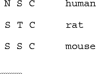
Figure 1 Alignment of human, rat and mouse NPS precursor proteins. NPS sequences are shaded, the putative processing site (KR) is underlined and the broken line indicates the presumed signal peptide.
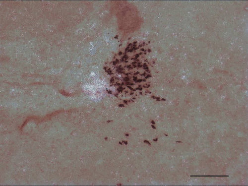
NPS gene expression has been analyzed by in situ hybridization in rat brain for both NPS precursor and NPS receptor (Xu et al. Citation2007). Our studies showed that NPS precursor mRNA is expressed discretely in a few brain areas, with the highest expression found in the pericoerulear area (), the principle sensory five nucleus of the trigeminal nerve and the lateral parabrachial nucleus. More detailed studies using double in situ hybridization showed that NPS-expressing cells in the LC area co-express neither tyrosine hydroxylase (TH; a marker for noradrenergic neurons), nor CRF (a marker for neurons in Barrington's nucleus). These results indicate that the cluster of NPS-expressing neurons defines a previously uncharacterized nucleus between the noradrenergic LC and Barrington's nucleus. We also showed that the majority of NPS-producing neurons in the LC area co-express vesicular glutamate transporters (a marker for glutamatergic neurons), while a small number contains choline acetyltransferase (a marker for cholinergic neurons) (Xu et al. Citation2007), indicating that NPS is primarily co-localized with excitatory transmitters.
Figure 2 Coronal section of the rat brainstem showing in situ hybridization for tyrosine hydroxylase (TH, brown) and NPS precursor (white grains) in the LC area. Antisense riboprobes for TH were labeled with digoxigenin while probes for NPS precursor were labeled using 35S-UTP. Hybridizations were carried out as described (Xu et al. Citation2004). TH signals were captured under brightfield illumination and a separate darkfield image was taken from the same section to capture the NPS precursor hybridization signals. Both images were combined using Photoshop and adjusted for brightness and contrast. In this section medial areas are on the left and lateral areas on the right side. Experimental conditions are described in detail in Xu et al. (Citation2004, Citation2007). Scale bar: 200 μm. (See colour online)
While NPS precursor mRNA is expressed in only a few brain regions, NPS receptor mRNA is widely expressed in the brain. Highest expression is found in the cortex, thalamus, hypothalamus, amygdala, parahippocampal formation including subiculum, but only low levels of expression are detected in the brainstem (Xu et al. Citation2007).
Pharmacological analysis was conducted using stable expression of human and mouse NPS receptors in HEK 293 T cells or CHO cells, respectively. Our results revealed that nanomolar doses of NPS produce a transient increase in intracellular-free Ca2+ and cAMP, which suggest that the NPS receptor couples to both Gq and Gs proteins, establishing that NPS might be an excitatory neurotransmitter. Half-maximal effective concentrations (EC50) for mobilization of Ca2+ were between 3 and 9 nM for human, rat, and mouse NPS, respectively (Xu et al. Citation2004; Reinscheid et al. Citation2005). Receptor binding of radiolabeled human NPS was saturable with high affinity (Kd = 0.33 ± 0.12 nM) and displaceable by increasing concentrations of cold NPS (IC50 = 0.42 ± 0.12 nM). High affinity binding and receptor activation in the low nanomolar range are typical for neuropeptides.
A number of single-nucleotide polymorphisms (SNPs) and several splice variants have been identified for the human NPS receptor. Some of these were suggested as a possible risk factor for asthma and other allergic diseases (Laitinen et al. Citation2004). Since physiological roles for any of these changes were unknown, we carried out a pharmacological analysis of one of the SNPs that produces one amino acid change (Asn107Ile) in the first extracellular loop of the receptor protein (SNP591694 A > T; ref SNP ID: rs324981), and a C-terminal splice variant of the receptor that was reported to be over-expressed in human asthmatic airway tissue (Laitinen et al. Citation2004). Our pharmacological experiments showed that the Asn107Ile polymorphism in the human NPS receptor results in a gain-of-function characterized by an increase in agonist potency at the Ile107 variant. The C-terminal splice variant, however, had little effect on the pharmacological profile (Reinscheid et al. Citation2005).
NPS promotes arousal and reduces stress related anxiety behavior
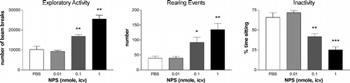
Searching for the physiological functions of the NPS system, we initiated a series of studies measuring behavioral responses in mice and rats (Xu et al. Citation2004). Male C57Bl/6 mice (age 10–12 weeks) were group-housed (four animals per cage) in a controlled environment (21°C ± 2; 60% relative humidity, 12 h light–dark cycle with lights on at 7 AM) and ad libitum access to food and water. Mice were habituated to the experimental room for at least 1 h and locomotion experiments were performed under normal fluorescent light illumination. Mice that received intracerebroventricular (i.c.v.) injection of NPS or vehicle were placed in a locomotor test chamber while exploratory activity, number of rearing events and periods of inactivity (percent time sitting) were measured. NPS-treated mice showed significant increases of locomotor activity lasting 1 h compared to vehicle-treated mice (). Since under these conditions the locomotor chambers represent a novel environment for the animals and exploration of novel places is a natural response, this result offered two possible hypotheses about the effect of NPS: (a) NPS could enhance responses to novelty, or (b) NPS could produce genuine arousal independent of novelty. In order to address these questions, we performed the same activity test with mice that had been habituated to the test chamber for 1 h before drug injection. Vehicle-treated habituated mice showed no further increase in exploratory activity, while NPS-treated habituated mice showed significantly enhanced locomotor activity and reduced inactivity (). These results suggested that low concentrations of NPS (0.1 and 1 nmol) produce profound arousal that is independent of novelty (Xu et al. Citation2004).
Figure 3 Exploratory activity in habituated male C57Bl/6 mice injected into a lateral cerebral ventricle with vehicle (PBS) or increasing doses of NPS during the first 60 min post injection. (A) Exploratory activity is defined as the total number of infrared beam breaks produced by the animals while moving horizontally or vertically (rearing). NPS dose-dependently increased this parameter. (B) NPS significantly stimulated rearing activity. (C) Inactivity is defined as an absence of beam breaks for a minimum duration of 2 s. NPS reduced periods of inactivity in a dose-dependent manner. Data are shown as means ± SEM from 9–10 animals per group. * p < 0.05, ** p < 0.01, *** p < 0.001 by ANOVA with Bonferroni post hoc test compared to vehicle-injected animals. Results were obtained by analysis of raw data from locomotion experiments described in Xu et al. (Citation2004) where detailed descriptions of methodology and experimental conditions can be found.
Since arousal is an important component of wakefulness, we next investigated whether NPS could modulate sleep/wake patterns in rats during their normal period of inactivity, i.e. during the light phase. Compared with vehicle-treated animals, NPS-injected rats displayed significant reductions in all stages of sleep during the first hour post-injection. Conversely, NPS induced long-lasting wakefulness duing a time when the animals would normally rest and usually spend more than half of their time asleep (Xu et al. Citation2004). This arousal-inducing effect of NPS might be at least partially mediated by NPS receptors expressed in thalamic midline nuclei, which have been identified as relays between arousal-inducing brainstem structures and the cortex (Van der Werf et al. Citation2002).
High levels of NPS receptor expression in amygdala and hypothalamus, which are brain areas commonly associated with emotional behaviors and stress responses (Berretta Citation2005), raised the possibility that NPS might also be involved in modulating behavioral responses to stress. In a battery of four different paradigms that measure innate fear and behavioral responses to novelty or unfamiliar objects (open field, elevated plus maze, light–dark box, marble burying), we found that NPS dose-dependently and consistently reduced measures of anxiety-like behavior to produce anxiolytic-like effects across all four tests.
Recently, NPS has also been shown to modulate feeding behavior. Central administration of 1 μg ( ≈ 0.5 nmol) NPS reduced food intake by more than 50% in fasted rats (Beck et al. Citation2005). Hyperactive and anorexigenic effects of NPS were also observed after central as well as local NPS injections into the hypothalamic paraventricular nucleus in rats, with possible concurrent activation of the HPA axis; central administration of NPS was found to increase secretion of ACTH and plasma corticosterone levels (Smith et al. Citation2006). In the same study, NPS was found to enhance CRF and AVP release from hypothalamic explants while not affecting neuropeptide Y (NPY) secretion, indicating that this effect is mediated by specific neuronal circuits. While at first glance, increases in stress hormone secretion appear contradictory to the anxiolytic effects of NPS, there are reports of other neuropeptides, for example NPY, that produce anxiolytic effects after central administration but also trigger release of stress hormones, such as CRF or ACTH, under certain conditions (Hanson and Dallman Citation1995; Brunton et al. Citation2006). These interesting findings about the interaction of NPS with other transmitter systems obviously warrant further research into how these circuits orchestrate specific behavioral outputs such as stress or feeding. It is also currently unclear if the anorexigenic effects of NPS are secondary to the behavioral arousal or if NPS is directly influencing feeding behavior. A possible neuronal substrate for such effects might be NPS receptors expressed in the arcuate nucleus, which is known to integrate a multitude of orexigenic and anorexigenic signals in the brain (Williams et al. Citation2001).
In summary, behavioral tests in rodents provide strong evidence that NPS can promote behavioral arousal accompanied by profound wakefulness and reduce stress-related anxiety responses at the same time. In addition, NPS might also produce anorexigenic effects.
Comparison with other neurotransmitters and neuropeptides
Previous studies revealed that NPS can induce arousal and produce anxiolytic effects at the same time. This is a quite unique pharmacologic profile when compared with other neurotransmitters or drugs that regulate wakefulness or anxiety ().
Table I. Comparison of various endogenous and exogenous ligands/drugs and their pharmacological effects on anxiety-related behaviors, sleep and food intake.
Psychostimulants such as methamphetamine or cocaine, for example, cause severe hyperlocomotion in rodents but have been reported to induce stress-related depression or anxiety in such animal models (Rogerio and Takahashi Citation1992; Paine et al. Citation2002; Sumnall et al. Citation2004; Hayase et al. Citation2005). It is also known that drugs used for treatment of stress responses, depression, or anxiety disorders, such as benzodiazepines and antidepressants, often cause sedation as major side effects (Bourin and Briley Citation2004). Preclinical studies with diazepam have shown that moderate to high doses clearly inhibit locomotor activity in experimental animals and this sedative effect of benzodiazepines is clinically widely used to induce sleep (Chaouloff et al. Citation1997).
Many neuropeptides are also known to modulate sleep/arousal, food intake, and stress responses (). Hcrt/ox, for example, induces wakefulness and stimulates food intake under certain conditions, but has been reported to either have no effect on anxiety-like behaviors (Hagen et al. Citation1999), or rather to produce anxiogenic effects (Suzuki et al. Citation2005). Ghrelin and NPY form a hypothalamic regulatory circuit together with hcrt/ox. Ghrelin and NPY are both orexigenic peptides. Ghrelin has recently been reported to suppress sleep (Szentirmai et al. Citation2006), while central administration of NPY causes sedation, presumably mediated by NPY Y5 receptors (Sorensen et al. Citation2004). In addition, ghrelin and NPY appear to modulate emotional behavior but their effects on anxiety are not conclusive or, in the case of NPY, might depend on particular receptor subtypes and anatomical substrates (Nakajima et al. Citation1998; Asakawa et al. Citation2001; Sudakov et al. Citation2001; Carlini et al. Citation2004; Sorensen et al. Citation2004; Karlsson et al. Citation2005). CRF, an anorexigenic neuropeptide expressed prominently in the paraventricular nucleus of the hypothalamus, is known to have an important role in stress mediation. When injected centrally, CRF causes both hyperlocomotion and anxiogenic stress-response behavior (Lowry and Moore Citation2006). Therefore, NPS represents an interesting example of a neurotransmitter that produces both arousing and anxiolytic-like effects. Expression of NPS receptors in multiple hypothalamic structures and the amygdala indicates possible interactions of NPS with other neuropeptide systems, forming a hypothalamic regulatory circuit for modulation of stress responses and potentially feeding behavior. Clearly, further research on the neuroanatomical substrates of such interactions is required to characterize the neuronal mechanisms and networks utilized by NPS in promoting anxiolytic-like effects.
In conclusion, NPS and its receptor constitute a neuropeptide system with a unique physiological spectrum by inducing arousal and wakefulness while also alleviating innate responses to stress and fear. Such a functional profile should be an interesting target for drug development and the availability of synthetic small molecule agonists and antagonists for the NPS receptor will certainly help to characterize further functions of the NPS system. The anatomical distribution of NPS receptors in the brain implies that the NPS system might interact closely with other neuropeptide systems in modulating stress responses, arousal and feeding behavior.
Acknowledgements
This work was supported in part by a grant from the National Institutes of Mental Health (NIMH) and a Young Investigator Award from the National Alliance for Research on Depression and Schizophrenia (NARSAD) to R.K.R and a grant from the Mitsubishi Pharma Research Foundation to N.O.
References
- Asakawa A, Inui A, Kaga T, Yuzuriha H, Nagata T, Fujimiya M, Katsuura G, Makino S, Fujino MA, Kasuga M. A role of ghrelin in neuroendocrine and behavioral responses to stress in mice. Neuroendocrinology 2001; 74: 143–147
- Beck B, Fernette B, Stricker-Krongrad A. Peptide S is a novel potent inhibitor of voluntary and fast-induced food intake in rats. Biochem Biophys Res Commun 2005; 332: 859–865
- Berretta S. Cortico-amygdala circuits: Role in the conditioned stress response. Stress 2005; 8: 221–232
- Bourin M, Briley M. Sedation, an unpleasant, undesirable and potentially dangerous side-effect of many psychotropic drugs. Hum Psychopharmacol 2004; 19: 135–139
- Brunton PJ, Bales J, Russell JA. Neuroendocrine stress but not feeding responses to centrally administered neuropeptide Y are suppressed in pregnant rats. Endocrinology 2006; 147: 3737–3745
- Carlini VP, Varas MM, Cragnolini AB, Schioth HB, Scimonelli TN, de Barioglio SR. Differential role of the hippocampus, amygdala, and dorsal raphe nucleus in regulating feeding, memory, and anxiety-like behavioral responses to ghrelin. Biochem Biophys Res Commun 2004; 313: 635–641
- Chaouloff F, Durand M, Mormede P. Anxiety- and activity-related effects of diazepam and chlordiazepoxide in the rat light/dark and dark/light tests. Behav Brain Res 1997; 85: 27–35
- Ford DE, Kamerow DB. Epidemiologic study of sleep disturbances and psychiatric disorders. An opportunity for prevention?. JAMA 1989; 262: 1479–1484
- Hagan JJ, Leslie RA, Patel S, Evans ML, Wattam TA, Holmes S, Benham CD, Taylor SG, Routledge C, Hemmati P, Munton RP, Ashmeade TE, Shah AS, Hatcher JP, Hatcher PD, Jones DN, Smith MI, Piper DC, Hunter AJ, Porter RA, Upton N. Orexin A activates locus coeruleus cell firing and increases arousal in the rat. Proc Natl Acad Sci USA 1999; 96: 10911–10916
- Hanson ES, Dallman MF. Neuropeptide Y (NPY) may integrate responses of hypothalamic feeding systems and the hypothalamo–pituitary–adrenal axis. J Neuroendocrinol 1995; 7: 273–279
- Hayase T, Yamamoto Y, Yamamoto K. Persistent anxiogenic effects of a single or repeated doses of cocaine and methamphetamine: Interactions with endogenous cannabinoid receptor ligands. Behav Pharmacol 2005; 16: 395–404
- Holmes A, Heilig M, Rupniak NM, Steckler T, Griebel G. Neuropeptide systems as novel therapeutic targets for depression and anxiety disorders. Trends Pharmacol Sci 2003; 24: 580–588
- Karlsson RM, Holmes A, Heilig M, Crawley JN. Anxiolytic-like actions of centrally-administered neuropeptide Y, but not galanin, in C57BL/6J mice. Pharmacol Biochem Behav 2005; 80: 427–436
- Laitinen T, Polvi A, Rydman P, Vendelin J, Pulkkinen V, Salmikangas P, Makela S, Rehn M, Pirskanen A, Rautanen A, Zucchelli M, Gullsten H, Leino M, Alenius H, Petays T, Haahtela T, Laitinen A, Laprise C, Hudson TJ, Laitinen LA, Kere J. Characterization of a common susceptibility locus for asthma-related traits. Science 2004; 304: 300–304
- Lowry CA, Moore FL. Regulation of behavioral responses by corticotropin-releasing factor. Gen Comp Endocrinol 2006; 146: 19–27
- Nakajima M, Inui A, Asakawa A, Momose K, Ueno N, Teranishi A, Baba S, Kasuga M. Neuropeptide Y produces anxiety via Y2-type receptors. Peptides 1998; 19: 359–363
- Nestler EJ, Barrot M, DiLeone RJ, Eisch AJ, Gold SJ, Monteggia LM. Neurobiology of depression. Neuron 2002; 34: 13–25
- Paine TA, Jackman SL, Olmstead MC. Cocaine-induced anxiety: Alleviation by diazepam, but not buspirone, dimenhydrinate or diphenhydramine. Behav Pharmacol 2002; 13: 511–523
- Reinscheid RK, Xu YL, Okamura N, Zeng J, Chung S, Pai R, Wang Z, Civelli O. Pharmacological characterization of human and murine neuropeptide S receptor variants. J Pharmacol Exp Ther 2005; 315: 1338–1345
- Reul JM, Holsboer F. Corticotropin-releasing factor receptors 1 and 2 in anxiety and depression. Curr Opin Pharmacol 2002; 2: 23–33
- Rogerio R, Takahashi RN. Anxiogenic properties of cocaine in the rat evaluated with the elevated plus-maze. Pharmacol Biochem Behav 1992; 43: 631–633
- Sakurai T, Amemiya A, Ishii M, Matsuzaki I, Chemelli RM, Tanaka H, Williams SC, Richardson JA, Kozlowski GP, Wilson S, Arch JR, Buckingham RE, Haynes AC, Carr SA, Annan RS, McNulty DE, Liu WS, Terrett JA, Elshourbagy NA, Bergsma DJ, Yanagisawa M. Orexins and orexin receptors: A family of hypothalamic neuropeptides and G protein-coupled receptors that regulate feeding behavior. Cell 1998; 92: 573–585
- Saper CB, Chou TC, Scammell TE. The sleep switch: Hypothalamic control of sleep and wakefulness. Trends Neurosci 2001; 24: 726–731
- Smith KL, Patterson M, Dhillo WS, Patel SR, Semjonous NM, Gardiner JV, Ghatei MA, Bloom SR. Neuropeptide S stimulates the hypothalamo–pituitary–adrenal axis and inhibits food intake. Endocrinology 2006; 147: 3510–3518
- Sorensen G, Lindberg C, Wortwein G, Bolwig TG, Woldbye DP. Differential roles for neuropeptide Y Y1 and Y5 receptors in anxiety and sedation. J Neurosci Res 2004; 77: 723–729
- Sudakov SK, Medvedeva OF, Rusakova IV, Terebilina NN, Goldberg SR. Differences in genetic predisposition to high anxiety in two inbred rat strains: Role of substance P, diazepam binding inhibitor fragment and neuropeptide Y. Psychopharmacology (Berl) 2001; 154: 327–335
- Sumnall HR, O'Shea E, Marsden CA, Cole JC. The effects of MDMA pretreatment on the behavioural effects of other drugs of abuse in the rat elevated plus-maze test. Pharmacol Biochem Behav 2004; 77: 805–814
- Sutcliffe JG, de Lecea L. The hypocretins: Setting the arousal threshold. Nat Rev Neurosci 2002; 3: 339–349
- Suzuki M, Beuckmann CT, Shikata K, Ogura H, Sawai T. Orexin-A (hypocretin-1) is possibly involved in generation of anxiety-like behavior. Brain Res 2005; 1044: 116–121
- Szelenberger W, Soldatos C. Sleep disorders in psychiatric practice. World Psychiatry 2005; 4: 186–190
- Szentirmai E, Hajdu I, Obal F, Jr., Krueger JM. Ghrelin-induced sleep responses in ad libitum fed and food-restricted rats. Brain Res 2006; 1088: 131–140
- Van der Werf YD, Witter MP, Groenewegen HJ. The intralaminar and midline nuclei of the thalamus. Anatomical and functional evidence for participation in processes of arousal and awareness. Brain Res Brain Res Rev 2002; 39: 107–140
- Williams G, Bing C, Cai XJ, Harrold JA, King PJ, Liu XH. The hypothalamus and the control of energy homeostasis: Different circuits, different purposes. Physiol Behav 2001; 74: 683–701
- Xu YL, Reinscheid RK, Huitron-Resendiz S, Clark SD, Wang Z, Lin SH, Brucher FA, Zeng J, Ly NK, Henriksen SJ, de Lecea L, Civelli O. Neuropeptide S: A neuropeptide promoting arousal and anxiolytic-like effects. Neuron 2004; 43: 487–497
- Xu YL, Gall CM, Jackson VR, Civelli O, Reinscheid RK. Distribution of Neuropeptide S receptor mRNA and neurochemical characteristics of Neuropeptide S expressing neurons in the rat brain. J Comp Neurol 2007; 500: 84–102