Abstract
Chronic subordinate colony (CSC) housing has been recently validated as a murine model of chronic psychosocial stress which induces alterations of stress-related parameters including decreased body-weight gain and an increased level of anxiety in comparison with single housed control (SHC) mice. By using immunohistochemical immediate early gene (IEG) mapping we investigated whether CSC housing causes alterations in neuronal activation patterns in limbic areas including the amygdala, hippocampus, septum and the periaqueductal gray (PAG) and hypothalamic paraventricular nucleus (PVN). While CSC housing increased basal Zif-268 expression in the nucleus accumbens shell compared to SHC, IEG responses to subsequent open arm (OA) exposure were attenuated in the ventral and intermediate sub-regions of the lateral septum, parvocellular PVN and the dorsal CA3 region of the hippocampus of CSC compared with SHC mice. In contrast, a potentiated c-Fos response in CSC mice was observed in the dorsomedial PAG after OA exposure. Confirming previous findings obtained on the elevated plus-maze, an enhanced anxiety-related behavior in CSC compared with SHC mice was also observed during OA exposure. In order to investigate the appropriate control conditions for CSC housing, group housed control (GHC) mice were additionally included in the behavioral testing. Interestingly, GHC as well as CSC mice showed significantly less risk assessment/exploratory behavior during OA exposure compared with SHC mice indicating that group housing itself is stressful for mice and not an adequate control for the CSC paradigm. Overall, CSC housing is an ethologically relevant chronic psychosocial stressor which results in an elevated sensitivity to a subsequent novel, aversive challenge. However, the CSC-induced increase in anxiety-related behavior was accompanied by differences in neuronal activation, compared with SHC, in defined sub-regions of brain areas known to be involved in the processing of emotionality and stress responses.
Introduction
A variety of adverse life events including psychosocial stress have been shown to predispose an individual to increased emotionality, such as increased anxiety-related behaviors in animals, and to a higher risk of developing anxiety or affective disorders in humans (Blanchard et al. Citation1998; Calvo-Torrent et al. Citation1999; Zurita et al. Citation2000; Hata et al. Citation2001; Dalla et al. Citation2005; Rygula et al. Citation2005; Matuszewich et al. Citation2007). The behavioral strategies shown during a challenging stimulus are modulated by prior history of perceived stress. Exposure of mice to various stressors including cat odor, inescapable foot-shocks, repeated cold-stress, social defeat or other kinds of psychosocial stress have been found to reduce their social interactions as well as the exploration time on the open arm (OA) during subsequent elevated plus-maze (EPM) testing, indicating increased levels of anxiety (Steenbergen et al. Citation1990; Zangrossi and File Citation1992; Rodgers and Cole Citation1993; Hata et al. Citation2001; McClung et al. Citation2004; Reber et al. Citation2007). Animals previously exposed to an acute or chronic homotypic and uncontrollable stressor also showed more passive coping strategies during a subsequent aversive stimulation by a heterotypic stressor (Armario et al. Citation1991; Molina et al. Citation1994; Zurita et al. Citation2000).
Social defeat has been established as an ethologically relevant animal model of psychosocial stress in most mammals (Blanchard et al. Citation1998). In this context, it has been hypothesized that the experience of social subordination rather than the social threat per se might be the crucial factor for defeat-induced behavioral maladaptations (Razzoli et al. Citation2006). Therefore, the chronic subordinate colony (CSC) housing recently established by Reber et al. (Citation2007), seems to be an adequate psychosocial stress paradigm for studying chronic stress-induced changes in emotionality. CSC housing results not only in reduced body weight gain, with increased adrenal and reduced thymus weights as signs of chronic stress, but also in increased anxiety-related behavior on the EPM (Reber et al. Citation2007; Reber, Birkeneder et al. 2007). However, the influence of chronic social stress on the differential patterns of neuronal activation in brain areas involved in the processing of emotionality and stress responses is still not clear. According to Martinez et al. (Citation1998), downregulation of neuronal activation after chronic social defeat in rats to levels found in unstressed controls could be observed in a number of regions, including e.g. lateral habenular, locus coeruleus or the central amygdala, and was defined as neuronal adaptation. Interestingly, especially within the lateral septum (LS) the type of chronic stressor seems to be important for the development of adaptation: While the LS has been shown to adapt to repeated social defeat (Martinez et al. Citation1998), repeated restraint stress does not induce adaptation within this region in contrast to most other stress-responsive cortical and limbic brain regions (Chen and Herbert Citation1995). However, the role of the LS in adaptational processes during chronic psychosocial stress has to be interpreted with caution as the c-Fos levels obtained under basal as well as repeated social stress conditions in previous studies (Martinez et al. Citation1998) were low and therefore slight differences in neuronal activation might not be detected. Importantly, there are brain regions showing an increased activation after exposure to chronic social defeat, including the bed nucleus of stria terminalis (BNST), paraventricular nucleus (PVN), medial amygdala (AMY) and dorsal raphe nucleus (DRN) (Martinez et al. Citation1998). The AMY, DRN, as well as the LS are considered to be part of limbic pathways and have been shown to be involved in regulation of anxiety (Graeff Citation1994). In this context, some reports have shown that especially the LS and its glucocorticoid receptors (GR) play an essential role in regulating the emotional consequences of stress, as intraseptal injection of the GR-antagonist mifepristone (RU486) normalizes the anxiogenic effect of social defeat (Calfa et al. Citation2006).
Using immunohistochemical immediate early gene (IEG) mapping, we aimed to determine, whether prior exposure of mice to the recently established chronic psychosocial stressor of CSC housing changes basal or acute stress-induced neuronal activation within brain areas involved in emotionality and stress responses (Duncan et al. Citation1996; Herman et al. Citation2005). As we expected a generally low basal c-Fos expression in the LS (Martinez et al. Citation2002), we additionally conducted Zif-268 staining within this nucleus which provides high sensitivity for minimal basal differences (Ziolkowska and Przewlocki Citation2002). Furthermore, both c-Fos and Zif-268 expression are well described IEGs activated in particular brain regions during acute stress (Watanabe et al. Citation1994), and therefore provide a means to observe changes in neuronal activity during subsequent heterotypic stress-exposure.
Experimental procedures
Animals
Male C57Bl/6 mice (Charles River, Sulzfeld, Germany) weighing 19–22 g (experimental mice n = 42) or 30–35 g (used as residents during the CSC procedure; n = 4) were individually housed in standard polycarbonate mouse cages (length: 22 cm, width: 16 cm and height: 12 cm) for at least one week before the experimental procedure started. All mice were kept under standard laboratory conditions (12-h light/dark cycle, lights on at 06:00 h, 22°C, 60% humidity) and had free access to tap water and standard mouse diet. All experimental protocols were approved by the Committee on Animal Health and Care of the local government, and conformed to international guidelines on the ethical use of animals. All efforts were made to minimize the number of animals used and their suffering.
Chronic subordinate colony (CSC) housing
The procedure of the CSC housing has been described recently (Reber et al. Citation2007; Reber, Birkeneder et al. 2007). Briefly, four experimental CSC mice were housed together with a larger dominant male in a polycarbonate observation cage (length: 38 cm, width: 22 cm, and height: 35 cm) for 19 consecutive days, in order to induce a chronic stressful situation. Before the CSC procedure, the future dominant males were tested for their aggressive behavior. Males that started to injure their opponents by harmful bites were not used. To avoid habituation, each dominant male was replaced by the dominant male of another colony on days 8 and 15. During the first 30 min after colony formation on day 1, 8, and 15 the mice were videotaped. Behavioral analyses showed that in all colonies the older and larger male mouse established a “dominant” status while it was chasing and attacking all four experimental CSC mice. These were all considered as “subordinates” based on their defensive behavior, including flight, retreat and submissive upright posture. Single housed controls (SHCs) were housed singly for the respective 19 consecutive days. Group housed controls (GHCs) were housed in groups of four mice per cage for 19 days and were only used for behavioral comparisons.
Open-arm (OA) exposure
OA exposure was carried out on day 20 of CSC exposure during the light phase between 0800 and 1300 h. Mice (CSC/SHC/GHC) were transported to the EPM test room on the evening of day 19. On day 20 each mouse of the respective groups was individually placed in the centre of an OA (30 × 5 cm, elevation 70 cm), with access to the neutral zone and the closed arms of the EPM prevented by a bar. The illumination on the OA was approximately 140 lux. The behavior of the mice was videotaped during the 10-min testing period. For analyses of the videotaped behavior using Eventlog 1.0 (EMCO Software) the OA was divided virtually by the tracking system into three equal segments: a distal, middle and proximal zone. The number of entries performed into the distal part of the OA, shown to represent anxiety-related behavior (Salome et al. Citation2004), the number and the time of head dips performed on the distal part of the OA, and the number of head dips on the entire OA, which are indicative of risk assessment/exploratory behavior (Ohl et al. Citation2001; Salome et al. Citation2006), and the total distance travelled on the OA, for assessment of locomotory behavior (Salome et al. Citation2006), were scored by a trained observer blind to the animals' treatment. Additionally, the number of grooming episodes (cleaning any parts of the body surface with the tongue, teeth, and/or forepaws) (da Cunha et al. Citation2007), which also appears to serve as an important indicator of anxiety and stress-related behavioral paradigm (Jaszberenyi et al. Citation2007), was monitored. Immediately after behavioral testing, the mice were singly housed until perfusion (2 h after OA exposure), to avoid disturbances of their CSC cage-mates. The OA was cleaned thoroughly before the first and after each trial. The mice assigned to the basal group (SHC basal/CSC basal) were also transported to the EPM test room but remained undisturbed in their home cages until perfusion.
Immunohistochemistry
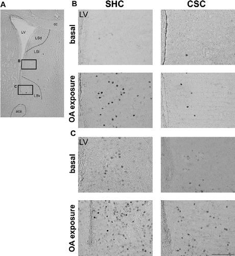
Two hours after OA exposure, mice (SHC and CSC) were deeply anesthetized with an overdose of sodium pentobarbital (200 mg/kg) injected i.p. and transcardially perfused with 20 mL of 0.9% saline followed by 20 mL of 4% paraformaldehyde in 0.1 mol/L phosphate buffered solution (PBS, pH 7.4). Brains were then removed and postfixed at 4°C overnight in 4% paraformaldehyde in phosphate buffered saline. Coronal sections (50 μm) were cut with a vibratome (Ted-Pella, Redding, CA, USA) and collected in immunobuffer (pH 7.4) comprising 0.1 mol/L NaCl, 5 mmol/L KCl, 8 mmol/L Na2HPO4, 15 mmol/L NaH2PO4, 10 mmol/L Tris–HCl, 0.3% Triton X-100, and 0.04% Thimerosal. The sections were processed for c-Fos- and Zif-268-like immunoreactivity as described previously (Singewald et al. Citation2003), diluted with a polyclonal primary antibody (c-Fos: 1:20000; sc-52 and Zif-268: 1:5000; sc-189/Santa Cruz Biotechnology, Santa Cruz, CA, USA) for 48 h and a biotinylated goat anti-rabbit secondary antibody (1:200; Vector Laboratories, Burlingame, CA, USA) for 24 h. An avidin–biotin–horseradish peroxidase procedure (Vectastain ABC Kit, Vector Laboratories) with 3,3′-diaminobenzidine as the chromogen was used to visualize Fos-positive cells. Cells containing a nuclear brown-black reaction product were considered as c-Fos- or Zif-268- positive cells. The anatomical localization of labeled cells was aided by use of adjacent Nissl stained sections and the illustrations in a stereotaxic atlas (Paxinos and Watson Citation1998). The numbers of c-Fos/Zif-268 positive cells were counted bilaterally in up to 2–3 sections (depending on the anterior–posterior extent of structures) in a tissue area of 0.02, 0.04, and 0.08 mm2 (depending on the size of brain area) or throughout the entire brain area (CeA, pyramidal cell layer, dentate gyrus) by an observer blind to the experimental groups. Representative photomicrographs of immunoreactivity in sub-regions of the LS are shown in .
Statistics
Statistical analysis of the behavioral data and changes in body weight gain of the three groups was performed using a one-way ANOVA (factor housing condition; SHC/GHC/CSC) followed by Bonferroni post hoc test when appropriate. Statistical analysis of c-Fos/Zif-268 data was performed using a two-way ANOVA (factor CSC; factor OA exposure) followed by Bonferroni post hoc test when appropriate. The level of significance was set at p ≤ 0.05. All values were expressed as means + SEM.
Results
Body weight gain
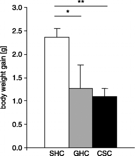
Immediately before (day 1) and after 19 days of CSC exposure (one day before the OA exposure) mice of all three groups (SHC, GHC, and CSC) were weighed to reveal possible differences in body weight gain (Δg) among the groups. Body weight gain was significantly affected by CSC housing (F2,37 = 16.289; p = 0.01). Post hoc analysis revealed that CSC (p = 0.01) and GHC (p = 0.02) mice showed a significantly decreased body weight gain during the 19 days compared with SHC mice ().
Figure 1 Effects of 19-day exposure to different housing conditions (SHCs, n = 17; GHCs, n = 9; CSC, n = 16) on body weight gain in male mice. Data represent means + SEM; *p ≤ 0.05; **p ≤ 0.01 vs. SHC group.
Behavior during OA exposure
CSC exposure affected the number of entries performed into the distal part of the OA (F2,23 = 5.792; p = 0.01), as well as the total number of head dips (F2,22 = 11.333; p = 0.01) and their duration (F2,23 = 9.643; p = 0.01) performed on the distal part of the OA (). Similarly, the number of head dips (F2,22 = 7.878; p = 0.01), the distance travelled (F2,22 = 10.918; p = 0.01), as well as grooming episodes (F2,17 = 3.914; p = 0.04) on the entire OA were found to depend on prior CSC exposure (). Post hoc analysis revealed that exposure to CSC for 19 days increased anxiety-related behavior, as indicated by a significant decrease in the number of entries performed into the distal part of the OA (p = 0.01) and a significant increase in the number of grooming episodes (p = 0.03) during OA exposure. Furthermore, the risk assessment/exploratory behavior was decreased in CSC compared with SHC mice, as indicated by a decrease in number (p = 0.01) and duration (p = 0.01) of distal head dips, and the number of total head dips (p = 0.01). Interestingly, in GHC mice all the mentioned parameters indicating risk assessment/exploratory behavior were also significantly decreased when compared with SHC mice (duration of distal head dips: p = 0.02; number of distal head dips: p = 0.02; number of total head dips: p = 0.03). Locomotor behavior was also found to be reduced in both the CSC and GHC mice, as indicated by a significantly decreased total distance travelled on the OA (CSC: p = 0.01; GHC: p = 0.02) when compared with SHC mice ().
Figure 2 Effects of 19-day exposure to different housing conditions (SHCs, n = 9; GHCs, n = 8; CSC, n = 8) on the behavioral responses of male mice during subsequent OA exposure (day 20). Anxiety-related behavior during 10 min of OA exposure is indicated by (A) number of entries into the distal zone, risk assessment/exploratory behavior is indicated by (B) number of head dips on the distal zone, (C) number of total head dips on the OA, (D) time [s] of distal head dips, and locomotor behavior is indicated by (E) distance [cm] travelled on the OA and (F) shows number of grooming episodes. Data represent means + SEM; *p ≤ 0.05; **p ≤ 0.01 vs. SHC group.
![Figure 2 Effects of 19-day exposure to different housing conditions (SHCs, n = 9; GHCs, n = 8; CSC, n = 8) on the behavioral responses of male mice during subsequent OA exposure (day 20). Anxiety-related behavior during 10 min of OA exposure is indicated by (A) number of entries into the distal zone, risk assessment/exploratory behavior is indicated by (B) number of head dips on the distal zone, (C) number of total head dips on the OA, (D) time [s] of distal head dips, and locomotor behavior is indicated by (E) distance [cm] travelled on the OA and (F) shows number of grooming episodes. Data represent means + SEM; *p ≤ 0.05; **p ≤ 0.01 vs. SHC group.](/cms/asset/7acac311-234b-42f3-b9ce-286b3e709694/ists_a_304376_f0002_b.gif)
c-Fos expression
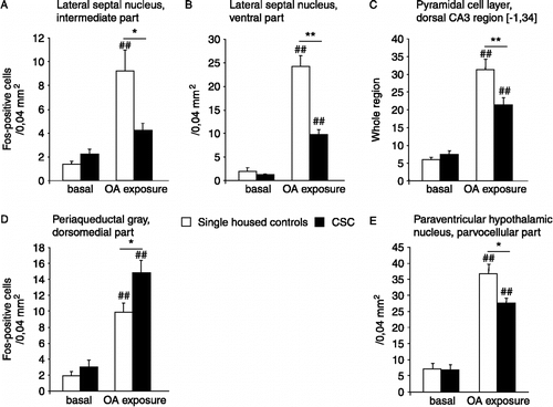
Mean numbers ± SEM of cells expressing c-Fos in all quantified brain regions are shown in . shows representative photomicrographs of c-Fos and Zif-268 labelling in sub-regions of the LS.
Table I. Effects of CSC exposure on c-Fos expression in all analyzed brain areas under basal conditions and in response to OA exposure (except brain regions separately illustrated in Figure 3). Used tissue area: 0.02, 0.04, and 0.08 mm2 (depending on size of brain area); (In central amygdala, pyramidal cell layer and dentate gyrus c-Fos positive cells were counted throughout the whole brain area). Values represent the means ± SEM of c-Fos positive cells; n=6–7 (basal groups), n=6–10 (OA exposure groups); ap≤0.05, bp≤0.01 vs. respective basal group.
Basal c-Fos expression
Basal c-Fos expression was generally low and did not differ between SHC and CSC mice in any of the investigated brain areas.
OA-induced c-Fos expression
A main effect of OA exposure on c-Fos expression was observed in the following brain areas: cingulate cortex (F1,28 = 21.374; p = 0.01); prelimbic cortex (F1,28 = 29.891; p = 0.01); infralimbic cortex (F1,27 = 51.693; p = 0.01); piriform cortex (F1,27 = 104.544; p = 0.01); nucleus accumbens [NAcc] shell (F1,28 = 19.448; p = 0.01); NAcc core (F1,28 = 9.982; p = 0.01); dorsal (F1,25 = 6.506; p = 0.02), intermediate (F1,24 = 25,838; p = 0.01), ventral (F1,24 = 142.638; p = 0.01) part of the lateral septum; medial septum (F1,28 = 32,925; p = 0.01); lateral posterior (F1,20 = 54.162; p = 0.01), medial anterior (F1,20 = 65.981; p = 0.01) part of the bed nucleus of the stria terminalis; parvocellular paraventricular hypothalamic nucleus [pPVN] (F1,25 = 121.760; p = 0.01); dorsomedial hypothalamic area (F1,29 = 26.175; p = 0.01); posteroventral (F1,27 = 534.637; p = 0.01), posterodorsal (F1,28 = 158.645; p = 0.01) medial amygdala; basolateral amygdala (F1,25 = 73.317; p = 0.01); anterior (F1,27 = 207.527; p = 0.01), posterolateral (F1,26 = 47.870; p = 0.01) cortical amygdala; CA1 layer dorsal (F1,28 = 16.079; p = 0.01); CA3 layer dorsal (F1,26 = 130.233; p = 0.01); CA1 layer ventral (F1,24 = 29.008; p = 0.01); CA3 layer ventral (F1,23 = 14.430; p = 0.01); dentate gyrus ventral (F1,26 = 6.600; p = 0.02); dorsomedial (F1,27 = 71.408; p = 0.01), dorsolateral (F1,22 = 15.787; p = 0.01), lateral (F1,26 = 92.496; p = 0.01), ventrolateral (F1,23 = 92.524; p = 0.01) periaqueductal gray [PAG]; dorsal raphe nucleus [DRN] (F1,27 = 51.693; p = 0.01). In addition, a main effect of CSC housing on c-Fos expression was observed in the following brain areas: dorsal (F1,25 = 6.506; p = 0.01), ventral (F1,24 = 29.418; p = 0.01) lateral septum; pPVN (F1,25 = 4.240 p = 0.05); CA3 layer dorsal (F1,26 = 4.315; p = 0.04); dorsomedial PAG [dmPAG] (F1,27 = 6.737; p = 0.02).
Statistical analysis further revealed a significant interaction of CSC housing and OA exposure on c-Fos expression in the intermediate (LSi) (factor CSC × OA: F1,24 = 7.721; p = 0.01) and ventral (LSv) (factor CSC × OA: F1,24 = 22.854; p = 0.01) part of the LS, the magnocellular PVN (mPVN) (factor CSC × OA: F1,26 = 6.184; p = 0.02) and the CA3 region of the dorsal pyramidal cell layer (factor CSC x OA: F1,26 = 8.539; p = 0.01). Post hoc analysis revealed a significantly increased c-Fos expression in OA-exposed mice within several of the investigated brain regions either in both the SHC and CSC housed mice or in only one of these groups when compared with respective unchallenged (basal) mice (; ). However, significant differences in c-Fos expression between SHC and CSC mice which were both exposed to the OA were only found in 5 brain regions (). Specifically, c-Fos expression of OA-exposed CSC mice was significantly lower in LSi (p = 0.02; ), LSv (p = 0.01; ), pPVN (p = 0.04; ), and CA3 region of the dorsal pyramidal cell layer (p = 0.01; ) and significantly greater in dmPAG (p = 0.02; ) when compared to respective OA-exposed SHC mice.
Figure 3 Effects of CSC housing in male mice on c-Fos expression under basal conditions and in response to OA exposure. Displayed are those five brain areas in which statistically significant differences after OA exposure were observed between the groups. Data represent means + SEM; ##p ≤ 0.01 vs. respective basal group; *p ≤ 0.05, **p ≤ 0.01 vs. respective SHC group.
Zif-268 expression
A main effect of OA exposure on Zif-268 expression was observed in the following brain areas: NAcc shell (F1,27 = 5.478; p = 0.03) and NAcc core (F1,28 = 14.665; p = 0.01); dorsal (F1,28 = 44.719; p = 0.01), intermediate (F1,28 = 201.540; p = 0.01), and ventral (F1,28 = 45.631; p = 0.01) lateral septum. In addition, a main effect of CSC on Zif-268 expression in the ventral LS [LSv] (F1,28 = 13.600; p = 0.01) was observed. Statistical analysis further revealed that Zif-268 expression in the NAcc shell and core was dependent on CSC housing and OA exposure (factor CSC × OA: F1,27 = 12.388; p = 0.01 [NAcc shell]; F1,28 = 6.031; p = 0.02 [NAcc core]). Post hoc analysis revealed an increased basal Zif-268 expression in the NAcc shell (p = 0.05) of CSC compared with SHC mice. Furthermore, a significantly increased Zif-268 expression within all of the investigated brain regions in the SHC group (p = 0.01) was found during OA exposure, while CSC housed mice only showed increased Zif-268 expression in the lateral septal subregions (p = 0.01) compared with respective unchallenged (basal) mice (). In addition, Zif-268 expression in LSv (p = 0.01) was lower in CSC compared with SHC mice after OA exposure ().
Table II. Effects of CSC exposure on Zif-268 expression in all analyzed brain areas under basal conditions and in response to OA exposure. Used tissue area: 0.04 mm2; Values represent the means ± SEM of Zif–268 positive cells; n=7 (basal groups), n=8 (OA exposure groups); ap≤0.05, bp≤0.01 vs. respective basal group; cp≤0.05, dp≤0.01 vs. respective SHC group.
Figure 4 Representative photomicrographs of immunoreactivity in sub-regions of the LS. (A) Low magnification overview of the LS at the level of +0.86 (bregma) based on the atlas of Paxinos and Watson (Citation1998). Boxed areas are shown at high magnification. Bright field photomicrographs in representative sections matched for comparable rostrocaudal levels: (B) c-Fos expression of SHC and CSC mice before (basal) and after OA exposure in the intermediate LS (LSi). (C) Zif-268 expression of SHC and CSC mice before (basal) and after OA exposure in the ventral LS (LSv). Scale bar = 100μm; aca, anterior commissure; cc, corpus callosum; LSd, dorsal lateral septum; and LV, lateral ventricle.
Discussion
The present study shows that exposure of male mice to chronic psychosocial stress, induced by 19 days of CSC housing, changed the animals' emotionality during subsequent exposure to a heterotypic stressor. In this context, CSC mice showed an increase in their anxiety-related behavior when exposed to an OA one day after termination of the chronic stress procedure compared with SHC mice. Interestingly, group-housing itself also tended to increase anxiety parameters in comparison with SHC. The observed behavioral differences between CSC and SHC mice were accompanied by distinct alterations in neuronal activation patterns in various brain regions involved in the processing of emotionality. Acute OA exposure revealed changes in neuronal responses in sub-regions of the LS, hippocampus, PVN and the PAG, while under basal conditions only changes in the NAcc shell were observed between CSC and SHC mice.
An established physiological marker of chronic stress is a reduction in body weight gain (Sachser and Lick Citation1989; Berton et al. Citation1998; Stefanski and Engler Citation1999). In agreement, CSC mice of the present study showed a reduced body weight gain. Furthermore, GHC mice also gained less weight during the 19-day period compared with SHC mice. This suggests that group housing per se poses a stressful condition for male mice; this is likely caused by one mouse out of the group of four obtaining dominant status and subordinating its cage mates. In support of this possibility, we found a greater variation of the body weight gain in GHC compared with CSC mice, as only three out of the four GHC mice showed a reduction in body weight gain comparable to CSC mice (data not shown). These similarities in reduced body weight gain between CSC and GHC mice provide a first hint for group housed male mice being to an extent socially stressed and, therefore, not providing an adequate and stress-free control group for mice in general. Indeed, GHC mice showed changes in emotionality including risk assessment/exploration and locomotor activity comparable to those seen in CSC mice although to a lesser extent. In detail, the number of head dips and the distance travelled on the OA, parameters related to risk assessment/exploratory (Ohl et al. Citation2001) and locomotor activity (Salome et al. Citation2006), respectively, were significantly decreased in both CSC and GHC compared with SHC mice.
These findings are in line with a reduction in risk assessment/exploratory behavior found in hyperanxious rats previously (Salome et al. Citation2004). In this context it should be mentioned that changes in the closed arm entries on the EPM (Reber et al. Citation2007; Reber, Birkeneder et al. 2007) or line crossings in the open field (Veenema et al. Citation2008), which are both known to be adequate parameters for assessment of locomotor activity, were not found in CSC mice. This indicates that the reduced distance travelled during OA exposure that was found in CSC and GHC compared to SHC mice does not just reflect pure locomotor activity but is influenced by emotionality.
In CSC mice, a robust increase in anxiety-related behavior during OA exposure was found as indicated by a decrease in the numbers of entries into the distal and, therefore, unprotected and more threatening (Salome et al. Citation2006) zone of the OA. In addition, an increase in self-grooming frequency, suggested to be an indicator of anxiety-related behavior (Jaszberenyi et al, 2007; Kalueff and Tuohimaa Citation2005) was observed in CSC mice only. Interestingly, it was recently shown that grooming co-occurs with increased anxiety in humans (Grant and Christenson Citation2007). Increased anxiety-related behavior in CSC mice has been repeatedly demonstrated in the classical EPM test (Reber et al. Citation2007; Reber, Birkeneder et al., 2007) or during open field exposure (Veenema et al. Citation2008) confirming the described CSC-induced changes in emotionality during a subsequent heterotypic challenge. However, the more pronounced increase in anxiety in CSC compared with GHC mice is possibly due to differences in the stability of the social hierarchy in these two housing conditions. In the CSC paradigm, the social hierarchy is disrupted on a weekly basis by exchanging the dominant male, whereas, in the GHC group, the hierarchy stays stable once it is established. Although there are also indications that single housing per se poses a chronic challenge for rats resulting in increased anxiety levels (Parker and Morinan Citation1986; Hall et al. Citation1998; Hellemans et al. Citation2004), by contrast, in male mice, group housing seems to be stressful. Indeed, most studies indicate that isolation itself does not cause any signs of stress in immune and endocrine function (Holson et al. Citation1991; Bartolomucci et al. Citation2003; Gasparotto et al. Citation2005). Taken together, these arguments speak in favour for the use of SHC mice to reveal CSC effects on IEG activation.
Several studies have been conducted in different species (rat, mice and hamsters) using repeated exposure to a homotypic stressor followed by a single exposure to a heterotypic stressor and monitoring changes in c-Fos activation patterns in several brain areas (Matsuda et al. Citation1996; Kollack-Walker et al. Citation1997; Chung et al. Citation2000; Girotti et al. Citation2006). While no adaptation in the LS has been found during repeated restraint stress (Chen and Herbert Citation1995) there is evidence for a down-regulated activation of the LS during repeated/chronic social stress (Kollack-Walker et al. Citation1997; Martinez et al. Citation1998). Given the generally low or absent basal c-Fos expression in the LS after CSC housing we additionally quantified the expression of the IEG Zif-268 in this brain region, as Zif-268 shows high expression levels also under basal conditions (Ziolkowska and Przewlocki Citation2002). Interestingly, CSC mice showed similar levels of Zif-268 expression in the LS compared to SHC mice, indicating that LS neurons might also adapt to the chronic social stress of CSC housing.
During OA exposure, we observed decreased c-Fos expression in the ventral and intermediate part of the LS in CSC compared with SHC mice. This is in accordance with a decrease in stress-induced c-Fos expression in the LS found in rats exposed to a learned helplessness paradigm (Steciuk et al. Citation1999). Furthermore, the LS was described previously to play a key role in anxiety-related responses (Sheehan et al. Citation2004), because increasing the activity of LS neurons was found to reduce fear and anxiety (Yadin et al. Citation1993). Hence, the attenuated LS activation observed in CSC mice after OA exposure might be related to the observed increases in their anxiety-related behavior. Interestingly, the lower Zif-268 expression in the LSv after OA exposure is in line with the respective c-Fos data, supporting the hypothesis that chronic homotypic social stress affects neuronal activity within the LS during subsequent exposure to a heterotypic stressor. There is general agreement that in the circumstances used here, activated c-Fos expression reflects neuronal activity, but not increased glial cell activity as shown by double staining for GFAP, a marker for astrocytes, and Fos protein in various brain areas of adult rodents (Herdegen et al. Citation1993; Hetzenauer et al. Citation2006). Hence alterations in c-Fos activation within this study are considered to mainly represent locally differential changes in neuronal activation.
Evaluation of Zif-268 immunoreactivity within the NAcc revealed increased neuronal activation following CSC housing. In line with this finding, mice exposed to predator odor were also shown to display increased expression of fos-related antigen in the NAcc shell (Hebb et al. Citation2004). These fox odor-exposed mice were further found to be more anxious during a subsequent light/dark box testing, again supporting the data of the present study. Interestingly, members of the EGR family of transcription regulatory factors (including Zif-268) are involved in long-term changes in synaptic strength and are sensitive to natural stimuli that are capable of inducing plasticity (O'Donovan et al. Citation1999) and hence functional or morphological remodeling (Bruel-Jungerman et al. Citation2007). These mechanisms may be associated with the observed increase in basal Zif-268 expression that is not further enhanced by subsequent heterotypic stressor exposure.
Highly anxious CSC mice showed an enhanced c-Fos response during OA exposure in the dmPAG when compared with SHC mice. This is in line with the increased c-Fos response in the dorsal PAG area described after acute airjet exposure in rats bred for high anxiety-related behavior (HAB) compared with rats bred for low anxiety-related behavior (LAB) (Salchner et al. Citation2006). This might indicate that increased levels of anxiety are generally linked to a hyperactivity of specific subregions within the PAG during an acute challenge. As this active process in the dmPAG seen in CSC mice after OA exposure is in accordance with the finding of increased aversion in animals and fear/anxiety in humans after activation of the dorsal PAG, the findings of the present study underline the importance of this structure in the change in emotionality after chronic psychosocial stress exposure.
As it is known that neurons of the pPVN primarily control hypothalamic-pituitary-adrenal (HPA) axis activity (Herman et al. Citation1996), the present finding of similar basal c-Fos expression in this brain region among the groups is in accordance with recent results showing that CRH mRNA and plasma CORT levels are unchanged after exposure to CSC housing (Reber et al. Citation2006). Also rats chronically stressed by intermittent cold exposures showed similar ACTH levels and c-Fos activation within the pPVN when compared with unstressed controls (Bhatnagar and Dallman Citation1998). However, while these chronically stressed rats showed increased c-Fos expression within the pPVN during subsequent heterotypic stressor exposure (restraint) (Bhatnagar and Dallman Citation1998), the CSC mice of the present study showed decreased c-Fos expression during heterotypic challenge in this brain region. Nonetheless, the present data are in line with a recent study describing an attenuated HPA-axis response to a novel heterotypic stressor during recovery from chronic variable stress (Ostrander et al. Citation2006), indicating that these effects are highly stressor dependent. Interestingly, evidence is accumulating that hypoactivity rather than hyperactivity of the HPA-axis is associated with various pathological states in humans (McEwen Citation1998; Duncko et al. Citation2006). Hence, CSC housing could represent a valuable animal model for HPA-axis dysfunctions characterized by hypoactivity during subsequent heterotypic challenge. In future studies, this hypothesis has to be tested by monitoring stress hormone levels during subsequent heterotypic stressor exposure in CSC mice. Interestingly, it has recently been proposed that the combination of maternal separation with CSC housing indeed serves as an animal model to study neuroendocrinologic alterations in hypocortisolemic disorders including anxiety (Veenema et al. Citation2008). In contrast to the differences in the pPVN c-Fos responses between CSC and SHC mice, neither CSC housing nor OA exposure were found to influence the activation pattern of mPVN neurons. While earlier studies describe a general adaptation of PVN neurons to repeated immobilization stress by using c-Fos and Zif-268 mRNA staining (Girotti et al. Citation2006), here we provide evidence for regional differences in the activation patterns of PVN sub-regions after chronic psychosocial stress.
The impaired IEG response to an acute challenge after chronic psychosocial stress in the CA3 region of the hippocampus may be explained by the well characterized effects of stress on retraction of dendritic spines (Conrad Citation2006), which is mainly restricted to this subfield (Donohue et al. Citation2006). However, we did not observe changes in c-Fos expression in the ventral part of the hippocampus, although its contribution to aspects of anxiety has been well described (Bannerman et al. Citation2004). Unfortunately, other studies using chronic stress paradigms did not include c-Fos quantification of sub-regions of the hippocampus (e.g. Martinez et al. Citation1998; Chung et al. Citation2000; Nikulina et al. Citation2004; Girotti et al. Citation2006), complicating interpretation of our findings with data from the literature.
The lack of altered activation in amygdaloid subnuclei or the DRN compared with clear alterations in the LS of CSC mice after OA exposure is not surprising, as this was also previously described in response to social defeat subsequent to chronic immobilization stress (Chung et al. Citation2000). Furthermore, the detection of altered amygdala responsivity depends on the type of challenge (Singewald Citation2007). By selecting a mild heterotypic stressor (exposure to the OA) we intended to rule out the possibility of masking possible differences in neuronal activation by a ceiling effect (i.e. maximal neuronal activation), which was recently discussed in relation to exposure to forced swimming (Muigg et al. Citation2007). However, exposure to the OA did not induce marked activity changes in the amygdala or DRN. Hence to conclude on possible effects of CSC housing in these areas, follow up studies using more severe challenges are necessary. However, it should be noted that an increase in anxiety-related behavior is not necessarily associated with an increase in c-Fos expression in parts of the DRN (Bouwknecht et al. Citation2007).
In conclusion, this report reinforces evidence from previous studies showing that CSC housing acts as an ethologically relevant chronic psychosocial stressor in male mice as reflected by reduced body weight gain and increased anxiety levels. The alteration in emotionality is accompanied by locally restricted changes in neuronal activation in defined sub-regions of brain areas known to be involved in processing of emotionality. Our results also show that group housing per se causes a state of psychosocial stress, although changes in emotionality on the OA were less pronounced compared with CSC housing. Therefore, group housing does not seem to be an appropriate control condition, at least in male mice. The results of the present study provide the basis for future studies into the adaptive neuronal mechanisms during chronic psychosocial stress.
Acknowledgments
This research was supported by the Deutsche Forschungsgemeinschaft (SOR, IDN) and Austrian Science Foundation FWF (NS).
We thank Dr. D. Slattery for proof-reading the manuscript.
Declaration of interest: The authors report no conflicts of interest. The authors alone are responsible for the content and writing of the paper.
References
- Armario A, Gil M, Marti J, Pol O, Balasch J. Influence of various acute stressors on the activity of adult male rats in a holeboard and in the forced swim test. Pharmacol Biochem Behav 1991; 39: 373–377
- Bannerman DM, Rawlins JN, McHugh SB, Deacon RM, Yee BK, Bast T, Zhang WN, Pothuizen HH, Feldon J. Regional dissociations within the hippocampus–memory and anxiety. Neurosci Biobehav Rev 2004; 28: 273–283
- Bartolomucci A, Palanza P, Sacerdote P, Ceresini G, Chirieleison A, Panerai AE, Parmigiani S. Individual housing induces altered immuno-endocrine responses to psychological stress in male mice. Psychoneuroendocrinology 2003; 28: 540–558
- Berton O, Aguerre S, Sarrieau A, Mormede P, Chaouloff F. Differential effects of social stress on central serotonergic activity and emotional reactivity in Lewis and spontaneously hypertensive rats. Neuroscience 1998; 82: 147–159
- Bhatnagar S, Dallman M. Neuroanatomical basis for facilitation of hypothalamic–pituitary–adrenal responses to a novel stressor after chronic stress. Neuroscience 1998; 84: 1025–1039
- Blanchard RJ, Hebert M, Sakai RS, McKittrick C, Henrie A, Yudko E, McEwen BS, Blanchard DC. Chronic social stress: Changes in behavioral and physiological indices of emotion. Aggress Behav 1998; 24: 307–321
- Bouwknecht JA, Spiga F, Staub DR, Hale MW, Shekhar A, Lowry CA. Differential effects of exposure to low-light or high-light open-field on anxiety-related behaviors: Relationship to c-Fos expression in serotonergic and non-serotonergic neurons in the dorsal raphe nucleus. Brain Res Bull 2007; 72: 32–43
- Bruel-Jungerman E, Davis S, Laroche S. Brain plasticity mechanisms and memory: A party of four. Neuroscientist 2007; 13: 492–505
- Calfa G, Volosin M, Molina VA. Glucocorticoid receptors in lateral septum are involved in the modulation of the emotional sequelae induced by social defeat. Behav Brain Res 2006
- Calvo-Torrent A, Brain PF, Martinez M. Effect of predatory stress on sucrose intake and behavior on the plus-maze in male mice. Physiol Behav 1999; 67: 189–196
- Chen X, Herbert J. Regional changes in c-fos expression in the basal forebrain and brainstem during adaptation to repeated stress: correlations with cardiovascular, hypothermic and endocrine responses. Neuroscience 1995; 64: 675–685
- Chung KK, Martinez M, Herbert J. c-Fos expression, behavioral, endocrine and autonomic responses to acute social stress in male rats after chronic restraint: Modulation by serotonin. Neuroscience 2000; 95: 453–463
- Conrad CD. What is the functional significance of chronic stress-induced CA3 dendritic retraction within the hippocampus?. Behav Cogn Neurosci Rev 2006; 5: 41–60
- da Cunha IC, Lopes AP, Steffens SM, Ferraz A, Vargas JC, de Lima TC, Marino Neto J, Paschoalini MA, Faria MS. The microinjection of AMPA receptor antagonist into the accumbens shell, but not into the accumbens core, induces anxiolysis in an animal model of anxiety. Behav Brain Res 2007; 188(1)91–99
- Dalla C, Antoniou K, Drossopoulou G, Xagoraris M, Kokras N, Sfikakis A, Papadopoulou-Daifoti Z. Chronic mild stress impact: Are females more vulnerable?. Neuroscience 2005; 135: 703–714
- Donohue HS, Gabbott PL, Davies HA, Rodriguez JJ, Cordero MI, Sandi C, Medvedev NI, Popov VI, Colyer FM, Peddie CJ, Stewart MG. Chronic restraint stress induces changes in synapse morphology in stratum lacunosum-moleculare CA1 rat hippocampus: A stereological and three-dimensional ultrastructural study. Neuroscience 2006; 140: 597–606
- Duncan GE, Knapp DJ, Johnson KB, Breese GR. Functional classification of antidepressants based on antagonism of swim stress-induced fos-like immunoreactivity. J Pharmacol Exp Ther 1996; 277: 1076–1089
- Duncko R, Makatsori A, Fickova E, Selko D, Jezova D. Altered coordination of the neuroendocrine response during psychosocial stress in subjects with high trait anxiety. Prog Neuropsychopharmacol Biol Psychiatry 2006; 30: 1058–1066
- Gasparotto OC, Lopes DM, Carobrez SG. Pair housing affects anxiety-like behaviors induced by a social but not by a physiological stressor in male Swiss mice. Physiol Behav 2005; 85: 603–612
- Girotti M, Pace TW, Gaylord RI, Rubin BA, Herman JP, Spencer RL. Habituation to repeated restraint stress is associated with lack of stress-induced c-fos expression in primary sensory processing areas of the rat brain. Neuroscience 2006; 138: 1067–1081
- Graeff FG. Neuroanatomy and neurotransmitter regulation of defensive behaviors and related emotions in mammals. Braz J Med Biol Res 1994; 27: 811–829
- Grant JE, Christenson GA. Examination of gender in pathologic grooming behaviors. Psychiatr Q 2007; 78: 259–267
- Hall FS, Huang S, Fong GW, Pert A, Linnoila M. Effects of isolation-rearing on voluntary consumption of ethanol, sucrose and saccharin solutions in Fawn Hooded and Wistar rats. Psychopharmacology (Berl) 1998; 139: 210–216
- Hata T, Nishikawa H, Itoh E, Funakami Y. Anxiety-like behavior in elevated plus-maze tests in repeatedly cold-stressed mice. Jpn J Pharmacol 2001; 85: 189–196
- Hebb AL, Zacharko RM, Gauthier M, Trudel F, Laforest S, Drolet G. Brief exposure to predator odor and resultant anxiety enhances mesocorticolimbic activity and enkephalin expression in CD-1 mice. Eur J Neurosci 2004; 20: 2415–2429
- Hellemans KG, Benge LC, Olmstead MC. Adolescent enrichment partially reverses the social isolation syndrome. Brain Res Dev Brain Res 2004; 150: 103–115
- Herdegen T, Sandkuhler J, Gass P, Kiessling M, Bravo R, Zimmermann M. JUN, FOS, KROX and CREB transcription factor proteins in the rat cortex: Basal expression and induction by spreading depression and epileptic seizures. J Comp Neurol 1993; 333: 271–288
- Herman JP, Ostrander MM, Mueller NK, Figueiredo H. Limbic system mechanisms of stress regulation: Hypothalamo–pituitary–adrenocortical axis. Prog Neuropsychopharmacol Biol Psychiatry 2005; 29: 1201–1213
- Herman JP, Prewitt CM, Cullinan WE. Neuronal circuit regulation of the hypothalamo–pituitary–adrenocortical stress axis. Crit Rev Neurobiol 1996; 10: 371–394
- Hetzenauer A, Sinnegger-Brauns MJ, Striessnig J, Singewald N. Brain activation pattern induced by stimulation of L-type Ca2+-channels: contribution of Ca(V)1.3 and Ca(V)1.2 isoforms. Neuroscience 2006; 139: 1005–1015
- Holson RR, Scallet AC, Ali SF, Turner BB. “Isollation stress” revisited: Isolation-rearing effects depend on animal care methods. Physiol Behav 1991; 49: 1107–1118
- Jaszberenyi M, Bagosi Z, Thurzo B, Foldesi I, Telegdy G. Endocrine and behavioral effects of neuromedin S. Horm Behav 2007; 52: 631–639
- Kalueff AV, Tuohimaa P. Mouse grooming microstructure is a reliable anxiety marker bidirectionally sensitive to GABAergic drugs. Eur J Pharmacol 2005; 508: 147–153
- Kollack-Walker S, Watson SJ, Akil H. Social stress in hamsters: defeat activates specific neurocircuits within the brain. J Neurosci 1997; 17: 8842–8855
- Martinez M, Calvo-Torrent A, Herbert J. Mapping brain response to social stress in rodents with c-fos expression: A review. Stress 2002; 5: 3–13
- Martinez M, Phillips PJ, Herbert J. Adaptation in patterns of c-fos expression in the brain associated with exposure to either single or repeated social stress in male rats. Eur J Neurosci 1998; 10: 20–33
- Matsuda S, Peng H, Yoshimura H, Wen TC, Fukuda T, Sakanaka M. Persistent c-fos expression in the brains of mice with chronic social stress. Neurosci Res 1996; 26: 157–170
- Matuszewich L, Karney JJ, Carter SR, Janasik SP, O'Brien JL, Friedman RD. The delayed effects of chronic unpredictable stress on anxiety measures. Physiol Behav 2007; 90: 674–681
- McClung CA, Ulery PG, Perrotti LI, Zachariou V, Berton O, Nestler EJ. DeltaFosB: A molecular switch for long-term adaptation in the brain. Brain Res Mol Brain Res 2004; 132: 146–154
- McEwen BS. Protective and damaging effects of stress mediators. N Engl J Med 1998; 338: 171–179
- Molina VA, Heyser CJ, Spear LP. Chronic variable stress or chronic morphine facilitates immobility in a forced swim test: Reversal by naloxone. Psychopharmacology (Berl) 1994; 114: 433–440
- Muigg P, Hoelzl U, Palfrader K, Neumann I, Wigger A, Landgraf R, Singewald N. Altered brain activation pattern associated with drug-induced attenuation of enhanced depression-like behavior in rats bred for high anxiety. Biol Psychiatry 2007; 61: 782–796
- Nikulina EM, Covington HE, 3rd, Ganschow L, Hammer RP, Jr, Miczek KA. Long-term behavioral and neuronal cross-sensitization to amphetamine induced by repeated brief social defeat stress: Fos in the ventral tegmental area and amygdala. Neuroscience 2004; 123: 857–865
- O'Donovan KJ, Tourtellotte WG, Millbrandt J, Baraban JM. The EGR family of transcription-regulatory factors: progress at the interface of molecular and systems neuroscience. Trends Neurosci 1999; 22: 167–173
- Ohl F, Toschi N, Wigger A, Henniger MS, Landgraf R. Dimensions of emotionality in a rat model of innate anxiety. Behav Neurosci 2001; 115: 429–436
- Ostrander MM, Ulrich-Lai YM, Choi DC, Richtand NM, Herman JP. Hypoactivity of the hypothalamo–pituitary–adrenocortical axis during recovery from chronic variable stress. Endocrinology 2006; 147: 2008–2017
- Parker V, Morinan A. The socially isolated rat as a model for anxiety. Neuropharmacology 1986; 25: 663
- Paxinos G, Watson C. The Rat Brain in Stereotaxic Coordinates. Academic Press, Sydney 1998
- Razzoli M, Roncari E, Guidi A, Carboni L, Arban R, Gerrard P, Bacchi F. Conditioning properties of social subordination in rats: Behavioral and biochemical correlates of anxiety. Horm Behav 2006; 50: 245–251
- Reber SO, Birkeneder L, Veenema AH, Obermeier F, Falk W, Straub RH, Neumann ID. Adrenal insufficiency and colonic inflammation after a novel chronic psycho-social stress paradigm in mice: Implications and mechanisms. Endocrinology 2007; 148: 670–682
- Reber SO, Obermeier F, Straub RH, Falk W, Neumann ID. Chronic intermittent psycho-social stress in mice increases the severity of an acute DSS-induced colitis and additionally impairs regeneration. Endocrinology 2006; 147(10)4968–4976
- Reber SO, Obermeier F, Straub RH, Veenema AH, Neumann ID. 2007. Aggravation of DSS-induced colitis after chronic subordinate colony housing (CSC) is partially mediated by adrenal mechanisms. Stress 2008; 11(3): 225–234.
- Rodgers RJ, Cole JC. Anxiety enhancement in the murine elevated plus maze by immediate prior exposure to social stressors. Physiol Behav 1993; 53: 383–388
- Rygula R, Abumaria N, Flugge G, Fuchs E, Ruther E, Havemann-Reinecke U. Anhedonia and motivational deficits in rats: Impact of chronic social stress. Behav Brain Res 2005; 162: 127–134
- Sachser N, Lick C. Social stress in guinea pigs. Physiol Behav 1989; 46: 137–144
- Salchner P, Sartori SB, Sinner C, Wigger A, Frank E, Landgraf R, Singewald N. Airjet and FG-7142-induced Fos expression differs in rats selectively bred for high and low anxiety-related behavior. Neuropharmacology 2006; 50: 1048–1058
- Salome N, Landgraf R, Viltart O. Confinement to the open arm of the elevated-plus maze as anxiety paradigm: Behavioral validation. Behav Neurosci 2006; 120: 719–723
- Salome N, Salchner P, Viltart O, Sequeira H, Wigger A, Landgraf R, Singewald N. Neurobiological correlates of high (HAB) versus low anxiety-related behavior (LAB): Differential Fos expression in HAB and LAB rats. Biol Psychiatry 2004; 55: 715–723
- Sheehan TP, Chambers RA, Russell DS. Regulation of affect by the lateral septum: Implications for neuropsychiatry. Brain Res Brain Res Rev 2004; 46: 71–117
- Singewald N. Altered brain activity processing in high-anxiety rodents revealed by challenge paradigms and functional mapping. Neurosci Biobehav Rev 2007; 31: 18–40
- Singewald N, Salchner P, Sharp T. Induction of c-Fos expression in specific areas of the fear circuitry in rat forebrain by anxiogenic drugs. Biol Psychiatry 2003; 53: 275–283
- Steciuk M, Kram M, Kramer GL, Petty F. Decrease in stress-induced c-Fos-like immunoreactivity in the lateral septal nucleus of learned helpless rats. Brain Res 1999; 822: 256–259
- Steenbergen HL, Heinsbroek RP, Van Hest A, Van de Poll NE. Sex-dependent effects of inescapable shock administration on shuttlebox-escape performance and elevated plus-maze behavior. Physiol Behav 1990; 48: 571–576
- Stefanski V, Engler H. Social stress, dominance and blood cellular immunity. J Neuroimmunol 1999; 94: 144–152
- Veenema AH, Reber SO, Selch S, Obermeier F, Neumann ID. 2008. Early life stress enhances the vulnerability to chronic psychosocial stress and experimental colitis in adult mice. Endocrinology 149(6): 2727–2736.
- Watanabe Y, Stone E, McEwen BS. Induction and habituation of c-fos and zif/268 by acute and repeated stressors. Neuroreport 1994; 5: 1321–1324
- Yadin E, Thomas E, Grishkat HL, Strickland CE. The role of the lateral septum in anxiolysis. Physiol Behav 1993; 53: 1077–1083
- Zangrossi H, Jr, File SE. Behavioral consequences in animal tests of anxiety and exploration of exposure to cat odor. Brain Res Bull 1992; 29: 381–388
- Ziolkowska B, Przewlocki R. Methods used in inducible transcription factor studies: focus on mRNA. Handbook of Chem Neuroanat 2002; 19: 1–38
- Zurita A, Martijena I, Cuadra G, Brandao ML, Molina V. Early exposure to chronic variable stress facilitates the occurrence of anhedonia and enhanced emotional reactions to novel stressors: reversal by naltrexone pretreatment. Behav Brain Res 2000; 117: 163–171