
Abstract
We studied mutation kinetics in ten relapsing and four non-relapsing patients with acute myeloid leukemia by whole exome sequencing at diagnosis to identify leukemia-specific mutations and monitored selected mutations at multiple time-points using IBSAFE droplet digital PCR. Five to nine selected mutations could identify and track leukemic clones prior to clinical relapse in 10/10 patients at the time-points where measurable residual disease was negative by multicolor flow cytometry. In the non-relapsing patients, the load of mutations gradually declined in response to different therapeutic strategies. Three distinct patterns of relapse were observed: (1) one or more different clones with all monitored mutations reappearing at relapse; (2) one or more separate clones of which one prevailed at relapse; and (3) persistent clonal hematopoiesis with high variant allele frequency and most mutations present at relapse. These pilot results demonstrate that IBSAFE analyses detect leukemic clones missed by flow cytometry with possible clinical implications.
The IBSAFE ddPCR MRD method seems applicable on virtually all newly diagnosed AML patients and was more sensitive than flow cytometry.
Monitoring a few mutations captured the kinetics of the evolving recurrent leukemia.
NPM1-mutation alone may not be a reliable MRD-marker.
Highlights
Introduction
Optimal management of patients with acute myeloid leukemia (AML) depends on accurate monitoring of measurable residual disease (MRD), after treatment. The presence of MRD predicts outcome, and guides treatment decisions [Citation1–3]. Most patients achieve complete remission (CR), but a significant number of patients nevertheless eventually relapse despite having one and often multiple instances of MRD-negativity. Therefore, improvements of MRD determination may optimize treatment and result in more cures. Current MRD methods include multicolor flow cytometry (MFC), real-time quantitative polymerase chain reaction (qPCR) with or without preceding reverse transcription (RT-qPCR), and more recently next-generation sequencing (NGS) and droplet digital PCR (ddPCR) [Citation1,Citation3–5]. MFC can be applied on most patients but suffers from limited sensitivity, around 0.1% leukemic cells among nucleated bone marrow cells, depending on the phenotypic aberrancies of the leukemic blasts as compared to the background phenotype of normal or regenerating bone marrow cells [Citation6]. RT-qPCR and qPCR are more sensitive than MFC [Citation1,Citation7] but can only be applied on leukemias carrying specific fusion genes such as t(8;21)(q22;q22); RUNX1-RUNX1T1 (Citation8) or a specific mutation such as those occurring in the nucleophosmin 1 (NPM1) gene [Citation9]. NGS has tremendously increased our knowledge about the molecular heterogeneity of AML [Citation10–13]. In the MRD setting, NGS has the advantage of tracking several mutations simultaneously and thus being applicable on nearly all AML patients; however, NGS has limited sensitivity and specificity with standard platforms. Computational error-correction and/or utilization of unique molecular indexes help to improve NGS limit of detection (LoD), however the approach is not widely used clinically [Citation14]. Another approach is to use the knowledge of the mutational content of an AML derived from standard NGS (e.g. panel sequencing, whole exome or whole genome sequencing), to choose mutations for follow-up by methods such as ddPCR. This method provides a new way to monitor several mutations simultaneously with higher sensitivity, indeed suitable for MRD assessment [Citation15–17].
The aim of this study was to investigate if pending relapses in AML can be identified by using an ultrasensitive molecular MRD approach targeting several mutations, thereby producing information on multiple putative subclones. We employed IBSAFE, an innovative method using a ddPCR platform with an alternative chemistry that allows for a lower LoD to 0.001% variant allele frequency (VAF) [Citation18–20]. To demonstrate the applicability of IBSAFE for MRD in AML, we analyzed ten relapsing and four non-relapsing AML patients.
Materials and methods
Patients and samples
Ten relapsing, defined by standard relapse definitions by the 2016 World Health Organization’s (WHO) criteria for AML, and four non-relapsing AML patients were selected and retrospectively tested for molecular MRD in bone marrow (BM) aspirates taken at two to twelve follow-up time-points between 145 and 2607 days after the diagnosis for the relapsing patients, and at three to five time-points between 176 and 895 days after the diagnosis for the non-relapsing patients (). Time-points were chosen after guidelines and clinical needs, and not by research purpose. End of follow-up was September 2019. The mean time from diagnosis to the first relapse was 498 days (range 145–2054). For the non-relapsing patients, the mean time from diagnosis to end of follow-up was 1630 days (range 1450–1750).
Table 1. Clinical information at diagnosis, NGS at first relapse, MFC-MRD and MRD for NPM1 (ddPCR and qPCR) until the first relapse.
For all patients, the diagnostic and follow-up samples were evaluated with morphology, flow cytometry, qPCR (if a NPM1 type A mutation was present at diagnosis) and IBSAFE ddPCR (except for patient #1; no immunophenotyping performed on the follow-up samples before relapse). In addition, whole exome sequencing (WES) was performed on all diagnostic samples and all relapse samples with one exception (#8; no WES at relapse). Follow-up samples were collected after two courses of cytoreductive chemotherapy, after completion of therapy, before stem cell transplantation (SCT), at suspicion of relapse and at various additional time-points.
Whole exome sequencing (WES)
The mutational profile of each leukemia was determined at diagnosis and at first relapse by WES using cultured skin fibroblasts as germline controls as previously described [Citation21]. Cutoff VAF for somatic variants was in general 5% in either the diagnostic or the relapse sample. The assignment of mutations to genes known to be recurrently mutated in AML or non-recurrently mutated genes was in accordance with recurrently mutated genes in The Cancer Genome Atlas Research Network data for AML [Citation11].
IBSAFE ddPCR
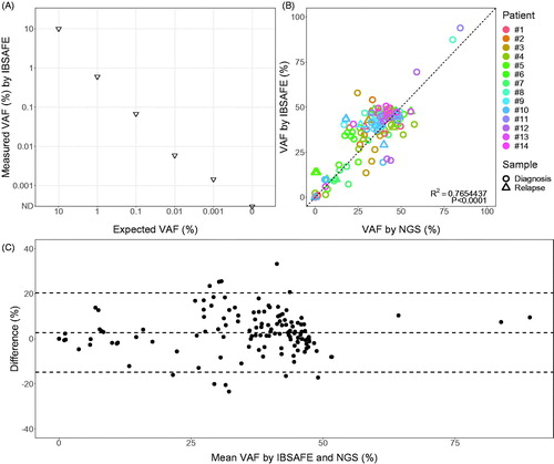
For molecular MRD detection we used the recently developed ultrasensitive mutation detection method IBSAFE, with an effective lower LoD down to approximately 0.001% VAF based on the amount of DNA analyzed per sample [Citation18–20]. In short, IBSAFE marries a two phase chemistry, linear copying and exponential signal generation, within the reaction droplet, thereby greatly enhancing true-positive signals and simultaneously reducing false-positive signals (described in the Supplementary Methods). An example dilution series from 10% VAF to 0.001% VAF as well as pure wild-type 0% VAF is shown in .
Figure 1. Performance of IBSAFE and comparison of IBSAFE and WES measured variant allele frequencies (VAFs) in diagnosis and relapse samples. (A) Dilution series for IBSAFE assay NPM1 type A for constructed samples with known VAFs at 10%, 1%, 0.1%, 0.01%, 0.001%, and 0%. (B) Scatterplot for agreement between the methods with Pearson’s coefficient of determination R2=0.77 and p-value <.0001, N = 144, (C) Bland Altman plot.
For the 14 patients, a total of 86 mutations (SNPs and small indels) were selected from WES data, and IBSAFE assays were developed for between 5–9 mutations for each patient. Candidate mutations were selected with priority toward mutations in genes known to be recurrently mutated in AML. In addition, some mutations present at both diagnosis and relapse as determined by WES were chosen. Finally, for a few patients, some mutations present only at relapse were selected to backtrack potential emerging clones.
Each IBSAFE assay was confirmed to have zero false-positive droplets using at least 59,000 haploid genome copies (180 ng) of negative control DNA (Promega), demonstrating an assay LoD of at least 0.0017% VAF. An example assay is shown in Supplementary Figure 1. IBSAFE analyses were performed on all diagnostic, follow-up, and relapse samples, using 60 ng of DNA per reaction and each reaction performed in duplicate thus enabling an effective LoD down to 0.003% VAF. In every IBSAFE run, positive (diagnostic or in a few cases relapse sample) and negative control (human male normal) DNA were used and confirmed test reliability.
Flow cytometry and qPCR for NPM1 type a mutations
Immunophenotyping and quantification of NPM1 type A mutations with qPCR was performed as previously described [Citation7].
Statistical analyses
To investigate the correlations between measuring the VAF of the mutations in the diagnostic and relapse samples with IBSAFE, WES and qPCR, Pearson’s correlation test and Bland-Altman plots were applied [Citation22].
Results
Diagnostic and monitored mutations
WES of the BM from 10 relapsing and four non-relapsing patients identified 10–30 somatic mutations at diagnosis (mean 18) in each patient, of which 0–7 (mean 3) were in genes known to be recurrently mutated in AML (). In total 12 of the patients had mutations in genes known to be recurrently mutated in AML. For 11 of the patients at least one of these recurrent mutations could be monitored by IBSAFE. In six of the relapsing patients, WES of the relapse sample detected between 2–22 new mutations (). For one patient, WES was not performed at relapse after allogeneic-SCT (allo-SCT) since the presence of donor cells prohibited detection of new mutations. For the remaining three patients, no new mutations could be identified due to poor sequence quality and therefore the WES data was only used to confirm presence of mutations identified at diagnosis.
Table 2. Number of mutations at diagnosis and first relapse.
On the basis of the WES results, 86 mutations, 81 of which were unique (NPM1 type A mutation was monitored in five patients, DNMT3A R882C in two), were monitored (Supplementary Table 1). Of these, 31 were mutations in genes known to be recurrently mutated in AML, including seven NPM1 mutations. Four mutations in genes known to be recurrently mutated in AML were identified by WES at relapse but absent at diagnosis in three patients (RUNX1, IDH1 and two different FLT3 mutations). Of these, FLT3 Y589H was monitored. Of note, scatterplot and Bland–Altman plot of VAFs as measured by IBSAFE and WES on the diagnostic and relapse samples displayed excellent agreement across a range of allele frequencies and considering the WES sequencing depth (R2=0.77, N = 144, p < .0001; ). A comparison of qPCR and IBSAFE data for the NPM1 type A mutation is shown in Supplementary Figure 2.
For all 10 relapsing patients, IBSAFE analysis revealed molecular evidence of persisting or emerging mutations at time-points prior to the clinical relapse (). Of all 66 follow-up time-points tested across the 10 patients, 35 time-points from 9 patients (2–7 time-points per patient, or 50–100% of all time-points tested for the patient) exhibited at least one IBSAFE-detected mutation detected at VAFs between 0.1% and 0.003%. In addition, all relapsing patients had at least one follow-up time-point with VAF >0.1% for at least one mutation detected before clinical relapse. Moreover, IBSAFE-based molecular MRD was more sensitive to identify residual disease as compared to MFC, with no time-point being MFC-positive and IBSAFE-negative (Supplementary Table 2).
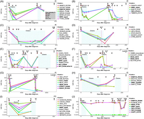
Figure 2. Monitoring leukemic mutations using ultrasensitive IBSAFE for ten relapsing AML patients (A) #4, (B) #9, (C) #6, (D) #8, (E) #7, (F) #5, (G) #1, (H) #3, (I) #10 and (J) #2. In each plot, the y-axis represents the detected variant allele frequency (VAF %) for each tracked mutation (key to the right; genes known to be recurrently mutated in AML in bold), and the x-axis indicates the days after diagnosis with therapies indicated by shading (chemotherapy) and clinical events indicated along the top of each plot. The inverted triangles indicate the flow cytometry MRD results, with the color-key indicated in the lower-right of plot (A). Dx: diagnosis; R: relapse; †: dead: SCT: stem cell transplantation; FU; follow-up; H: harvest; ND: not detected (VAF below lower effective limit of detection of 0.003% determined by input DNA quantity).
Patterns of mutations for relapsing patients
Interestingly, distinct patterns of emerging and retreating mutations could be discerned from the IBSAFE results. In Pattern 1, featuring four patients, one or several clones were apparent during follow-up with all the monitored mutations reappearing at relapse (; ). Some mutations were undetectable at certain time-points, whereas others were present at all time-points at low levels, possibly representing minor pre-leukemic clones. Two of these patients (#4 and #9) displayed mutations in genes known to be recurrently mutated in AML (DNMT3A, NPM1 or TET2) (). In these two patients, selection of clones containing mutations in DNMT3A, ANAPC2, CLSTN3 and GPR98 () and TET2 and TMEM35 (), respectively, was evident after induction therapy. For patient #4, a subclone containing two additional mutations (EPHA3 and TTYH1) at VAFs 1–2% was present at diagnosis (). Except for patient #8 (), MFC-MRD was not detected before the relapse in this group. This patient had a positive MFC-MRD at day 92 (0.2–0.3%), 18 days before the SCT with all monitored mutations detectable. After SCT, MFC-MRD was negative but 4/5 mutations were still detectable, and the patient soon exhibited clinical relapse about two months later.
In Pattern 2 containing four patients (), the emerging relapsing leukemia carried only some of the mutations monitored, both in recurrently as well as non-recurrently mutated genes (). All patients in this group displayed at least two mutations in known AML-associated genes including NPM1, IDH2, WT1, NRAS, KRAS, FLT3, PTPN11, RUNX1, or DNMT3A. After three courses of chemotherapy, at least one mutation was detectable by IBSAFE in all four patients despite negative MFC-MRD when available, with TRIM42 in patient #7 consistently higher than 0.1% VAF. MFC-MRD was positive (>0.1%) in two of the patients after completion of chemotherapy with concomitant VAF >0.1% for several mutations (patient #5, ; patient #3, ). At least one clone disappeared in all four patients in response to therapy as evidenced by the diminishing or undetectable amounts of mutant DNA.
In Pattern 3 with the remaining two relapsing patients (), there was no distinct decrease of the mutation allele frequencies before SCT for two or more of the mutations despite morphological remission and negative MFC-MRD, demonstrating the pre-leukemic nature of the regenerating hematopoiesis. In patient #10 (), nine mutations were monitored. Two mutations were detected at 17% VAF (CACNA9 and CPA4) after induction therapy, preceding the relapse about two months later, after completion of therapy despite MFC-MRD negativity <0.1% and morphological remission. Patient #2 () showed clonal hematopoiesis and an U2AF1 mutation with high residual VAF, apparently unresponsive to chemotherapy, but in complete morphological and immunophenotypical (<0.1%) remission until the relapse at day 736. At the first relapse, both the mutational profile (loss of 19 mutations and gain of six new mutations by WES) and the immunophenotype changed significantly (). Only after the second relapse (150 days after the first relapse) treated with SCT, a significant decrease of the mutational load for the monitored mutations was seen, but the VAFs of monitored mutations never fell below 0.2%.
Patterns of mutations for patients in complete remission
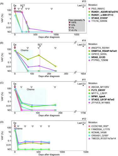
Among the non-relapsing patients (), patients #13 and #14 underwent allo-SCT and displayed a similar pattern of monitored mutations before transplantation with some persistent mutations with high VAF despite morphological and immunophenotypical (<0.1%) remission, suggesting pre-leukemic hematopoiesis refractory to conventional cytoreductive therapy (). This pattern was reminiscent of that of the relapsing patients #10 and #2 () described above. After allo-SCT the mutations gradually disappeared and were unmeasurable at the last follow-up time-point except for 0.01% VAF of the DNMT3A mutation in patient #14 (). In patient #11 and #12 all monitored mutations declined after conventional cytoreductive therapy; some of them disappeared completely whereas others seemed to stabilize at low VAF levels between 0.01% and 0.08% ().
Figure 3. Monitoring leukemic mutations using ultrasensitive IBSAFE for four non-relapsing AML patients (A) #13, (B) #14, (C) #11 and (D) #12. In each plot, the y-axis represents the detected variant allele frequency (VAF %) for each tracked mutation (key to right; genes known to be recurrently mutated in AML in bold), and the x-axis indicates the days after diagnosis with therapies indicated by shading (chemotherapy) and clinical events indicated along the top of each plot. The inverted triangles indicate the flow cytometry MRD results, with the color-key indicated in the lower-right of plot (A). Dx: diagnosis; FU: follow-up; SCT: stem cell transplantation; ND: not detected (VAF below 0.003%).
NPM1-mutations in relapsing and non-relapsing patients
Six out of ten patients in the relapsing group had an NPM1 mutation (type A in patients #2, #4, #7 and #9; type D in #10; and type DD5 in #3). All of these but patient #2 experienced an NPM1-positive relapse. For patient #2 the NPM1 type A mutation was undetectable by IBSAFE and qPCR at all follow-up time-points after day 73, including the complete morphological and immunophenotypical remission time-points and the relapse time-points () and (). Patient #7 (in complete morphological and immunophenotypical remission after completion of therapy) had no detectable NPM1 type A mutation by IBSAFE at any time-point before the NPM1 positive relapse () but a quantifiable signal by qPCR at the last time-point (0.011%) and a detectable but not quantifiable signal for the remaining two time-points. Patient #4 and #9 () had detectable NPM1 mutations by IBSAFE as well as qPCR at all follow-up time-points except the last time-point, when it was undetectable by IBSAFE and weakly positive (0.0028%, #4) or detectable but not quantifiable (<0.0024%, #9) by qPCR. Both these patients were in complete morphological and immunophenotypical remission after completion of therapy. For the type D and DD5 NPM1 mutation-positive patients (#10 and #3) no qPCR data exist. For the type D NPM1 mutation-positive patient (#10) only one follow-up sample exists between diagnosis and relapse, day 63. At this time-point MFC-MRD was negative, but IBSAFE-MRD was positive (0.08% VAF). For the type DD5 NPM1 mutation-positive patient (#3), three time-points between the diagnosis and relapse were tested. IBSAFE detected MRD in all these three time-points, whereas MFC was only positive at the last time-point before the relapse.
In the non-relapsing group, one patient (#11) had a NPM1 mutation (type A; ) detectable at low VAF <0.1% with IBSAFE and qPCR after induction therapy, day 28, that disappeared during follow-up (). MFC-MRD was negative at all time-points ( and ).
Discussion
Because of the clonal complexity of AML, the assessment of MRD in routine practice is difficult. Minor subclones present at diagnosis may evolve and escape detection by MFC or targeted qPCR-MRD. The aim of this study was to investigate if relapses in AML can be identified and predicted by using a sensitive molecular MRD approach (IBSAFE) targeting several mutations, thereby producing information on multiple putative subclones. Our proof-of-concept study demonstrates the ability of the IBSAFE method to do this. For all ten relapsing patients, a few selected mutations were able to track early recurrence of leukemic clones. In a clinical routine setting, the most commonly used MRD-method is MFC except for RNA based methods for NPM1 mutated leukemias and for some fusion genes [Citation23]. Currently, a cutoff of 0.1% residual leukemic blasts after two courses of therapy, as determined by MFC, has important implications for risk stratification and therapy decisions [Citation24]. Three of the relapsing patients in this study (#3, #5 and #8) displayed MFC-MRD levels >0.1% after completion of therapy. In concordance, these three patients also had persisting mutations >0.1% VAF as determined by IBSAFE. IBSAFE also showed a better sensitivity than standard MFC-MRD for all patients in this study. Among the ten relapsing patients, MFC-MRD was positive at 4/30 follow-up time-points (from day 25 until the last time-point before the first relapse) while IBSAFE-MRD was detectable for at least one mutation at 31/31 follow-up time-points (). These results are important, because many patients eventually relapse despite MFC-MRD levels below 0.1%, emphasizing the problem of false-negative MFC-MRD and therefore the need for new MRD-strategies to more accurately predict recurrence.
For comparison, we also monitored four non-relapsing patients without clinical, morphological or immunophenotypical signs of residual disease. Two of these patients showed a persisting but stabilized subclone with low VAF at the last follow-up () of which one patient () exclusively in non-recurrently mutated genes. The significance of the persisting mutations in the non-relapsing patients is unclear. They could represent pre-leukemic stem cells or progenitors that have not yet acquired all mutations needed for progression, or sub-clinical leukemia where overt relapse has not yet occurred. Alternatively, they are passenger mutations of clonal hematopoiesis [Citation25].
Our approach allowed for the deciphering of three different mutational patterns in the follow-up samples from the relapsing patients. In Pattern 3 (N = 2; ), no distinct decrease of the VAF before relapse was observed for two or more of the mutations despite morphological remission and negative MFC-MRD, indicating the pre-leukemic nature of the regenerating hematopoiesis. It is tempting to speculate that patients with this pattern are at risk for relapse. However, the presence of some of these mutations do not necessarily signalize impending relapse. For example, DNMT3A and TET2 mutations, as in patient #2, can persist at variable VAF levels in the follow-up samples in patients in CR without impact on prediction of relapse [Citation24,Citation26]. Moreover, it is well known that the number of somatic mutations rises with increasing age. In some individuals, these mutations form clonal hematopoiesis that arises when a single hematopoietic stem cell contributes disproportionately to the population of mature blood cells [Citation27,Citation28]. The presence of clonal hematopoiesis is associated with an increased risk of developing a myeloid neoplasm, but the vast majority of individuals with age-related clonal hematopoiesis do not develop AML [Citation29–33]. Such clonal mutations are sometimes called pre-leukemic and include genes such as DNMT3A, TET2, IDH1/2, ASXL1, and IKZF1 (29). Other recurrent mutations such as those occurring in spliceosome genes (e.g. U2AF1; patient #2) are more often predictable of evolution to AML, [Citation34]. Hence, with respect to MRD-assessment, the presence of mutations such as those in DNMT3A, ASXL1, and TET2 are often of limited value for prediction of relapse. Nevertheless, even after exclusion of DNMT3A and TET2 mutations, MRD was detectable at all follow-up time-points. Likely, both the nature (e.g. preleukemic versus non-preleukemic mutations), and the kinetics of the monitored mutations are important biological determinants for reemerging AML [Citation35].
In 2011 Krönke et al. showed that 9% of all NPM1 mutated patients at diagnosis had no detected transcript levels at relapse [Citation36]. In a recently published study from the Munich Leukemia Laboratory, 13% of leukemias harboring a NPM1-mutation at diagnosis relapsed with a NPM1-wildtype leukemia [Citation37]. More recent studies have also indicated that markers such as mutated NPM1 may not always be stable over the course of disease and that relapses sometimes emanate from NPM1-wildtype clones [Citation29,Citation32]. In a recently published case report, featuring a case resembling our patient #2 (), the authors describe loss of the NPM1 mutation found at diagnosis, a persisting DNMT3A mutation, and a late relapse [Citation38].
Our results support the limitations of employing NPM1 mutation as the sole marker of disease. In two of our six NPM1 positive relapsing patients (33%) the NPM1 mutation was not a reliable marker of residual disease. Patient #7, () had NPM1 negative MRD and patient #2 () had a NPM1 negative relapse.
The difference between the results from qPCR and IBSAFE might at least partly be explained by the amount of input DNA (600–)1000 ng/test compared to 120 ng/test. It is important to point out that although several mutations were monitored for each patient, there may have been additional relevant subclones to follow. In addition, acquired new mutations after therapy will not be detected with the approach of the present study. Indeed, backtracking of mutations from the relapse of two patients, #1 and #4 showed emerging clones containing these mutations (). Nevertheless, the pattern of persisting and emerging clones in the relapse group suggest that it may suffice to select a limited number of mutations for powerful MRD-assessment.
In conclusion, this pilot study demonstrates the feasibility of the IBSAFE method to measure MRD with high sensitivity and on essentially any newly diagnosed adult with AML where there are no fusion genes that are recommended for MRD follow-up. The method allows for a lower LoD to 0.003% VAF, based on available input DNA, to follow several mutations and track different emerging clones. Developed IBSAFE assays can rapidly be applied on follow-up samples and easily utilized for other patients carrying the same mutation. In addition to the established recurrent mutations, personalized assays (due to the mutational heterogeneity of AML) may also be developed for individual AML patients based on their specific mutational profiles. The prognostic relevance of such monitoring should be evaluated in large prospective studies.
Author contributions
LP, YC, ME and LHS conceived the study. LP and ME provided bone marrow samples. GJ and VL provided clinical information. LP and ME analyzed the MFC-MRD and qPCR data. LP, YC, RR, AMG, and LHS performed IBSAFE analyses. LP, YC, ME and LHS wrote the report. All authors approved the final version of the report for publication.
GLAL-2019-1075-File011.pdf
Download PDF (39.8 KB)GLAL-2019-1075-File010.pdf
Download PDF (120.5 KB)GLAL-2019-1075-File009.docx
Download MS Word (15 KB)GLAL-2019-1075-File008.docx
Download MS Word (33.3 KB)GLAL-2019-1075-File007.docx
Download MS Word (17 KB)Acknowledgements
The authors would like to thank Christina Orsmark-Pietras, Henrik Lilljebjörn and Thoas Fioretos for performing the sequencing analyses and analyzing the WES data. The authors would also like to thank Li Zhou, Heike Kotarsky, Kerstin Torikka and Marianne Rissler for excellent technical support.
Disclosure statement
YC, AMG, RR, and LHS have ownership interest (including stock, patents, etc.) in SAGA Diagnostics AB. LP, VL, GJ and ME report no conflict of interest.
Additional information
Funding
References
- Grimwade D, Freeman SD. Defining minimal residual disease in acute myeloid leukemia: which platforms are ready for “prime time”? Blood. 2014;124(23):3345–3355.
- Ivey A, Hills RK, Simpson MA, et al. Assessment of minimal residual disease in standard-risk AML. N Engl J Med. 2016;374(5):422–433.
- Kayser S, Walter RB, Stock W, et al. Minimal residual disease in acute myeloid leukemia–current status and future perspectives. Curr Hematol Malig Rep. 2015;10(2):132–144.
- Ommen HB. Monitoring minimal residual disease in acute myeloid leukaemia: a review of the current evolving strategies. Ther Adv Hematol. 2016;7(1):3–16. Feb
- Sung PJ, Luger SM. Minimal residual disease in acute myeloid leukemia. Curr Treat Options Oncol. 2017;18(1):1.
- Jaso JM, Wang SA, Jorgensen JL, et al. Multi-color flow cytometric immunophenotyping for detection of minimal residual disease in AML: past, present and future. Bone Marrow Transplant. 2014;49(9):1129–1138.
- Pettersson L, Leveen P, Axler O, et al. Improved minimal residual disease detection by targeted quantitative polymerase chain reaction in Nucleophosmin 1 type a mutated acute myeloid leukemia. Genes Chromosomes Cancer. 2016;55(10):750–766. Oct
- Leroy H, de Botton S, Grardel-Duflos N, et al. Prognostic value of real-time quantitative PCR (RQ-PCR) in AML with t(8;21). Leukemia. 2005;19(3):367–372. Mar
- Falini B, Mecucci C, Tiacci E, et al. Cytoplasmic nucleophosmin in acute myelogenous leukemia with a normal karyotype. N Engl J Med. 2005;352(3):254–266.
- Bullinger L, Dohner K, Dohner H. Genomics of acute myeloid leukemia diagnosis and pathways. JCO. 2017;35(9):934–946.
- Ley TJ, Cancer Genome Atlas Research Network, Miller C, Ding L, et al. Genomic and epigenomic landscapes of adult de novo acute myeloid leukemia. N Engl J Med. 2013; 368(22):2059–2074.
- Grimwade D, Ivey A, Huntly BJ. Molecular landscape of acute myeloid leukemia in younger adults and its clinical relevance. Blood. 2016;127(1):29–41.
- Papaemmanuil E, Gerstung M, Bullinger L, et al. Genomic classification and prognosis in acute myeloid leukemia. N Engl J Med. 2016;374(23):2209–2221.
- Roloff GW, Lai C, Hourigan CS, et al. Technical advances in the measurement of residual disease in acute myeloid leukemia. J Clin Med. 2017;6(9):pii: E87.
- Cruz NM, Mencia-Trinchant N, Hassane DC, et al. Minimal residual disease in acute myelogenous leukemia. Int J Lab Hem. 2017;39(Suppl 1):53–60.
- Mencia-Trinchant N, Hu Y, Alas MA, et al. Minimal residual disease monitoring of acute myeloid leukemia by massively multiplex digital PCR in patients with NPM1 mutations. J Mol Diagn. 2017;19(4):537–548.
- Wertheim GBW, Bagg A. NPM1 for MRD? Droplet like it’s hot! J Mol Diagn. 2017;19(4):498–501.
- Arildsen NS, Martin de la Fuente L, Masback A, et al. Detecting TP53 mutations in diagnostic and archival liquid-based Pap samples from ovarian cancer patients using an ultra-sensitive ddPCR method. Sci Rep. 2019;9(1):15506.
- Fornvik D, Aaltonen KE, Chen Y, et al. Detection of circulating tumor cells and circulating tumor DNA before and after mammographic breast compression in a cohort of breast cancer patients scheduled for neoadjuvant treatment. Breast Cancer Res Treat. 2019;177(2):447–455.
- Isaksson S, George AM, Jonsson M, et al. Pre-operative plasma cell-free circulating tumor DNA and serum protein tumor markers as predictors of lung adenocarcinoma recurrence. Acta Oncologica. 2019;58(8):1079–1086.
- Lazarevic V, Orsmark-Pietras C, Lilljebjorn H, et al. Isolated myelosarcoma is characterized by recurrent NFE2 mutations and concurrent preleukemic clones in the bone marrow. Blood. 2018;131(5):577–581.
- Bland JM, Altman DG. Statistical methods for assessing agreement between two methods of clinical measurement. Lancet. 1986;327(8476):307–310.
- Ehinger M, Pettersson L. Measurable residual disease testing for personalized treatment of acute myeloid leukemia. APMIS. 2019;127(5):337–351.
- Schuurhuis GJ, Heuser M, Freeman S, et al. Minimal/measurable residual disease in AML: a consensus document from the European LeukemiaNet MRD Working Party. Blood. 2018;131(12):1275–1291.
- Parkin B, Londono-Joshi A, Kang Q, et al. Ultrasensitive mutation detection identifies rare residual cells causing acute myelogenous leukemia relapse. J Clin Investig. 2017;127(9):3484–3495.
- Jongen-Lavrencic M, Grob T, Hanekamp D, et al. Molecular minimal residual disease in acute myeloid leukemia. N Engl J Med. 2018;378(13):1189–1199.
- Steensma DP, Bejar R, Jaiswal S, et al. Clonal hematopoiesis of indeterminate potential and its distinction from myelodysplastic syndromes. Blood. 2015;126(1):9–16.
- Zink F, Stacey SN, Norddahl GL, et al. Clonal hematopoiesis, with and without candidate driver mutations, is common in the elderly. Blood. 2017;130(6):742–752.
- Corces-Zimmerman MR, Hong WJ, Weissman IL, et al. Preleukemic mutations in human acute myeloid leukemia affect epigenetic regulators and persist in remission. PNAS. 2014;111(7):2548–2553.
- Jaiswal S, Fontanillas P, Flannick J, et al. Age-related clonal hematopoiesis associated with adverse outcomes. N Engl J Med. 2014;371(26):2488–2498.
- Shlush LI, Mitchell A, Heisler L, et al. Tracing the origins of relapse in acute myeloid leukaemia to stem cells. Nature. 2017;547(7661):104–108.
- Shlush LI, Zandi S, Mitchell A, et al. Identification of pre-leukaemic haematopoietic stem cells in acute leukaemia. Nature. 2014;506(7488):328–333.
- Sykes SM, Kokkaliaris KD, Milsom MD, et al. Clonal evolution of preleukemic hematopoietic stem cells in acute myeloid leukemia. Exp Hematol. 2015;43(12):989–992.
- Abelson S, Collord G, Ng SWK, et al. Prediction of acute myeloid leukaemia risk in healthy individuals. Nature. 2018;559(7714):400–404.
- Rothenberg-Thurley M, Amler S, Goerlich D, et al. Persistence of pre-leukemic clones during first remission and risk of relapse in acute myeloid leukemia. Leukemia. 2018;32(7):1598–1608.
- Kronke J, Schlenk RF, Jensen KO, et al. Monitoring of minimal residual disease in NPM1-mutated acute myeloid leukemia: a study from the German-Austrian acute myeloid leukemia study group. J Clin Oncol. 2011;29(19):2709–2716.
- Hollein A, Meggendorfer M, Dicker F, et al. NPM1 mutated AML can relapse with wild-type NPM1: persistent clonal hematopoiesis can drive relapse. Blood Adv. 2018;2(22):3118–3125.
- Bacher U, Porret N, Joncourt R, et al. Pitfalls in the molecular follow up of NPM1 mutant acute myeloid leukemia. Haematologica. 2018;103(10):e486–e8.