
Abstract
Chimeric antigen receptor T (CAR-T) cells are a promising approach in hematopoietic malignancies. We evaluated the safety and efficacy of a combination of humanized anti-BCMA and murine anti-CD38 CAR-T cell therapy in patients with relapsed or refractory multiple myeloma (R/RMM). Twenty-two R/RMM patients, with a median age of 56 years and a median number of previous therapies of 8, were included in the study. Both CAR-T cells infusion doses were 2.0 × 10⁶/kg. The overall response rate (ORR) was 90.9%, with 12 patients (54.5%) achieving a strict complete response/complete response (sCR/CR). The 24-month overall survival (OS) rate was 56.6%, and the progression-free survival (PFS) rate was 48.7%. Cytokine release syndrome (CRS) of grades 1–2 occurred in 16 patients (72.7%) and of grade ≥3 in six patients (27.3%). Immune effector cell-associated neurotoxic syndrome (ICANS) of grades 1–2 occurred in three patients (13.6%). The combination therapy is potential in R/RMM patients.
Trial registration: The patients were enrolled in clinical trials registered as ChiCTR1800017051.
Introduction
Multiple myeloma (MM) is a malignant, plasma cell clonal disease. MM has the second-highest incidence among hematologic tumors [Citation1]. Although the efficacy of MM treatment has been significantly improved by the application of proteasome inhibitors, immunomodulators, and hematopoietic stem cell transplantation, the prognosis of some R/RMM patients remains particularly poor [Citation2]. Many clinical trials have used chimeric antigen receptor (CAR) T cells for R/RMM. The therapeutic targets include B cell maturation antigen (BCMA), CD19, CD38, and CD138, among others [Citation3–8]. To date, CAR-T cells targeting BCMA have shown excellent results in initial clinical trials [Citation9–17]. BCMA belongs to the tumor necrosis factor receptor superfamily and is mainly expressed on normal plasma cells and malignant plasma cells (PCs), as well as on a small proportion of mature B cells and plasma cell-like dendritic cells. However, BCMA is not expressed on hematopoietic stem cells or non-hematopoietic stem cells. Because BCMA is expressed on most MM cells, it is considered an ideal target for CAR-T cell therapy in R/RMM [Citation18,Citation19]. Although anti-BCMA CAR-T cell therapy has a high overall response rate in R/RMM, some patients still fail to respond or relapse within a short period of time after therapy. CD38 is a transmembrane glycoprotein that mediates signal transduction in lymphocytes and bone marrow cells. CD38 is highly expressed on myeloma cells, but its expression level on normal lymphocytes, myeloid cells, and some tissues of non-hematopoietic origin is relatively low, which makes it a potential target for the treatment of multiple myeloma [Citation6]. In a clinical trial, anti-CD38 CAR-T cell therapy for R/RMM achieved satisfactory efficacy without irreversible side effects [Citation6]. This clinical trial enrolled 22 patients with R/RMM who received humanized anti-BCMA CAR-T cells in combination with murine anti-CD38 CAR-T cell therapy, and we analyzed the efficacy and safety of this therapeutic strategy in these patients.
Patients and methods
Study design and participants
We conducted a phase 2, open-label, single-arm, single-center clinical trial at Tianjin First Center Hospital in China (ChiCTR1800017051) and enrolled 22 patients with R/RMM admitted to the Department of Hematology between August 2018 and December 2019. All patients were diagnosed with MM according to the IMWG classification and were not previously treated with daratumumab (CD38 monoclonal antibody). All 22 R/RMM patients or their representatives provided informed consent before enrollment. The objective of this clinical trial was to assess both short-term and long-term efficacy along with the side effects of the therapy.
Eligibility criteria included patients aged between 18 and 80 years, Karnofsky score of ≥60, meeting the international myeloma working group diagnostic criteria for relapsed/refractory multiple myeloma, having measurable or evaluable lesions, and main tissues and organs functions as follows: (1) liver function—ALT/AST <3 times the upper limit of normal (ULN) and bilirubin ≤34.2 µmol/L; (2) renal function—creatinine <220 µmol/L; (3) lung function—indoor oxygen saturation ≥95%; (4) heart function—left ventricular ejection fraction (LVEF) ≥40%. Key exclusion criteria included pregnancy (positive urine/blood pregnancy test) or breastfeeding, uncontrollable infectious diseases within four weeks of enrollment, active hepatitis B/C, or HIV infection (for detailed inclusion and exclusion criteria, please refer to the China Clinical Trial Registration Center).
Generation and detection of humanized anti-BCMA CAR-T cells and anti-CD38 CAR-T cells
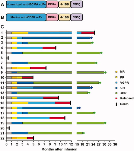
Peripheral blood mononuclear cells (PBMCs) were collected from all enrolled R/RMM patients. First, CD3+ T cells were extracted from PBMCs using CD3+ microbeads (Miltenyi Biotec, Cambridge, MA, USA) and then expanded by Dynabeads® Human T-Expander CD3/CD28 (Thermo Fisher Scientific, Waltham, MA, USA). Next, the cells were transduced with a lentiviral vector encoding the humanized anti-BCMA CAR gene (Creative Biolabs, Inc., Shirley, NY, USA) or murine anti-CD38 CAR gene (Creative Biolabs, Inc., Shirley, NY, USA) constructed separately. The humanized anti-BCMA CAR-T cells expressed a humanized anti-BCMA antigen-binding region (scFv), CD8 alpha hinge and transmembrane domain, and a 4–1BB-CD3ζ costimulatory activation domain. The murine anti-CD38 CAR-T cells expressed the murine anti-CD38 antigen-binding region (scFv), CD8 alpha hinge and transmembrane domain, and a 4–1BB-CD3ζ costimulatory-activation domain (). After 12 days of culture, transduction efficiency/gene expression was analyzed using flow cytometry (FCM), which detected the ratio of both kinds of CAR-T cells (PE, BD Biosciences) to CD3+ T cells (FITC, BD Biosciences).
Figure 1. Structures of CAR-T cells and response to combination therapy. (A,B) The construct of anti-BCMA/CD38 CAR-T. (C) Response assessment after combination therapy.
Lymphodepletion and anti-BCMA CAR-T and anti-CD38 CAR-T cell infusions
All enrolled R/RMM patients received FC (fludarabine 30 mg/m2 and cyclophosphamide 300 mg/m2) for lymphodepleting chemotherapy from days −4 to −2. Humanized anti-BCMA CAR-T cells (2.0 × 10⁶/kg) and murine anti-CD38 CAR-T cells (2.0 × 10⁶/kg) were infused on day 0 (the interval time between the infusions of both kinds of CAR-T cells was 4 h).
Objectives and clinical response criteria
The primary endpoint of our study was the overall response rate; secondary endpoints included overall survival (OS), progression-free survival (PFS), the incidence of adverse events (AEs), cytokine levels, and duration of expression of both kinds of CAR-T cells. The patients were followed up from the date of CAR-T cell infusion until death. Therapy responses were assessed at 2 weeks, 1 month, 2 months, 3 months, 6 months, 12 months, and every 6 months after 1 year after CAR-T cell infusion. Clinical response and disease progression were assessed according to the IMWG Uniform Response Criteria for Multiple Myeloma [Citation20]. The overall response rate (ORR) was defined as the proportion of patients experiencing a stringent complete response (sCR), complete response (CR), very good partial response (VGPR), and partial response (PR). Minimal residual disease (MRD) was detected using next-generation sequencing with a minimum cutoff of 10−6 nucleated cells.
Observation of adverse events after CAR-T cell infusion
AEs were monitored according to the National Cancer Institute’s Common Terminology Criteria for Adverse Events (NCI-CTCAE v. 4.03), and cytokine release syndrome (CRS) and immune effector cell-associated neurotoxic syndrome (ICANS) were assessed according to ASTCT Consensus Grading for CRS and ICANS [Citation21]. Secretion levels of cytokines, including interleukin-1β (IL-1β), IL-6, IL-2R, tumor necrosis factor-α (TNF-α), IL-8, IL-10, C-reactive protein (CRP), and ferritin, were measured on days 0, 4, 7, 14, 21, and 28 by enzyme-linked immunosorbent assay (ELISA) using the double-antibody one-step sandwich method.
Expansion and persistence of anti-BCMA CAR-T and anti-CD38 CAR-T cells
CAR-T cells were detected in the peripheral blood on days 0, 4, 7, 14, 21, and 28. Flow cytometry was used to measure the percentage of CAR-T cells in peripheral blood mononuclear cells. The absolute number of CAR-T cells in peripheral blood was determined by multiplying the percentage of CAR-T cells by the total white cell number.
Statistical analysis
The Kaplan–Meier method was used to calculate the progression-free survival and overall survival of the patients. Statistical data were analyzed using SPSS 19.0 and GraphPad Prism 7.0; categorical variables were examined using t-tests or χ2 tests. The difference was considered statistically significant if p < 0.05.
Results
Characteristics of the patients
The medical history of the R/RMM patients at the time of enrollment is provided in . The patients enrolled in this clinical trial had a median age of 56 years (47–68 years). The median number for previous therapy was 8; 100% of the patients were resistant to bortezomib, and 81.8% were resistant to lenalidomide. Additionally, at least one high-risk cytogenetic abnormality was found in 86.4% of the subjects, with 50% having deletions of 17p or mutations in TP53. High risk was detected by FISH and defined as the following abnormalities: del(17p), t(4;14), or t(14;16). Patients with only minimal residual disease (MRD) in bone marrow (BM) were not enrolled in our trial. During enrollment, the median proportion of myeloma cells in BM was 32.9% (10.0–86.0%). The median follow-up time was 24 months (0.5–33 months). All 22 patients recruited showed high BCMA and CD38 expression levels on MM cells in BM, as analyzed by FCM.
Table 1. Baseline characteristics in 22 relapsed/refractory multiple myeloma patients.
Transduction efficiency, amplification, and infusion of CAR-T cells
The median BCMA-CAR and CD38-CAR transduction efficiencies of 22 patients were 45.7 ± 6.1 and 41.6 ± 5.1%, respectively. The median anti-BCMA CAR-T and anti-CD38 CAR-T cell numbers for all patients on the harvest day were 28.1 ± 3.5 × 10⁶ and 21.3 ± 3.3 × 10⁶ cells/kg, respectively. The median CD4+/CD8+ ratios were 2.1 and 1.9. For both CAR-T cell infusion doses, 2.0 × 10⁶/kg was used. Notably, we did not detect any bacteria, mycoplasma, fungi, or endotoxins in the final products.
Clinical responses
In the 22 evaluated patients, 20 (90.9%) showed an ORR to therapy: 12 patients (12/22, 54.5%) achieved sCR/CR, five (5/22, 22.7%) achieved VGPR, and three (3/22, 13.6%) achieved PR (). The median response time to achieve the best effect was 3.5 months (2.0–5.0 months). After achieving the best response, 10 (10/22, 45.5%) patients were found to be MRD negative, all of whom achieved sCR/CR; 12 patients (12/22, 54.5%) maintained sCR/CR for more than 12 months, and six (6/22, 27.3%) for more than 24 months ().
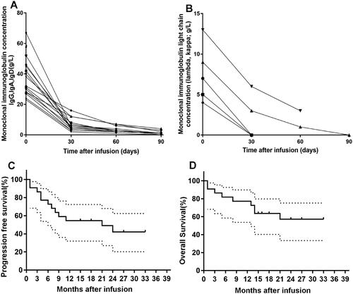
Monoclonal immunoglobulins and light chains in the peripheral blood were assessed in the 22 patients, and levels were found to be significantly decreased after treatment compared to baseline (). Based on Kaplan–Meier analysis, the progression-free survival (PFS) rate of all 22 patients was 48.7%, and the overall survival (OS) rate was 56.6% at 24 months ().
Figure 2. Changes in monoclonal immunoglobulin and light chain from baseline and survival in R/RMM to CAR-T. (A) The change in plasma M-protein concentration from baseline after CAR-T cell infusion. (B) The change in light chain concentration from baseline after CAR-T cell infusion. (C) The curve shows the time to progression after infusion of CAR-T cells. (D) The curve shows overall survival data censored at the time of the last follow-up.
We analyzed the BM immunophenotype of eight patients who exhibited disease progression after CAR-T cell therapy. No plasma cells were detected in the bone marrow on the best response after CAR-T cell therapy. However, plasma cells were BCMA/CD38 positive on progression. There was no emergence of antigen loss variants.
Adverse effects
AEs occurred in all 22 patients (). The most common (≥50.0%) AEs were CRS (100.0%), fever (100.0%), leukopenia (100.0%), and elevated D-dimer (54.5%). Leukopenia (36.4%), fever (36.4%), anemia (27.3%), and CRS (27.3%) were the most common events observed among ≥3 grade AEs.
Table 2. Adverse event associated with treatment.
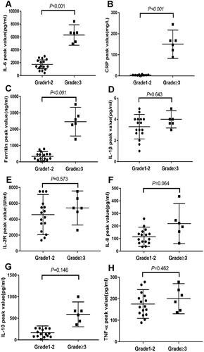
CRS was found in all the patients (), grade 1–2 CRS in 16 (16/22, 72.7%) and grade ≥3 CRS in six (6/22, 27.3%). The median occurrence time of CRS after CAR-T cell infusion was 4.5 days (2–14 days), and the median duration of CRS was 7.5 days (3–24 days). The cytokines IL-6, CRP, and ferritin were closely related to grade ≥3 CRS, and high peaks were observed in the blood ().
Figure 3. Serum cytokines associated with CRS severity. The chart shows the peak levels of serum cytokines over baseline in the first 30 days after infusion of CAR-T cells in patients with grade 1–2 CRS (n = 16) compared with patients with grade ≥3 CRS (n = 6). (A–C). Peak levels of IL-6, CRP and ferritin in the 1–2 grade CRS group were higher than those in the ≥3 grade CRS group (p < 0.001). (D–H) There was no significant difference in peak levels of IL-1β, IL-2R, IL-8, IL-10, and TNF-α between 1–2 grade and the ≥3 grade CRS groups (p = 0.643, p = 0.573, p = 0.064, p = 0.146, p = 0.462).
Sixteen patients with grade 1–2 CRS received antipyretic drugs as treatment for AEs. However, six patients with grade ≥3 CRS required methylprednisolone and tocilizumab during therapy (). Patients 6 and 20 were diagnosed with grade 5 CRS after CAR-T cell infusion. In addition to high fever and dyspnea, they displayed grade 3 liver dysfunction, grade 4 renal dysfunction, severe electrolyte metabolism disorders, and heart failure. Despite administering glucocorticoids and tocilizumab and providing plasma exchange, supportive treatments, and artificial ventilation therapy, these two patients died of respiratory failure and heart failure within days of receiving CAR-T cell infusion.
Table 3. Management of patients with CRS ≥ grade 3.
Hematological adverse events occurred in all patients. Grade 3–4 leucopenia and neutropenia were detected in eight patients (8/22, 36.4%); grade 3–4 anemia was observed in six patients (6/22, 27.3%) (). The median occurrence time for grade 3–4 leukopenia after CAR-T cell infusion was 6.5 days (3–10 days), and the median recovery time was 6 days (3–12 days). All cytopenia events occurred within 28 days after CAR-T cell infusion, whereas no delayed cytopenia was observed at 1, 2, or 3 months after CAR-T cell infusion. Four patients developed bacterial infections between 7 and 23 days after CAR-T cell infusion and were successfully treated with antibiotics and supportive therapy. None of the 22 patients was diagnosed with invasive fungal disease.
Three of the 22 patients using this combination therapy were diagnosed with grade 1 ICANS (). These patients developed headaches, dizziness, confusion, bradyphrenia, brain edema, somnolence, and tremors. However, the ICANS alleviated rapidly after supportive treatment. We did not observe delayed neurotoxicity within 90 days after CAR-T infusion in patients with a high tumor burden.
Cardiovascular complications occurred in six patients, including hypotension, heart failure, and arrhythmia. Except for Patients 6 and 20, the other four patients’ symptoms alleviated rapidly after supportive treatment ().
Before CAR-T cell infusion, CRS classification was related to the number of bone marrow clonal plasma cells (p < 0.001). Additionally, other factors, including age, KPS score, ISS stage, lactate dehydrogenase level, M protein level, hemoglobin level, high-risk cytogenetics, and previous treatments, did not affect the severity of CRS ().
Table 4. Factors possibly associated with CRS.
Expansion of anti-BCMA CAR-T and anti-CD38 CAR-T cells
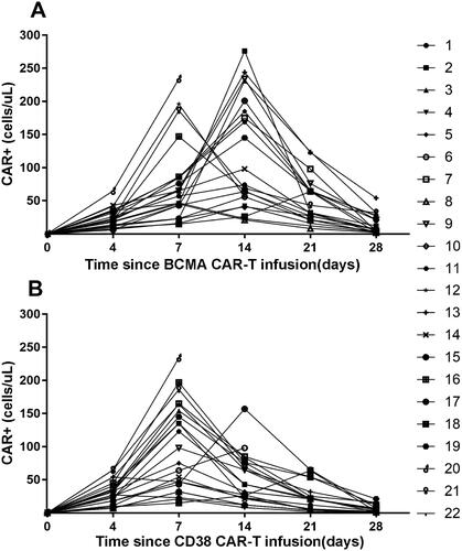
During the first 28 days after infusion with the CAR-T cells, the absolute number of both types expanded, with different peak times and degrees. The average number of anti-BCMA CAR-T cells and anti-CD38 CAR-T cells at the peak was 146 cells/µL () and 110 cells/µL (). The expansion peak of anti-BCMA CAR-T cells appeared at 7–14 (9.0) days after infusion (), while anti-CD38 CAR-T cells appeared at 4–7 (4.9) days (). Anti-CD38 CAR-T cell expansion occurred before that of anti-BCMA CAR-T cells, but the absolute number was lower ().
Figure 4. Expansion of CAR-T cells after infusion. CAR-T cell levels over time in peripheral blood are depicted, as measured by quantitative PCR for CAR sequence. (A) The absolute number of BCMA CAR+ cells/µl in peripheral blood within 28 days of infusion. (B) The absolute number of CD38 CAR+ cells/µl in peripheral blood within 28 days of infusion.
Discussion
CAR-T cell therapy for R/RMM has shown considerable results in previous clinical trials [Citation22,Citation23]. However, some patients relapse during follow-up. In this study, we combined humanized anti-BCMA CAR-T cells with murine anti-CD38 CAR-T cells for the treatment of patients with R/RMM. The median time to achieve the best response was three months, which was later than that seen in a previous study [Citation11]. Notably, patients with a high tumor burden and high-risk cytogenetics were not excluded from that study. In contrast, the median malignant plasma cell percentage in the bone marrow in our study was 32.9%, and 86.4% of patients had high-risk cytogenetic abnormalities.
In an earlier study that used BCMA CAR-T cell treatment for R/RMM [Citation14], a 48% ORR was reported in only three patients (12.00%) along with progression-free survival of more than 11 months. Another study using BCMA CAR-T cell treatment for R/RMM reported an ORR of 84.85% [Citation11]. At a median follow-up of 11.3 months, 48.45% of patients showed no disease progression. The ORR in our study was 90.9%, with 54.5% of patients achieving sCR/CR and 45.5% of patients being MRD negative. In another combination therapy study of combined anti-CD19 and anti-BCMA CAR T cell infusion [Citation24], an ORR of 95.24% was obtained, with 42.86% of patients maintaining sCR or CR for more than half a year and 19.05% maintaining it for more than one year. Although our study showed a similar ORR as that observed for the previous combination therapy, 54.5% of patients in our study maintained sCR/CR for more than one year, and 27.3% of patients maintained sCR/CR for more than 2 years.
Anti-CD38 CAR-T cells lyse the CD38+ portion of CD34+ hematopoietic progenitor cells, monocytes, and natural killer cells and lyse T/B lymphocytes to some extent [Citation6]. Therefore, anti-CD38 CAR-T cells will induce a certain degree of adverse events. Due to the high tumor load and anti-CD38 CAR-T cell infusion, AEs related to CAR-T treatment were observed in all of our patients. This was similar to the clinical outcomes in the study of CD19 CAR-T therapy for B cell acute lymphoblastic leukemia, in which a high tumor burden resulted in severe CRS [Citation25,Citation26]. CRS is essentially an acute explosive inflammatory syndrome characterized by a rapid rise in a large number of cytokines and a series of clinical symptoms [Citation27]. Plasmapheresis may reduce the inflammatory factor load quickly control symptoms. Although CRS of grade ≥3 occurred in six patients, four patients recovered with glucocorticoids, tocilizumab, plasmapheresis, and supportive treatment. Patient 6 had a history of atrial fibrillation and Patient 20 had a history of heart failure. Cardiac function deteriorated in both after CAR-T cell infusion, and they died due to respiratory failure and heart failure at 10 and 15 days after CAR-T cell infusion despite being transferred to the ICU for salvage treatment.
AEs are a common event in the hematologic system with leukopenia and anemia, which was higher in our study than in previous studies [Citation9]. Although the incidence of grade 3–4 leukopenia was high, no severe infections were observed during the period of agranulocytosis when using this combination therapy. Only four patients developed a bacterial infection and later recovered with antibiotic treatment. Additionally, no fungal or viral infections were observed in our study. Anemia and thrombocytopenia also improved after supportive treatments.
None of the 22 patients showed central nervous system infiltration before CAR-T cell infusion; three patients who had grade 1 ICANS after CAR-T cell infusion presented only mild headache, dizziness, and confusion, which resolved after symptomatic treatment. The incidence of cardiovascular system-related AEs was higher in our study than in another clinical study [Citation28]. These AEs mainly included hypotension, heart failure, and arrhythmias, which might have been related to the high tumor burden in BM and usage of two kinds of CAR-T cell infusion.
We analyzed the correlation between CRS and levels of cytokines in the peripheral blood and found IL-6, CRP, and ferritin to be related to the occurrence and severity of CRS, similar to previous studies [Citation14]. The factors that influenced CRS classification in our study were only related to the number of bone marrow clonal plasma cells before CAR-T cell infusion, which was different from a previous study [Citation29] and perhaps related to the high tumor burden in BM.
In summary, humanized anti-BCMA CAR-T cells combined with murine anti-CD38 CAR-T cell therapy constitute a potential therapy with a long-term response in R/RMM patients. Further research is continuing to expand the sample size. This combination therapy is a promising treatment option for R/RMM patients.
Ethical approval
This study was approved by the Medical Ethics Committee of the Department of Hematology, Tianjin First Center Hospital (Tianjin, China) (Approved No 2015002X). The patients agreed to participate in our clinical trials. They agreed to the use of their specimens and data for our study.
Author contributions
ZMF: concept and design. ZH: drafting or revision of the manuscript. LM, YT, LJY, and XX: acquisition of data. YT: analysis and interpretation of data. All authors: writing, review, and/or revision of the manuscript. ZMF: study supervision.
Acknowledgments
We thank patients for their participation in our experimental studies and clinical trials.
Disclosure statement
The authors have no conflicts of interest to report.
References
- Palumbo A, Anderson K. Multiple myeloma. N Engl J Med. 2011;364(11):1046–1060.
- Kumar SK, Dimopoulos MA, Kastritis E, et al. Natural history of relapsed myeloma, refractory to immunomodulatory drugs and proteasome inhibitors: a multicenter IMWG study. Leukemia. 2017;31(11):2443–2448.
- Mikkilineni L, Kochenderfer JN. Chimeric antigen receptor T-cell therapies for multiple myeloma. Blood. 2017;130(24):2594–2602.
- Maruyama D. B-Cell maturation antigen(BCMA)-targeting therapy in multiple myeloma. Gan to Kagaku Ryoho. 2020;47:778–782.
- Mueller KT, Maude SL, Porter DL, et al. Cellular kinetics of CTL019 in relapsed/refractory B-cell acute lymphoblastic leukemia and chronic lymphocytic leukemia. Blood. 2017;130(21):2317–2325.
- Drent E, Groen RW, Noort WA, et al. Pre-clinical evaluation of CD38 chimeric antigen receptor engineered T cells for the treatment of multiple myeloma. Haematologica. 2016;101(5):616–625.
- Tian C, Yang H, Zhu L, et al. Anti-CD138 chimeric antigen receptor-modified T cell therapy for multiple myeloma with extensive extramedullary involvement. Ann Hematol. 2017;96(8):1407–1410.
- Sun C, Mahendravada A, Ballard B, et al. Safety and efficacy of targeting CD138 with a chimeric antigen receptor for the treatment of multiple myeloma. Oncotarget. 2019;10(24):2369–2383.
- Brudno JN, Maric I, Hartman SD, et al. T cells genetically modified to express an anti-B-Cell maturation antigen chimeric antigen receptor cause remissions of poor-prognosis relapsed multiple myeloma. J Clin Oncol. 2018;36(22):2267–2280.
- Ali SA, Shi V, Maric I, et al. T cells expressing an anti-B-cell maturation antigen chimeric antigen receptor cause remissions of multiple myeloma. Blood. 2016;128(13):1688–1700.
- Raje N, Berdeja J, Lin Y, et al. Anti-BCMA CAR T-cell therapy bb2121 in relapsed or refractory multiple myeloma. N Engl J Med. 2019;380(18):1726–1737.
- Friedman KM, Garrett TE, Evans JW, et al. Effective targeting of multiple B-cell maturation antigen-expressing hematological malignances by anti-B-cell maturation antigen chimeric antigen receptor T cells. Hum Gene Ther. 2018;29(5):585–601.
- Lam N, Trinklein ND, Buelow B, et al. Anti-BCMA chimeric antigen receptors with fully human heavy-chain-only antigen recognition domains. Nat Commun. 2020;11(1):283.
- Cohen AD, Garfall AL, Stadtmauer EA, et al. B cell maturation antigen-specific CAR T cells are clinically active in multiple myeloma. J Clin Invest. 2019;129(6):2210–2221.
- Zhao WH, Liu J, Wang BY, et al. A phase 1, open-label study of LCAR-B38M, a chimeric antigen receptor T cell therapy directed against B cell maturation antigen, in patients with relapsed or refractory multiple myeloma. J Hematol Oncol. 2018;11(1):141.
- Demel I, Bago JR, Hajek R, et al. Focus on monoclonal antibodies targeting B-cell maturation antigen (BCMA) in multiple myeloma: update 2020. Br J Haematol. 2021;193(4):705–722.
- Feng D, Sun J. Overview of anti-BCMA CAR-T immunotherapy for multiple myeloma and relapsed/refractory multiple myeloma. Scand J Immunol. 2020;92(2):e12910.
- Carpenter RO, Evbuomwan MO, Pittaluga S, et al. B-cell maturation antigen is a promising target for adoptive T-cell therapy of multiple myeloma. Clin Cancer Res. 2013;19(8):2048–2060.
- Tai YT, Mayes PA, Acharya C, et al. Novel anti-B-cell maturation antigen antibody-drug conjugate (GSK2857916) selectively induces killing of multiple myeloma. Blood. 2014;123(20):3128–3138.
- Rajkumar SV, Dimopoulos MA, Palumbo A, et al. International myeloma working group updated criteria for the diagnosis of multiple myeloma. Lancet Oncol. 2014;15(12):e538–e548.
- Lee DW, Santomasso BD, Locke FL, et al. ASTCT consensus grading for cytokine release syndrome and neurologic toxicity associated with immune effector cells. Biol Blood Marrow Transplant. 2019;25(4):625–638.
- Susanibar Adaniya SP, Cohen AD, Garfall AL. Chimeric antigen receptor T cell immunotherapy for multiple myeloma: a review of current data and potential clinical applications. Am J Hematol. 2019;94(S1):S28–S33.
- Timmers M, Roex G, Wang Y, et al. Chimeric antigen receptor-modified T cell therapy in multiple myeloma: beyond B cell maturation antigen. Front Immunol. 2019;10:1613.
- Yan Z, Cao J, Cheng H, et al. A combination of humanised anti-CD19 and anti-BCMA CAR T cells in patients with relapsed or refractory multiple myeloma: a single-arm, phase 2 trial. Lancet Haematol. 2019;6(10):e521–e529.
- Maude SL, Frey N, Shaw PA, et al. Chimeric antigen receptor T cells for sustained remissions in leukemia. N Engl J Med. 2014;371(16):1507–1517.
- Lee DW, Kochenderfer JN, Stetler-Stevenson M, et al. T cells expressing CD19 chimeric antigen receptors for acute lymphoblastic leukaemia in children and young adults: a phase 1 dose-escalation trial. Lancet. 2015;385(9967):517–528.
- Lee DW, Gardner R, Porter DL, et al. Current concepts in the diagnosis and management of cytokine release syndrome. Blood. 2014;124(2):188–195.
- Tang F, Lu Y, Ge Y, et al. Infusion of chimeric antigen receptor T cells against dual targets of CD19 and B-cell maturation antigen for the treatment of refractory multiple myeloma. J Int Med Res. 2020;48(1):300060519893496.
- Xu J, Chen LJ, Yang SS, et al. Exploratory trial of a biepitopic CAR T-targeting B cell maturation antigen in relapsed/refractory multiple myeloma. Proc Natl Acad Sci USA. 2019;116(19):9543–9551.