
ABSTRACT
Cerium-doped Silymarin nanoparticles (Ce-Sil NPs) were synthesised through green methods employing Melothria maderaspatana aqueous extract in a one-pot synthesis. The confirmation of the biosynthesis was evident through a colour change from dark black to brown. Ce-Sil NPs underwent characterisation using UV-Vis, FT-IR, XRD, SEM and EDAX. UV–Visible spectrophotometry detected Ce-Sil NPs at 350 nm. FT-IR analysis revealed the presence of four functional groups in the synthesised nanoparticles: Nitrile, Alcohol (ROH), C-O-C and N-H. XRD analysis indicated that Ce-Sil NPs were more crystalline (82.5%) and less amorphous (17.5%). SEM and EDX spectra offer distinct signals used to detect the morphology of Ce-Sil NPs. Ce-Sil NPs shown noteworthy antibacterial activity when tested against clinical pathogens such as E. coli, MRSA, Pseudomonas aeruginosa and Enterococcus faecalis. Furthermore, at 50 μg/ml concentration, Ce-Sil NPs exhibited a maximum cancer cell death rate (40.6%). These findings suggest that the synthesised Ce-Sil NPs hold promise as potential drugs for future treatments against bacterial infections and cancer.
GRAPHICAL ABSTRACT
Introduction
Melothria maderaspatana, commonly known as Maderaspatana or Indian Mouse Melon, belongs to the Cucurbitaceae family, and is found in tropical and subtropical countries in Asia, including India, Sri Lanka and Nepal [Citation1]. Among the Cucurbitaceae family is Melothria maderaspatana, Cucumis maderaspatana or Mukia scabrella plant, utilised for therapy in Siddha for a long period of illnesses [Citation2]. The Melothria maderaspatana plant, an annual vine that can grow up to 2–3 metres in length and bear tiny, edible fruits with a mildly sour taste and resemblance to miniature watermelons or cucumbers, is also edible. Melothria maderaspatana has been used as a traditional medicine for a variety of therapeutic purposes, such as the treatment of fever, cough, diabetes, arthritis and skin disorders [Citation3]. Numerous pharmacologic advantages of the plant have been demonstrated, including anti-inflammatory, antioxidant, antibacterial and anticancer effects [Citation4]. The plant leaf extract has been shown to have hepatoprotective, antiarthritic and immunomodulatory actions [Citation5]. Melothria maderaspatana exhibits antihypertensive, antioxidant and antihyperlipidemic properties. The fruit of the plant is used in treating dysuria, polyuria, piles and tuberculosis [Citation2]. Higher plant extracts have been used to treat infectious diseases throughout the past few years, according to research published in the literature [Citation6]. Cerium is a rare earth element with unique chemical and physical properties, including antioxidant, anti-inflammatory and anticancer properties. Cerium is used to modify the naturally occurring Silymarin, which is derived from the milk thistle plant and has been used to cure liver problems and a variety of illnesses [Citation7]. Silymarin has a flavonoid compound with anti-inflammatory, antioxidant and hepatoprotective properties [Citation8].
Doping is defined as the intentional incorporation of foreign elements to modify the characteristics of an empty crystal lattice of another element [Citation9]. It is a common technique for enhancing the physical, chemical, optical, electrical and biological properties of nanoparticles. Doping various metals has increased their biological activities, such as their ability to fight germs. In drug delivery applications, metal oxides are doped with various metals to enhance their characteristics. Because it may also affect the production of reactive oxygen species (ROS), doping is also used to fight against cancer. The techniques used to dope the nanoparticles include sol-gel, microwave-assisted, green synthesis, laser ablation and solvothermal procedures [Citation10]. In a cerium doping procedure, the addition of cerium ions to silymarin boosts its biological activities such as antibacterial activity. In the field of nanomedicine, CeO2 NPs are currently being researched due to their potential applications in drug delivery, biosensing and medicine. CeO2 nanoparticles are also accessible; they are inexpensive, biocompatible, low in toxicity and usually stable [Citation11]. Synthesising metal oxides and metal nanoparticles using the photosynthesis method is called ‘Green synthesis’ associated with numerous advantages of this approach, including its low cost and potential for large-scale commercial manufacturing. In the present research, Melothria maderaspatana mediated Cerium-doped Silymarin nanoparticles were synthesised by the one-pot synthesis method and characterised Ce-Sil NPs by FT-IR, UV-vis Spec, SEM, EDX and XRD. These Ce-Sil NPs revealed significantly higher activities for antibacterial and anticancer activities.
Materials and reagents
Cerium oxide (CeO2) was purchased from SRL (Sisco Research Laboratory, India), and Silymarin (Sil) was purchased from Loba Chemie Pvt. Ltd. Nutrient agar, Mueller–Hinton (MH) agar, was purchased from Hi-Media (Mumbai, India) and used. The four bacterial cultures were obtained from the Department of Microbiology at Saveetha Dental College.
Sample collection
Melothria maderaspatana leaves were collected in Poonamallee, Chennai, Tamil Nadu, and Dr N. SIVA, Assistant Professor, Department of Botany, Raja Doraisingam Government Arts College Sivagangai, Tamil Nadu, authenticated the sample’s taxonomic identification.
Preparation of aqueous extract and Synthesis of Ce-Sil NPs
Melothria maderaspatana leaves were washed with distilled water and air-dried. The dried sample was ground into powder form using a grinder mixture. Two hundred millilitres of distilled water was added to 10 g of Melothria maderaspatana powder, extracted by decoction methods. The solution was filtered using Whatman No. 1 filter paper, and the extract was stored at 4°C.
One hundred and fifty millilitres of aqueous extract was taken into the conical flask, and 25 ml of 25 mM of cerium oxide (CeO2) and 25 ml of 25 mM of Silymarin were each loaded into separate burettes using the titration method. Cerium oxide and Silymarin were added dropwise into 150 ml of aqueous extract. Following the addition, the mixture was placed in a shaker for overnight incubation. The colour change of synthesised nanoparticles was observed at 0 and 24 h time interval. The colour of the plant extract was changed from dark black to brown. Then, the solution was centrifuged at 4,500 RPM for 30 min. Following this, the pellet was collected, and it was dissolved in distilled water. Then, it was recentrifuged at 4,500 RPM for 30 min. After the centrifugation process, the pellet was transferred into the plate, and it was placed in a hot air oven at 60°C for 24 h. Finally, the pellet was stored in an air tight container at room temperature for further studies.
Characterisation
UV–visible spectroscopy was used to examine the starting point and optical characteristics of the nanoparticle synthesis, using a Thermo Scientific Evolution 600 UV–vis spectrometer and 1 cm quartz cuvettes in the range of 200–800 nm. FTIR examination in the 4000–400 cm−1 range was performed using a Bruker FTIR spectrophotometer to investigate the role of different biomolecules that act as capping, reducing and stabilising agents in the synthesis of Cerium doped Silymarin. XRD is commonly applied for analysing molecular and crystal structures, qualitative identification and resolution of active compounds, and different molecules, measurements of crystallinity, isomorphous substitution and particle size. The phytochemical characteristics of the crystal lattice are represented by the abundance of diffraction peak values that are produced when X-ray radiation is reflected on any particle. XRD is used to investigate the structural properties of different materials, inorganic catalysts, superconductors, biomolecules, glasses and polymers. Employing Scanning Electron Microscopy (SEM) techniques on a (JSM-7001F, JEOL, Tokyo, Japan) working at an accelerating voltage of 20 kV, the surface morphology of the synthesised Ce-Sil NPs was investigated. The elements of the doped nanoparticles were examined using an Energy Dispersive X-ray (EDX) spectrometer (JSM-7001F, JEOL, Tokyo, Japan) heated at 10°C/min in a nitrogen environment.
Agar well diffusion method
The Ce-Si NPs were tested for antibacterial activity using a well diffusion process. In Muller-Hinton broth, bacterial cultures were subcultured, and the mixture was incubated for 14 h at 37°C. Next, using a sterile cotton swab, the cultures were swabbed onto petri plates that contained Muller Hinton agar. The agar plates were pierced with 6 mm diameter wells, and various concentrations (20, 40, 60 and 80 μg/ml) of Ce-Sil NPs solution were added to the wells, and streptomycin was utilised as a control. After the plates were incubated, each well’s zone of inhibition was assessed. The zone of inhibition (ZOI) of biosynthesised Ce-Sil NPs against E. coli, Pseudomonas aeruginosa, Enterococcus faecalis and MRSA bacterial strains was determined.
Anticancer activity
The anti-cancer activity of Ce-Sil NPs against cervical cancer cells (HeLa cell line) was assessed using the MTT assay. In 96-well microtiter plates, cells were seeded at a confluency rate of 70%. Following treatment with Ce-Sil NPs at several doses (10–50 µg/ml), the cells were incubated for 24 h in a CO2 incubator. Subsequently, 5 mg/ml of MTT was added to each well, and the plate was once again placed in the incubator for an additional 3–4 hours under dark conditions. Discard the solution and further dissolve the crystal using DMSO and measure the colour produced in the plate at 590 nm.
Statistical analysis
All data were subjected to statistical analysis and were reported as mean ± standard deviations for triplicate experiments. Using MS-Excel 2016, statistical significance at p < 0.05 was analysed by one-way ANOVA.
Results and discussion
Synthesis of Ce-Sil NPs
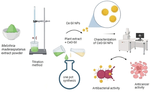
Cerium-doped Silymarin Nanoparticles (Ce-Sil NPs) were synthesised along with the aqueous extract of Melothria maderaspatana by the one-pot synthesis method. Green synthesised Ce-Sil NPs were visually confirmed by the colour change from dark black to brown colour, as shown in (). Melothria maderaspatana contains various types of phytochemicals it is able to reduce Cerium oxide (CeO2) into Cerium nanoparticles (CeNPs). Similar research showed that the green synthesis of Cerium oxide nanoparticles was carried out by Calotropis procera floral extract. Initially, Ce3+ ions are reduced by these phytochemicals to zero-valent Ce atoms. The conversion of cerium ions to cerium oxide is one of the potential reaction mechanisms for the phytochemicals found in the floral extract. Additionally, the phytochemicals in the floral extract serve as a capping and stabilising agent when oxide nanoparticles are formed [Citation12].
Figure 1. Melothria maderaspatana mediated synthesis of Ce-Sil NPs by the One-Pot synthesis method.

Characterisation of Ce-Sil NPs
UV–Spectrophotometer
Synthesised Ce-Si NPs absorbance peaks are around 310–350 nm. It revealed a major peak obtained at 350 nm. Thus, the presence of Ce-Sil NPs was confirmed. Another study showed that the UV–Vis absorption spectra of biosynthesised CeO2-NPs from the Hibiscus Sabdariffa flower extract exhibited an absorption peak at 377 nm [Citation13]. These results confirm that the synthesised nanocomposites containing Cerium and Silymarin results clearly show peaks as per the observation by UV–Vis Spectroscopy ().
Figure 2. UV–vis absorption spectrum of the Ce-Sil NPs suspension.

Fourier transformed infrared spectroscopy (FT-IR)
The synthesised Ce-Sil NPs were characterised by FT-IR spectroscopy to identify the functional groups present in the synthesised nanocomposites. It showed more than four different functional groups present in the synthesised nanocomposites. It revealed the peak values of 2340.31, 1644.2, 1068.55 and 719.45 cm−1. These peak values correspond to functional groups like the Nitrile group, Alcohol (ROH), C-O-C group and N-H group, respectively (). An analogous investigation demonstrated that the FT-IR spectra of CeO2-NPs were obtained at around 2340–2370 and 1000–1500 cm−1. These bands correspond to carbonate-type species connected to the oxide particle surface, such as ambient CO2, which interacts with the cerium cations. It showed a very broad and intense band near 3400 cm−1 is assigned to the collective in-phase symmetric (O–H) mode of hydroxylmolecules [Citation14]. This study assesses the presence of numerous functional groups present in the synthesised CeO2NPs.
Figure 3. Fourier transformed infrared spectra of the synthesised Ce-Sil NPs.

X-Ray diffraction analysis (XRD)
Based on this study, the synthesised Ce-Sil NPs are more crystalline (82.5%) and less amorphous (17.5%). As a result, the synthesised Ce-Sil NPs are highly crystalline and stable in nature (). Ce-Sil NPs were characterised using XRD to obtain closer insight into the crystal structure. These Ce-Sil NPs exhibited sharp peaks at 28.71°, 33.24°, 47.59° and 56.38° that may be indexed with (111), (200), (220) and (311) planes, respectively. A similar study revealed that the X-ray diffraction peaks of CeO2 NPs produced with leaf extract from G. superba were displayed. The angles (2θ) of 28.51, 33.06 and 47.42 correspond to the (111), (200) and (220) planes of the CeO2 NPs, where the XRD peaks are positioned [Citation15].
Figure 4. X-ray powder diffraction spectra of Melothria Maderaspatanus mediated Ce-Sil NPs.

Scanning electron microscope (SEM)
SEM images confirmed the surface morphology of the synthesis of Ce-Sil NPs at different magnifications. The Ce-Sil NPs showed spherical to irregular shapes were agglomerated size ranges from 70 to 110 nm (). A previous study prepared CeO2 NPs with leaf extract, which revealed uniform spherical aggregated structures due to high surface activity and large specific surface area [Citation16].
Figure 5. Scanning electron microscope images of prepared Ce-sil NPs with different diameter distribution.

Energy-dispersive X-ray analysis (EDX)
EDX was used to determine and identify the composition of the elements present in Ce-Sil NPs. The EDX results showed that Ce (58.7%), O (26.7%) and C (12.1%) signals were observed, which confirmed the Ce-Sil NPs occurrence (). In a similar study, the EDX analysis of CeO2-NPs from Rheum turkestancium synthesised through Green synthesis confirmed the presence of Ce (92.04%) and O (7.96%) elements in the prepared sample [Citation17].
Figure 6. EDX images of synthesised Ce-Sil NPs.

Antibacterial efficacy of Ce-Sil NPs
The antibacterial activity of the synthesised Ce-Sil NPs were evaluated against different strains. The zone of inhibition of Ce-Sil NPs is denoted in . The antibacterial activity of Ce-Sil NPs showed higher activity for Enterococcus faecalis, MRSA and Pseudomonas aeruginosa, and significant antibacterial activity for Escherichia coli. Previous studies of CeO2 NPs synthesised from Morinda citrifolia showed significantly higher antibacterial property for Gram-positive S. aureus and Gram negative Pseudomonas aeruginosa, E. coli [Citation11]. Another study reported that CeO2 nanoparticles exhibit effective bactericidal activity due to Reactive Oxygen Species generation and electrostatic interaction [Citation18]. The ROS group includes hydroxyl radicals, superoxides and hydrogen peroxide. These radicals have the ability to cause serious damage to DNA, leading to cell death. The excess ROS production may lead to oxidative damage to the cellular membrane [Citation19]. The rough edges of CeO2 NPs directly interact with the surface of the bacteria and also can cause cell damage immediately [Citation20]. According to the present study, Ce-Sil NPs show significant antibacterial activity, and they can be used as a potential antibacterial agent for future use.
Figure 7. Antibacterial activity of Ce-Sil NPs.

Anticancer activity of Ce-Sil NPs
The results of the anticancer activity of Ce-Sil NPs are shown in . It showed lower cell viability at the maximum concentration (50 μg/ml) and higher cell viability was observed at the minimum concentration (10 μg/ml). The IC50 value of Ce-Sil NP was about 44.6 μg/ml. Previous studies indicate that the CeCh-NPs’ cytotoxic effects revealed that they selectively inhibited A549 cells (lung cancer cell line) (IC50: 50.65 µg/ml) compared with HFF cells (IC50: 131.108). The effects of cerium oxide nanoparticles with chitosan polymer were studied, and it was shown that the nanoparticles suppress cancerous cells by causing oxidative stress in apoptosis-related genes, producing cytotoxicity and controlling their expression in cancer cells. CeCh-NPs’ characteristics suggest that they could potentially serve as an efficient agent for cancer treatment [Citation21]. Similarly, our synthesised Ce-Sil NPs have the ability to inhibit cervical cancer cell proliferation.
Figure 8. Anticancer activity of Ce-sil NPs for HeLa cells. Data are presented in mean ± S.D. from three independent experiments. Statistical significance considered at p < 0.05 among various concentrations.

Conclusion
In our study, Melothria maderaspatana-mediated Ce-Sil NPs were successfully synthesised using the one-pot synthesis method, and the morphological structures were determined using characterisation methods such as UV-vis, FT-IR, SEM, EDX and XRD. The results showed that the produced Ce-Sil NPs revealed the potential antibacterial activity against multidrug-resistant Pseudomonas aeruginosa, Enterococcus faecalis and MRSA and moderate antibacterial activity against Escherichia coli. The biosynthesised Ce-Sil NPs showed significant anti-cancer activity for Hela cells in minimal dosage. The present study suggested that the synthesised Melothria maderaspatana-mediated Ce-Sil NPs may be used as an effective drug for the treatment of bacterial infections and cancer therapy in the future.
Author contributions
Conceptualisation: M.S.; literature search: P.A.M., P.G., R.S., D.S.C., D.C.V. and M.S.; data extraction: M.S., P.G. and R.S.; formal analysis: D.S.C., M.S. and D.C.V.; original draft preparation: R.S., D.S.C. and M.S.; manuscript review and editing: M.S. and P.A.M.; supervision: M.S. All authors have read and agreed to the published version of the manuscript.
Acknowledgments
The authors would like to acknowledge Saveetha Institute of Medical and Technical Sciences for providing research facilities, encouraging successful completion of this research.
Disclosure statement
No potential conflict of interest was reported by the author(s).
References
- Devi GK, Sathishkumar K. Synthesis of gold and silver nanoparticles using plant extract and its anticancer activity. IET Nanobiotechnol. 2017;11(2):143–9. doi: 10.1049/iet-nbt.2015.0054
- Raja B, Pugalendi K. Evaluation of antioxidant activity of Melothria maderaspatana in vitro. Open Life Sci. 2010;5(2):224–230. doi: 10.2478/s11535-010-0005-5
- Balaraman AK, Singh J, Dash S, et al. Antihyperglycemic and hypolipidemic effects of melothria maderaspatana and coccinia indica in Streptozotocin induced diabetes in rats. Saudi Pharm J. 2010;18(3):173–178. doi: 10.1016/j.jsps.2010.05.009
- Habeeb Rahuman HB, Dhandapani R, Narayanan S, Palanivel V, Paramasivam R, Subbarayalu R, et al. Medicinal plants mediated the green synthesis of silver nanoparticles and their biomedical applications. IET Nanobiotechnol. 2022;16(4):115–144.
- Veeramani C, Aristatile B, Pushpavalli G, et al. Effects of Melothria maderaspatana leaf extract on antioxidant status in sham-operated and uninephrectomized DOCA-salt hypertensive rats. Saudi J Biol Sci. 2011;18(1):99–105. doi: 10.1016/j.sjbs.2010.05.002
- Devi GK, Kumar KS, Parthiban R, et al. An insight study on HPTLC fingerprinting of mukia maderaspatna: mechanism of bioactive constituents in metal nanoparticle synthesis and its activity against human pathogens. Microb Pathog. 2017;102:120–132. doi: 10.1016/j.micpath.2016.11.026
- Koltai T, Fliegel L. Role of silymarin in cancer treatment: facts, hypotheses, and questions. J Evid Based Integr Med. 2022;27:2515690X211068826. doi: 10.1177/2515690X211068826
- Hölzl J. Biosynthesis and (14C) labelling of flavonolignans (silymarin) by Silybum marianum (Haller) (author’s transl). Z Naturforsch C. 1974;29(1):82–83. https://www.ncbi.nlm.nih.gov/pubmed/4276432
- Rekha K, Nirmala M, Nair MG, et al. Structural, optical, photocatalytic and antibacterial activity of zinc oxide and manganese doped zinc oxide nanoparticles. Physica B Condens Matter. 2010;405(15):3180–3185. doi: 10.1016/j.physb.2010.04.042
- Shenoy RUK, Rama A, Govindan I, et al. The purview of doped nanoparticles: insights into their biomedical applications. OpenNano. 2022;8(100070):100070. doi: 10.1016/j.onano.2022.100070
- Elderdery AY, Alzahrani B, Alabdulsalam AA, et al. Structural, optical, antibacterial, and anticancer properties of cerium oxide nanoparticles prepared by green synthesis using leaves extract. Bioinorg Chem Appl. 2022;2022:6835625. doi: 10.1155/2022/6835625
- Muthuvel A, Jothibas M, Mohana V. Green synthesis of cerium oxide nanoparticles using Calotropis procera flower extract and their photocatalytic degradation and antibacterial activity. Inorg Chem Commun. 2020;119:108086. doi: 10.1016/j.inoche.2020.108086
- Kweyama Z. Green synthesis of europium (III) oxide nanoparticles using hibiscus sabdariffa flower extract. 2018. https://books.google.com/books/about/Green_Synthesis_of_Europium_III_Oxide_Na.html?hl=&id=ZpTrvgEACAAJ
- Altaf M, Manoharadas S, Zeyad MT. Green synthesis of cerium oxide nanoparticles using Acorus calamus extract and their antibiofilm activity against bacterial pathogens. Microsc Res Tech. 2021;84(8):1638–1648. doi: 10.1002/jemt.23724
- Arumugam A, Karthikeyan C, Haja Hameed AS, et al. Synthesis of cerium oxide nanoparticles using Gloriosa superba L. leaf extract and their structural, optical and antibacterial properties. Mater Sci Eng C Mater Biol Appl. 2015;49:408–415. doi: 10.1016/j.msec.2015.01.042
- Senthilkumar RP, Bhuvaneshwari V, Ranjithkumar R, et al. Synthesis, characterization and antibacterial activity of hybrid chitosan-cerium oxide nanoparticles: as a bionanomaterials. Int j biol macromol. 2017;104(Pt B):1746–1752. doi: 10.1016/j.ijbiomac.2017.03.139
- Sabouri Z, Sabouri M, Amiri MS, et al. Plant-based synthesis of cerium oxide nanoparticles using rheum turkestanicum extract and evaluation of their cytotoxicity and photocatalytic properties. Mater Technol. Published online July 3, 2022;37(8):555–568. doi: 10.1080/10667857.2020.1863573
- Zhang M, Zhang C, Zhai X, et al. Antibacterial mechanism and activity of cerium oxide nanoparticles. Sci China Mater. 2019;62(11):1727–1739. doi: 10.1007/s40843-019-9471-7
- Rana SB, Singh RPP. Investigation of structural, optical, magnetic properties and antibacterial activity of Ni-doped zinc oxide nanoparticles. J Mater Sci Mater Electron. 2016;27(9):9346–9355. doi: 10.1007/s10854-016-4975-6
- Theuretzbacher U. Surface chemistry of cerium oxide nanocubes: toxicity against pathogenic bacteria and their mechanistic study. J Ind Eng Chem. 2014;20(5):3513–3517. doi: 10.1016/j.jiec.2013.12.043
- Abbasi N, Homayouni Tabrizi M, Ardalan T, et al. Cerium oxide nanoparticles-loaded on chitosan for the investigation of anticancer properties. Mater Technol. 2022;37(10):1439–1449. doi: 10.1080/10667857.2021.1954279