

Abstract
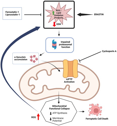
Ferroptosis has been characterized as a form of iron-dependent regulated cell death accompanied by an accumulation of reactive oxygen species and lipid oxidation products along with typical morphological alterations in mitochondria. Ferroptosis is activated by diverse triggers and inhibited by ferrostatin-1 and liproxstatin-1, apart from iron chelators and several antioxidants, and the process is implicated in multiple pathological conditions. There are, however, certain ambiguities about ferroptosis, especially regarding the final executioner of cell death subsequent to the accumulation of ROS. This study uses a typical inducer of ferroptosis such as erastin on SH-SY5Y cells, and shows clearly that ferroptotic death of cells is accompanied by the loss of mitochondrial membrane potential and intracellular ATP content along with an accumulation of oxidative stress markers. All these are prevented by ferrostatin-1 and liproxstatin-1. Additionally, cyclosporine A prevents mitochondrial alterations and cell death induced by erastin implying the crucial role of mitochondrial permeability transition pore (mPTP) activation in ferroptotic death. Furthermore, an accumulation of α-synuclein occurs during erastin induced ferroptosis which can be inhibited by ferrostatin-1 and liproxstatin-1. When the knock-down of α-synuclein expression is performed by specific siRNA treatment of SH-SY5Y cells, the mitochondrial impairment and ferroptotic death of the cells induced by erastin are markedly prevented. Thus, α-synuclein through the involvement of mPTP appears to be the key executioner protein of ferroptosis induced by erastin, but it needs to be verified if it is a generalized mechanism of ferroptosis by using other inducers and cell lines.
Graphical Abstract
Acknowledgements
The authors acknowledge the infrastructural and administrative support received from the management at Maharishi Markandeshwar (Deemed to be) University, Ambala, India and Dr B C Roy Multispeciality Medical Research Centre, IIT Kharagpur, India. The graphical abstract was drawn with the help of Biorender software (https://www.biorender.com).
Disclosure statement
No potential conflict of interest was reported by the author(s).
Data availability statement
Detailed raw data may be available from the corresponding author upon reasonable request.