Abstract
Here we describe the application of microparticles (MPs) for the delivery and release of the drug a benzopsoralen. We also evaluated the intracellular distribution and cellular uptake of the drug by using an encapsulation technique for therapeutic optimization. MPs containing the compound 3-ethoxycarbonyl-2H-benzofuro[3,2-f]-1-benzopyran-2-one (psoralen A) were prepared by the solvent evaporation technique, and parameters such as particle size, drug encapsulation efficiency, effect of the encapsulation process on the drug's photochemistry, zeta potential, external morphology, and in vitro release behavior were evaluated. The intracellular distribution of MPs as well as their uptake by tissues were monitored. Size distribution studies using dynamic ligh scattering and scanning electron microscopy revealed that the MPs are spherical in shape with a diameter of 1.4 μ m. They present low tendency toward aggregation, as confirmed by their zeta potential (+10.6 mV). The loading efficiency obtained was 75%. As a consequence of the extremely low diffusivity of the drug in aqueous medium, the drug release profile of the MPs in saline phosphate buffer (pH 7.4) was much slower than that obtained in the biological environment. Among the population of peritoneal phagocytic cells, only macrophages were able to phagocytose poly-d,l-lactic-co-glycolic acid (PLGA) MP. The use of psoralen A in association with ultraviolet light (360 nm) revealed morphological characteristics of cell damage such as cytoplasmic vesiculation, mitochondria condensation, and swelling of both the granular endoplasmatic reticulum and the nuclear membrane. These results indicate that PLGA MP could be a promising delivery system for psoralen in connection with ultraviolet irradiation therapy (PUVA).
The association of psoralen administration with ultraviolet A irradiation (UVA, 320–400 nm) is commonly referred as PUVA therapy (Canton et al. Citation2002). PUVA therapy was first reported for the treatment of several skin diseases and can be traced back to ancient Egypt, India, and Greece, where plant extracts containing psoralen were applied on the skin in association with solar light (Machado et al. Citation2001; Roop et al. Citation2004). The efficacy of this treatment was soon confirmed by controlled clinical trials in a large series of patients. These original findings contributed to the development of photomedicine and established the clinical efficiency of PUVA for more than 30 skin disorders, especially severe psoriasis (Middelkamp-Hup et al. Citation2004).
The planar structure of psoralens () facilitates their intercalation between nucleotide base pairs in DNA and upon photoexcitation, photocycloaddition reactions result in the covalent modifications of DNA (Wenjian et al. Citation2002). This may be one of the reasons why high-dose PUVA exposure has been correlated with an increased risk of epithelial skin cancer, which is a limitation for the use of PUVA therapy for skin disorders (Middelkamp-Hup et al. Citation2004). To avoid or minimize the risk of psoralen intercalation with DNA pairs, the substitution of hydrogen in positions 3, 4, 4′, and 5′ () by bulky and electron withdrawing groups has been proposed (Dall'Acqua, Vedaldi, and Caffieri Citation1996; Dalla Via et al. Citation2003, Oliveira et al. Citation2003, Citation2004).
In our study the potential of biodegradable microparticles (MP) were investigated not only for controlled drug release, but also for target drug delivery of psoralen A, a strategy that has proven effective in increasing therapeutic effects and reducing side effects of other drugs (Okada and Toguchi Citation1995; Srinath and Diwan Citation1998). The poly(d,l-lactic-co-glycolic acid) (PLGA) copolymer is a synthetic biodegradable and biocompatible polymer that has reproducible, slow drug release characteristics in vivo and has attracted a lot of attention because of its versatility, especially with regard to its nonimmunogenic properties and capacity to encapsulate both hydrophilic and lipophilic drugs (Tsung and Burgess Citation2001).
In the present work, PLGA MPs were tested as drug carriers for the delivery of psoralen A in PUVA treatment. Our aim was to study the effect of the MPs formulation parameters and physical properties (e.g., surface charge, particle size) on the cellular uptake of MPs and their distribution in various cellular compartments (e.g., endolysosomes, cytoplasm, nucleus, etc.). The understanding of the intracellular and tissue distribution of MPs also is useful for the elucidation of enhanced therapeutic efficacy.
MATERIAL AND METHODS
Poly(D,L-lactic-co-glycolic acid) (PLGA, 50:50, Mw 17kDa) was obtained from Sigma Chemical. (St. Louis, MO, USA); poly(vinyl alcohol) (PVA, 13-23 kDa, 87-89% hydrolyzed) was supplied by Aldrich (Milwaukee, WI, USA); analytical grade dichloromethane was supplied by VETEC (Rio de Janeiro, Brazil). All other chemicals were of analytical grade and used without further purification. The compound 3-ethoxy carbonyl-2H-benzofuro[3,2-f]-1-benzopiran-2-one (psoralen A) was synthesized and generously supplied by Dr. Oliveira-Campos, from Minho University, Minho, Portugal.
Formulation of the Polymeric Psoralen MPs
MPs were produced by the solvent evaporation procedure described by Gomes et al. (Citation2005). Typically, the organic phase consisted of PLGA 50:50 polymer (0.1 g) and psoralen A (10 μM) dissolved in CH2Cl2 (10 mL). The dispersed phase was dropped into the homogeneous aqueous phase (aqueous phase [100 mL] containing 3% [w/v] of hydrolyzed PVA [88%] as dispersing agent), stirred at 13,500 rpm for 3 min, with ice cooling. Solvent evaporation was carried out by gentle magnetic stirring at room temperature, for of 3–5 hr. MPs were recovered by centrifugation for 5 min at 10,000 rpm, at 4°C. They were then washed (three times) with distilled water (10°C) and lyophilized. MPs without psoralen A were prepared by the same procedure.
Drug Entrapment Efficiency
The amount of psoralen A entrapped within the MP was evaluated spectrophotometrically by measuring the amount of nonentrapped drug recovered in the external aqueous phase after centrifugation. Aliquots (2 mL) of each aqueous sample were evaporated to dryness by purging with nitrogen. The MP suspension was reconstituted by adding methylene chloride (2 mL) and assayed in triplicate, by measuring the absorbance at 350 nm read with a spectrophotometer (Shimadzu UV-250 1 PC). The drug entrapment efficiency was expressed as the percentage of entrapped psoralen with respect to the theoretical value; drug loading was calculated as the amount of psoralen entrapped per gram of polymer. Because the indirect method used to evaluate the amount of nonentrapped psoralen in the aqueous phase was correlated with the direct method after dissolution of MP in methylene chloride, the drug content was indirectly estimated within the aqueous phase, which is an easier and faster method.
In Vitro Drug Release
MPs (10 mg) containing psoralen A were incubated in triplicate and incubated in 10 mL of release medium (PBS buffer, pH 7.4) under stirring (150 rpm) at 37°C. At determined intervals, the MP suspension was centrifuged at 10,000 rpm, for 10 min. The supernatant (10 mL) was withdrawn and replaced with 10 mL of fresh release medium. This procedure was repeated daily over a period of 7 days. The amount of psoralen released was determined by measuring the drug concentration in the supernatant, using the procedure described in the previous section.
Particle Size and Surface Charge (Zeta Potential)
Particle size and size distribution were determined by photon correlation spectroscopy (PCS) using the quasielastic light scattering technique, in a Zetasizer 3000 equipment from Malvern Instrument (Worcstershire, UK) equipped with a 10 mW He-Ne 633 nm laser beam, at 25°C and at a scattering angle of 90°. For the particle size analysis, a dilute suspension (1.0 mg/mL) of MP was prepared in double distilled water and sonicated in an ice bath for 30 sec. The zeta potential of the MPs in PBS buffer, (0.1mM, pH 7.4 1.0 mg/mL) was determined by using ZetaPlus™ in the zeta potential analysis mode.
Spectrophotometric Assay for Psoralen A
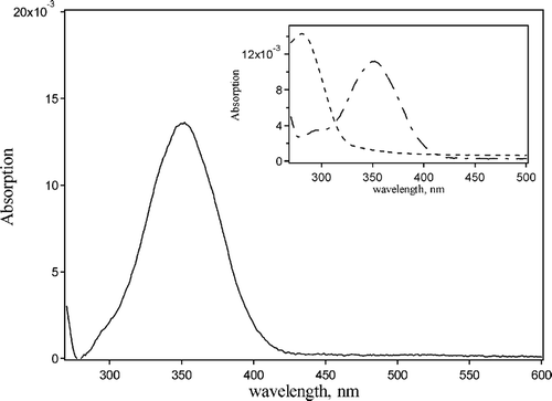
The absorption attributed to psoralen A incorporated into the MPs was determined by deconvoluting the absorption spectrum into its three parts using a program based on the self-modeling factor analysis (SM-FA) method to determine the number of components contributing to the absorption spectra and the spectrum of each component (Lunardi et al. Citation2003). These components were glycerol and MP-loaded with psoralen A. The resulting spectrum was the absorption of the drug alone in the psoralen-doped particle.
Microparticle Morphology: SEM Analysis
The size and shape of the MPs were obtained by using scanning electron microscopy (SEM). Samples were washed with sterile distilled water and fixed in 2.5% (v:v) glutharaldehyde (GA) in water for 2 hr. Samples were then washed with water, dehydrated in a graded ethanol series, and critical-point dried. Since they lack electrical conductivity, the MPs were coated with a thin layer of gold before SEM analysis. The diameter of the MPs was measured using a ruler and the mean value was found by using the SEM scale. An electronscan (Philips ESEM 2020) operating at 5 kV was used for these measurements.
Transmission Electron Microscopy (TEM) of MPs in Exudate Rat Cells
Peritoneal exudate cells from male Wistar rats (mean weight ∼ 150 g, n = 6) were used in the experiments. The rats were intraperitoneally injected with PBS buffer (20 mL), and their abdomen was massaged for 5 min to free adherent macrophages. The resulting cell suspension was withdrawn by a syringe through a small incision made in the abdominal wall. The lavage fluid was centrifuged (400 g; 10 min), and the cell pellet was ressuspensed in PBS buffer and divided into 4 aliquots of 2 mL each. Time-dependent assays were carried out by incubating samples with PLGA MPs (300 μ g/mL) for 15, 30, and 120 min. Negative controls were performed by incubating cells in the absence of MP for 120 min. After incubation, the cells were washed twice with PBS solution.
For the TEM studies, the samples were fixed in a 2.5% glutaraldehyde prepared in sodium cacodylate buffer (0.1 M, pH 7.4) for 2 hr, at 25°C. The cells were then postfixed in 1% osmium tetroxide in the same buffer for 1 hr, then dehydrated in a graded acetone series, and embedded in epoxy resin. Ultrathin sections were contrasted with alcoholic 2% uranyl acetate and 5% lead citrate. Ultrastructural examination was performed using a transmission electron microscope (Philips CM 100).
Cell Irradiation
The exudate peritoneal cells were incubated with psoralen A–loaded MP (10 μM), in Hank's solution for 15, 30, and 120 min. The photochemical experiments were carried out in a 1-cm pathlength spectrophotometric cell. A 400-W mercury arc lamp was used as the irradiation source. A pass-band filter (Ocean Optics U360) filtered the radiation, ensuring the samples were irradiated with light of 360 ± 10 nm (maximum transmittance 69%). Typical irradiances of 0.030 to 0.035 W/cm2 were used to deliver a fluency of 1.0 J/cm2. The irradiances were quantified using a PMA 2100 solar light radiometer. After light treatment, the cells were kept in the dark for 3 hr. Nonirradiated cells were kept in the dark for the same period of time. The ambient light was < 0.001 mW/cm2. Both irradiated and nonirradiated cells were processed by TEM.
Data Processing
Experiments were carried out in triplicate. Results are expressed as the mean standard deviation (s.d.), and statistical analysis was performed using a two-tailed Student's t-test.
RESULTS AND DISCUSSION
Polymeric Psoralen Microparticles
The solvent evaporation method is widely used for the preparation of polymeric MPs (Garcia et al. Citation1999; Gomes et al. Citation2005; Rosca, Watari, and Uo Citation2004) and hydrophobic drugs can be encapsulated in a polymer matrix by means of this method. Various formulation factors and characteristics of the MPs play a key role in biological applications. This study focuses mainly on the evaluation of the role of their physicochemical properties on the cellular uptake of MPs and their distribution in various cellular compartments. describes the physical characterization of these MPs (with and whithout psoralen A), evaluating particle size, encapsulation efficiency, and zeta potential.
TABLE 1 Mean diameter zeta potential and encapsulation efficiency of microparticles prepared by the solvent evaporation method
The drug entrapment efficiency parameter was calculated from the experimentally determined actual drug loading of the MP and the theoretical drug loading. The values obtained by this method vary between 72.0% and 78.0% incorporation. In general, high microencapsulation of hydrophobic drugs such as psoralen A is relatively easy with hydrophobic polymers such as PLGA because of limited drug loss to the aqueous phase. The improvement obtained in the encapsulation efficiency of psoralen A is due to the use of 3.0% PVA, which is contrary to that observed by Schlicher et al. (Citation1997). According to Kompella, Bandi, and Ayalasomayajula (Citation2001), PVA stabilizes the oil/water (o/w) emulsion globules, thereby reducing diffusion of the drug into the aqueous medium.
Zeta Potential
Particle size is an important parameter for drug delivery. The smaller the particle size, the better the cellular uptake of the particle. Some studies with MPs have been carried out in biomedical and biotechnological areas, since their particle size (ranged from 10 to 4000 nm) are acceptable for intravenous (i.v.) injection (Kreuter Citation2004). In our work, we developed some modifications to the solvent evaporation method proposed by Garcia et al. (Citation1999), aiming at the production of smaller MPs, with a more uniform size. This modified method is highly reproducible, producing small MPs with a mean diameter in the range of 1370 (± 53) nm (drug-free) and 1402 (± 37) nm (when loaded with psoralen A), as analyzed by dynamic light scattering. The MPs exhibited a unimodal size distribution. For all the MP formulations obtained, the polydispersivity index was lower than 0.7, which was considered satisfactory. According to the literature (Jeon et al. Citation2000), the administration of particles with a diameter of several micrometers seems to be inefficient as a drug delivery system (DDS) because of its accumulation in lung capillaries and its difficult removal from the endothelial reticulum system (Jeon et al. Citation2000). Our modifications to the solvent evaporation method resulted in the preparation of smaller particles (1500 nm), thereby making them useful as a DDS.
The MPs colloidal stability was analyzed by measuring the MP zeta potential. In theory, more pronounced zeta potential values, either positive or negative, tend to stabilize particle suspension, since the electrostatic repulsion between particles with the same electric charge prevents aggregation of the spheres (Kumar, Bakowsky, and Lehr Citation2004). The particles consisting of PLGA-free dye were negatively charged (−3.2mV at pH 7.4) whereas the zeta potential measured for the MPs loaded with psoralen A was +10.6mV. In general, this value is considered to be associated with a stable colloid (Ruan and Feng Citation2003). The significant variation in zeta potential can be explained by residual PVA that is still present on the surface of the particles even after three washings, which affects the number of carboxylate-end groups.
On the other hand, the positive zeta potential value obtained for MPs loaded with psoralen A may be attributed to an almost uniform positive charge density over the molecule, as observed in the mapped potential energy surface and calculated from the optimized structure of the drug by quantum mechanical calculation (Machado Citation2004). Therefore, this positive zeta potential indicates that psoralen A also may be located on the external surface of the PLGA MPs. This feature should influence the release of psoralen A into the medium.
In Vitro Drug Release
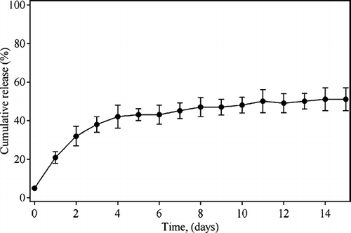
In vitro drug release from encapsulated MPs was determined to obtain quantitative information on the profile of psoralen A release into the disperse system over a 15-day period, which may be correlated with the carrier microstructure.
Psoralen A presents a biphasic pattern (), in which the burst effect in the initial phase of the release might be attributed to the presence of nonentrapped psoralen A molecules adsorbed on the surface of the MPs, or to psoralen A molecules that are accessible to the release medium through pores and channels formed during the preparation of the MPs. This initial psoralen A release profile reaches 38% after 3 days. In the second phase (4–15 days), the psoralen A release from the PLGA MPs was reduced due to the MPs low porosity that results in longer diffusion distances for psoralen A through the MP matrix. The permeability for penetrating water also is reduced, leading to slow PLGA degradation. In this step, the exhaustion of psoralen A molecules near the surface of the MPs is observed. However, the psoralen A molecules completely entrapped in the PLGA matrix cannot be released until the polymer matrix starts to lose its integrity. At this point, psoralen A molecules are then accessible to the aqueous release medium.
Spectrophotometric Assay for Psoralen A
The measurement of the spectroscopic properties of a molecule (absorption and fluorescence spectra) is critical for the understanding of its photophysical and photochemical properties. The absorption spectrum of monomeric psoralen A in a methylene chloride dilute solution exhibitis a typical absorption band in the ultraviolet (UV) region at 351 nm, as previously reported when using chloroform as solvent (Machado et al. Citation2001). When loaded into PLGA MPs, psoralen A exhibits a hypsochromic shift, with a maximum at 349 nm (). In a more recent theoretical-experimental study, Machado and co-workers (Citation2001) showed that the position of this peak is dependent on solvent polarity and has an oscillator strength and molar absorptivity (log ε > 4) compatible with a π, π* electronic transition. However, subsequent work has shown that this is a π, π* S0→S2 transition (Machado Citation2004).
FIG. 2 Absorption spectrum of psoralen-loaded microparticle. The spectrum is deconvoluted into three components: (–)-deconvoluted absorption of psoralen A. Inset: (- - -)-glycerol and (—)-PLGA microparticle-psoralen in glycerol.

The absorption spectrum of the MPs loaded with psoralen A in solution shows an overlap of bands in the UV region, with considerable light scattering due to the MPs suspended in water. The use of a deconvolution technique (Lunardi et al. Citation2003) minimizes this problem, giving the absorption spectrum of the psoralen A-MP alone ().
The light scattering in the UV region by the MP in water is mainly due to the different refraction indexes of PLGA and water. This is evident by the rapid rise of the measured absorption spectrum in the range between 230 and 300 nm, caused by light scattering. Placing the MPs in a high refractive index solvent, like glycerol, minimizes this scattering.
Psoralen A-MP: Ultrastructural Characterization and Cellular Uptake
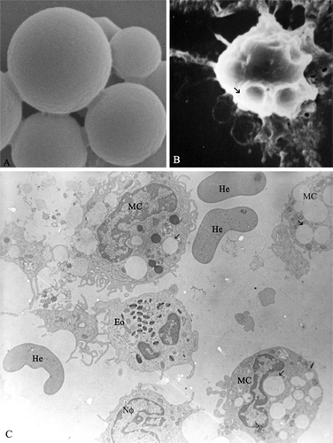
SEM is an excellent tool to observe the external morphology of the PLGA MPs, and shows micrographs of MPs loaded with psoralen A magnified at 20,000×. In , the MPs are shown to be well formed and nonaggregated. They are spherical in shape and present very few pores on the external surface. No significant difference between the loaded PLGA MPs and the empty MPs (used as control) were found.
FIG. 3 Ultrastructural characterization of PLGA-microparticle loaded with psoralen A. (A) Representative SEM shows the surface of PLGA microparticle loaded with psoralen A (30000×). (B) PLGA-microparticle (SEM) (→)-phagocited by macrophage cell after incubation for 120 min (5000×). (C) Representative TEM of microparticle in incubation for 120 min peritoneal exudate cell population: neutrophil-Nϕ, eosinophil-Eo, hemacea-He, and selective phagocytose by macrophage-MC (3200×).

shows that macrophage cells incubated with MP for 120 min completely phagocyted these particles, which appear as an extension of the cell surface (arrow). Among the population of phagocytic peritoneal exudate cells (neutrophils [No], eosinophil [Eo], and macrophage [Mc]), only macrophage cells phagocytose PLGA MPs ().
Ultrastructure: Time Dependence and PUVA Studies
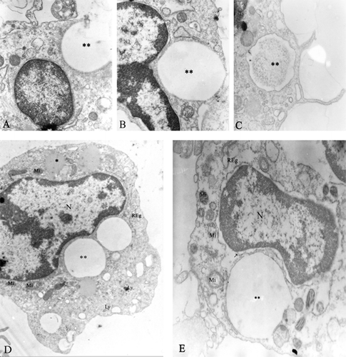
Ultrastructural studies using rat peritoneal exudate cells incubated with PLGA MPs demonstrated that MPs are phagocyted by macrophages by the apparently conventional receptor-mediated phagocytosis in which phagocyte pseudopods move around the MPs until they fuse at the distal tip (). The MPs contour boundary is clearly observed without any specific electron-dense marker (, , ). In a time dependent process, these results indicate that the phagocytosis process begins after 15 min of incubation (), followed by internalization of the particle within 30 min (). After 120 min, PLGA MPs of different sizes are located inside the macrophages ().
FIG. 4 Phagocytose of PLGA-microparticle loaded with psoralen A by macrophage peritoneal exudate. (A) PLGA microparticle (**) interacting with cellular membrane in the process of phagocytosis (15 min incubation). (B) Phagosome with PLGA microparticle (30 min incubation). (C) Psoralen A after microparticle digestion by macrophage (120 min incubation). (D) PLGA microparticle inside the macrophage cell, without irradiation (incubated for 120 min), mitochondria-Mi, lysosomes-Ly, nucleus (N), and granular endoplasmatic reticulum-Reg. (E) Photo-damage (PUVA) in macrophage cell with microparticle after 120 min incubation: cytoplasmic vesiculation, mitochondria condensation (Mi), and a swelling of granular endoplasmatic reticulum (Reg) and nuclear membrane (arrow) (12000×).

In a control cell (without irradiation) incubated with psoralen A-MPs for 120 min is observed. PLGA MPs are present in the cell cytoplasm before and after digestion. As can be observed, the ultrastructures of mitochondria (Mi), lysosomes (Ly), and granular endoplasmatic reticulum (Reg) remained unchanged is the presence of psoralen A-MPs.
After irradiation (), TEM analysis of the PUVA-treated cells revealed morphological characteristics of cell damage such as cytoplasmic vesiculation, mitochondria condensation, and a swelling of the granular endoplasmatic reticulum (arrow) and nuclear membrane. Then 3 hr after irradiation, no condensation and margination of the nuclear chromatin were observed, which is in contrast to the case of cultured JB6 cells using 8-MOP as drug (Santamaria et al. Citation2002). However, the degradation mechanism by which the cell damage occurs must be different from that proposed for 8-MOP, because psoralen A sensitizes singlet oxygen with a quantum efficiency near unity, even in media with different polarities (Machado et al. Citation2001; Machado Citation2004). The structure of psoralen A was designed to avoid drug intercalation with DNA and other biological substrates (Oliveira et al. Citation2003). Here we observed that although the use of psoralen A led to photodamage as seen by the ultrastructural analysis, we did not observe morphological features of apoptosis prominent in PUVA-treated cells, such as chromatin condensation at the nuclear periphery.
CONCLUSION
MPs of biodegradable poly (D,L-lactide-co-glycolide) were successfully prepared using the solvent evaporation technique. This technique proved to be simple and reproducible for the preparation of MPs, and high levels (up to 75%) of drug incorporation were obtained by this method. The release profile of psoralen A was characterized by an initial burst, followed by a slow release, probably due to the low porosity of the MPs, as observed by SEM. These MPs have a narrow size distribution of approximately 1.4 μm, and a zeta potential of +10.6 mV, a value that is considered to be associated with a stable colloidal environment. In peritoneal exudates, a great variety of cells are able to promote phagocytosis. However, the PLGA MPs were only phagocytosed by macrophage cells, in a time-dependent process that finished after 120 min. Our results indicate that the properties of psoralen A were not affected when it was encapsulated in PLGA MPs, and that after PUVA treatment, cell damage without chromatin condensation was observed.
The authors thank Monika Iamondi and Antonio Teruyoshi Yabuky from the Laboratory of Electron Microscopy-ICB-UNESP - Rio Claro-SP-Brazil, for technical assistance. We also thank Prof. Richard J. Ward for careful reading of the manuscript. This study was supported by CNPq and FAPEMIG (grants303911/2003-4, 302679/2002-2, CEX85/02) and FAPESP MEDZ (01/06781-7). MEDZ is a CNPq research fellow.
REFERENCES
- Canton M., Caffieri S., Dall'Acqua F., Di Lisa F. PUVA-induced apoptosis involves mitochondrial dysfunction caused by the opening of the permeability transition pore. FEBS Lett. 2002; 522: 168–172, [INFOTRIEVE], [CSA]
- Dall'Acqua F., Vedaldi D., Caffieri S. The Fundamental Bases of Phototherapy. OEMF, MilanoItaly 1996
- Dalla Via L., Uriarte E., Quezada E., et al. Novel pyrone side tetracyclic psoralen derivatives: synthesis and photobiological evaluation. J. Med. Chem. 2003; 46: 3800–3810, [INFOTRIEVE], [CSA], [CROSSREF]
- Garcia J. T., Farina J. B., Munguia O., Llabrés M. Comparative degradation study of biodegradable microspheres of poly(dl-lactide-co-glycolide) with poly(ethyleneglycol) derivates. J. Microencaps. 1999; 16: 83–94, [CSA], [CROSSREF]
- Gomes A. J., Lunardi L. O., Marchetti J. M., et al. Photobiological and ultrastructural studies of nanoparticle of poly(lactic-co-glycolic acid)-containing bacteriochlorophyll-a as a photosensitizer useful for PDT treatment. Drug Del. 2005; 12: 1–6, [CSA], [CROSSREF]
- Jeon H.-J., Jeong Y.-I., Jang M.-K., et al. Effect of solvent on the preparation of surfactant-free poly(DL-lactide-co-glycolide) nanoparticles and norfloxacin release characteristics. Int. J. Pharm. 2000; 207: 99–108, [INFOTRIEVE], [CSA], [CROSSREF]
- Kompella U. B., Bandi N., Ayalasomayajula S. P. Poly(lactic acid) nanoparticles for sustained release of budesonide. Drug Del. Techn. 2001; 1: 28–34, [CSA]
- Kreuter J. Influence of the surface properties on nanoparticle-mediated transport of drugs to the brain. J. Nanosci. Nanotechn. 2004; 4: 484–488, [CSA], [CROSSREF]
- Kumar M. N. V. R., Bakowsky U., Lehr C. M. Preparation and characterization of cationic PLGA nanospheres as DNA carriers. Biomaterials 2004; 25(10)1771–1777, [CSA], [CROSSREF]
- Lunardi C. N., Tedesco A. C., Kurth T. H., Brinn I. M. The complex between 9-(n-decanyl)acridone and bovine serum albumin. Part 2. What do fluorescence probes probe?. Photochem. Photobiolog. Sci. 2003; 2: 954–959, [CSA], [CROSSREF]
- Machado A. E. H. Presentation in VIII Congresso Latinoamericano de Fotoquímica y Fotobiologia (ELAFOT), La Plata, Argentina, 2004, Personal Communication
- Machado A. E. H., Miranda J. A., Oliveira-Campos A. M. F., et al. Photophysical properties of two new psoralen analogs. J. Photochemi. Photobiol. A: Chem. 2001; 146: 75–81, [CSA], [CROSSREF]
- Middelkamp-Hup M. A., Pathak M. A., Parrado C., et al. Orally administered polypodium leucotomos extract decreases psoralen-UVA-induced phototoxicity, pigmentation, and damage of human skin. J. Am. Acad Dermatol. 2004; 50(1)41–49, [INFOTRIEVE], [CSA], [CROSSREF]
- Okada H., Toguchi H. Biodegradable microspheres in drug delivery. Crit. Rev. Therapeut. Drug Carrier Sys. 1995; 12(1)1–99, [CSA]
- Oliveira A. M. A. G., Raposo M. M. M., Oliveira-Campos A. M. E., et al. Synthesis of psoralen analogues based on dibenzofuran. Helvet. Chim. Acta 2003; 86: 2900–2907, [CSA], [CROSSREF]
- Oliveira A. M. A. G., Raposo M. M. M., Oliveira-Campos A. M. F., et al. Fries rearrangement of dibenzofuran-2-yl ethanoate under photochemical and Lewis-acid-catalysed conditions. Tetrahedron 2004; 60: 6145–6154, [CSA], [CROSSREF]
- Roop S., Guy J., Berl V., et al. Synthesis and photocytotoxic activity of new □-methylene-□butyrolactone-psoralen heterodimers. Bioorg. Med. Chem. 2004; 12: 3619–3625, [CSA], [CROSSREF]
- Rosca I. D., Watari F., Uo M. MP formation and its mechanism in single and double emulsion solvent evaporation. J. Control. Rel. 2004; 99: 271–280, [CSA], [CROSSREF]
- Ruan G., Feng S.-S. Preparation and characterization of poly(lactic acid)-poly(ethyleneglycol)-poly(lactic acid) (PLA-PEG-PLA) microspheres for controlled release of paclitaxel. Biomaterials 2003; 24: 5037–5044, [INFOTRIEVE], [CSA], [CROSSREF]
- Santamaria A. B., Davis D. W., Nghiem D. X., et al. p53 and Fas ligand are required for psoralen and UVA-induced apoptosis in mouse epidermal cells. Cell Death Differentiation 2002; 9: 549–560, [CSA], [CROSSREF]
- Schlicher E. J. A. M., Postma N. S., Zuidema J., et al. Preparation and characterization of poly(d,l-lactic-co-glycolic acid) microspheres containing desferrioxamine. Int. J. Pharmaceut. 1997; 153: 235–245, [CSA], [CROSSREF]
- Srinath P., Diwan P. V. Pharmacodynamic and pharmacokinetic evaluation of lipid microspheres of indomethacin. Acta Helvet. 1998; 73: 199–203, [CSA], [CROSSREF]
- Tsung M. J., Burgess D. J. Preparation and characterization of gelatin surface modified PLGA microspheres. AAPS Pharm Sci. 2001; 3: 1–11, [CSA], [CROSSREF]
- Wenjian W., Wlaschek M., Hommel C., et al. Psoralen plus UVA (PUVA) induced premature senescence as a model for stress-induced premature senescence. Exp. Gerontol. 2002; 37: 1197–1201, [CSA], [CROSSREF]

![DIAG. 1. Representation of the typical psoralen (A) and psoralen A (3-ethoxy carbonyl-2H-benzofuro[3,2-f]-1-benzoyiran-2-one) (B).](/cms/asset/a33b6956-e0a4-4fdc-a997-e0b647d31f92/idrd_a_164015_uf0001_b.gif)
