Abstract
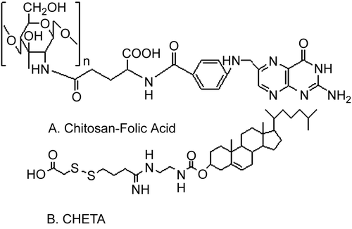
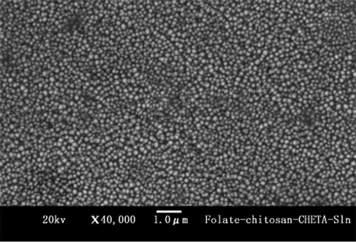
Compared to viral carriers, non-viral gene delivery systems showed good biocompatibility and safety, but low transfection efficiencies. Fortunately, the mechanism of folic acid uptake by cells to promote targeting and internalization could improve transfection rates. In this study, folate-chitosan and one kind of cholesterol derivatives CHETA (Cholest-5-en-3β-yl[2-[[4-[(carboxymethyl)dithio]-1-iminobutyl]amino]ethyl] carbamate, C36H61N3O4S2) were synthesized to prepare the charge changing Solid Lipid Nanoparticles (Folate-chitosan−CHETA−Sln) by a reverse micelle-double emulsion method. The resulted particles showed the distributions of size and zeta potential were 254.5 ± 20 nm and −40.5 ± 0.8 mV, respectively. The image observed by scanning electron microscopy (SEM) showed that Folate-chitosan−CHETA−Sln was spherical in shape. Moreover, after reaction with a disulfide reducing agent dithiothreitol (DTT), the zeta potential changed from negative to positive (20.5 ± 1.9 mV). The results of transfection showed that Folate-chitosan–CHETA−Sln enhanced the reporter gene expression against a folate receptor over-expressing cell line (SKOV3 cells) compared to a folate receptor deficient cell line (A549 cells) and did not induce obvious cytotoxicity against HEK 293 cells. In addition, the presence of serum did not affect the transfectivity of Folate-chitosan−CHETA−Sln complexes. In conclusion, Folate-chitosan−CHETA−Slns with proper physical characteristics and high transfection efficiency might act as a novel non-viral gene delivery system.
Introduction
A safe and efficient gene transfer system is necessary for successful tumor gene therapy. There have been many efforts to develop an effective intracellular delivery system for therapeutic macromolecules including peptides, proteins, antisense oligonucleotides, and plasmid DNA (CitationBenns et al., 2001; CitationJeong, Kim, & Park, 2003). Gene delivery vehicles are generally classified into two categories: recombinant viruses and synthetic vectors. Viral vectors, particularly adenovirus (CitationNathwani et al., 2002) and lentivirus (CitationCheung et al., 2002), have demonstrated a high level of transgene expression. However, significant concerns exist about clinical applications of viral vectors (CitationCichon et al., 1999) such as host immune response against the vectors (CitationJooss & Chirmule, 2003), intrinsic toxicity of the viral proteins, and limited packaging size. The advantages of using synthetic vectors include no-restriction on size of DNA that they can carry, ability to administer repeatedly with minimal host immune response, targetability, stability in storage, and easy production in large quantities. Synthetic vectors can be divided into two categories: cationic polymers and cationic lipid carriers. Cationic polymer/DNA complexes tend to be more stable than cationic lipid/DNA complexes (CitationPack et al., 2005).
Chitosan, the second most abundant polysaccharide in nature, has attracted particular interest as a biodegradable material for drug and gene delivery systems (CitationMansouri et al., 2006). Chitosan is soluble in acid solutions (pH < 5.5) and could form complexes with anionic macromolecules to yield nanoparticles, microparticles, hydrogels, foams, and fibers (CitationLiu & Yao, 2002). It has been widely used in pharmaceutical and medical areas for its favorable biological properties such as biodegradability, biocompatibility, low toxicity, hemostatic, bacteriostatic, fungistatic, and anti- cholesteremic properties, as well as reasonable cost (CitationHejazi & Amiji, 2003). Due to its unique cationic nature, chitosan is able to interact electrostatically with negatively charged polyions such as indomethacin, sodium hyaluronate, pectin, and acacia polysaccharides; it also can interact with negatively charged DNA in a similar fashion (CitationMacLaughlin et al., 1998). Moreover, chitosan can condense DNA effectively and protect DNA from nuclease degradation (CitationMao et al., 2004). However, the transfection efficiency of chitosan-DNA nanoparticles is still very low in comparison with viral vectors.
Receptor recognizable ligands, such as monoclonal antibody, transferring, and epidermal growth factor, could be tethered to the conjugates or the complexes to improve the specificity of the intracellular delivery systems to specific target cells (CitationQian et al., 2002; CitationSapra & Allen, 2003). Folate receptors (FR) are known to be over-expressed on many human cancer cell surfaces (such as SKOV3 cells) (CitationAntony, 1996), but rarely found on normal cell surfaces, or they are localized to the apical surfaces of polarized epithelia (CitationLu & Low, 2003). Folic acid (FA) is appealing as a ligand for targeting cell membrane and allowing nanoparticle endocytosis via the folate receptor (FR) for higher transfection yields. It is well known that folate conjugates, which are covalently derivatized via its g-carboxyl moiety, could retain the high affinity ligand binding property of folate (Kd ~ 10−9 M), and the kinetics of cellular uptake of conjugated folate compounds by folate receptors is similar to that of free folate. Recycling of folate receptors in target cells could lead to further accumulation of folate conjugates (CitationWang et al., 1997; CitationBenns et al., 2001). There have been numerous studies to develop cancer-targeted delivery of anti-cancer drugs, proteins, and genes via a folate receptor-mediated endocytosis system (CitationZhou et al., 2002; CitationKe, Mathias, & Green, 2003).
It is well known that for common cells the surface charge is negative, therefore positive charged drug delivery vehicles may be more attractive to cells. However, the most components of serum protein are negative charged at physical pH and positive charged complexes might aggregate in serum following intravenous injection. We have developed a charge changing cationic liposome made of CHETA (CitationZhong et al., 2007). CHETA consists of a hydrophobic cholesterol tail group (X) and a hydrophilic head group (Y+-S-S-Z−), of which the head group incorporates both positively and negatively charged regions connected by a disulfide bond. The disulfide bond is susceptible to cleavage (X-Y+-S-S-Z− → X-Y+-SH + HS-Z−) by components of the intracellular membrane such as protein disulfide isomerase or thioredoxin reductase (TrxR) (CitationLeamon, 2002). Since the content of TrxR in tumor cells is 10-times higher than in normal tissues (CitationLincoln et al., 2003; CitationTurunen et al., 2004) when the negative charged complexes reached the surface of tumor cells, the disulfide bond would be cleaved, which results in the removal of the negatively charged region from the head group and the formation of positive charged complexes. This strategy may enhance the cell infusion and therefore increase the transfectivity.
Based on the above-mentioned reasons, the aim of the present work was to use folate-chitosan and CHETA to prepare the charge changing solid lipid nanoparticles, a folate receptor mediated intracellular gene delivery system (Folate-chitosan−CHETA−Sln). In order to achieve these goals, CHETA was synthesized and folate was conjugated to chitosan. Through a reverse micelle-double emulsion strategy, DNA with or without folate-chitosan and CHETA were formulated according to the optimized ratio by Box-Wilson’s central composite design (CCD) optimization method (CitationZhong et al., 2007) to produce Folate-chitosan−CHETA−Sln, Folate-chitosan−Sln, and CHETA−Sln, respectively. MTT assay was employed to investigate the cellular toxicity of the preformed complexes against HEK293 cells. Both LacZ and eGFP were used as reporter genes to assess the transfectivity of these complexes against different cell lines of a folate receptor over-expressing cell line (SKOV3 cells) and a folate receptor deficient cell line (A549 cells) with or without the presence of serum.
Materials and methods
Chitosan (with 90% degree of deacetylation (DD) and a molecular weight (Mw) of 50 kDa) used for synthesizing folate-chitosan was purchased from BoAo Biochemical Company (Shanghai, China). The plasmid pORF LacZ (3.54 kb) encoding the X-galactosidase reporter gene was purchased from Invivogen (USA) and recombinant plasmid pEGFP-TKAFP encoding the enhanced green fluorescence protein reporter gene was a gift from Dr Ji Liu (Sichuan University). β-gal assay kit was from Invitrogen (USA), BCA Protein Assay Kit was purchased from Pierce (USA). Folate was purchased from Sangon (Shanghai, China). 1-(3-dimethylaminopropyl)-3-ethylcarbodiimide hydrochloride (EDC), chitosanase, dimethylsulfoxide (DMSO) and 3-(4, 5-dimethylthiazol-2-yl)-2,5-diphenyltetrazolium bromide (MTT) were purchased from Sigma-Aldrich (St. Louis, MO).
The conjugation of folate-chitosan and synthesis of CHETA
CHETA () was synthesized according to our previous work (CitationZhong et al., 2007) and the synthesis procedure of folate-chitosan was as follows: Chitosan (Mw: 50 kDa) was dissolved in acetate buffer (0.1 M, pH 4.7) to get a concentration of 1% (w/v). A solution of 1-(3-dimethylaminopropyl)-3-ethylcarbodiimide hydrochloride (EDC, 5 mg/ml) and folate (5 mg/ml) in anhydrous DMSO was prepared and stirred at room temperature until folate was well dissolved (1 h). It was then added to the previous chitosan solution. The resulting mixture was stirred at room temperature in the dark for 24 h. It was brought to pH 9.0 by drop-wise addition of diluted aqueous NaOH and dialyzed first against phosphate buffer pH 7.4 for 3 days and then against water for 3 days (dialysis tubing with MWCO 50,000; Pierce, Rockford, IL). The polymer was isolated by lyophilization (CitationDubé et al., 2002; CitationChan et al., 2007) and the last product was further identified by 1H NMR and MS (ESI).
Preparation of CHETA−Sln, Folate-chitosan−Sln, and Folate-chitosan−CHETA−Sln
Folate-chitosan−CHETA−Sln, Folate-chitosan−Sln, and CHETA−Sln were prepared by a reverse micelle-double emulsion technique. Typically, a folate-chitosan-DNA solution was prepared as follows: 125 μg DNA (pORF-eGFP or pORF-LacZ) and 500 μg folate-chitosan (1:4, w/w) were dissolved in 1 ml 5% glucose solution, separately. Then, equal volumes of them were mixed together and the mixture was incubated at room temperature for 30 min to produce f olate-chitosan-DNA complexes (inner aqueous phase), and, then, the solution was added to 1 ml ethyl acetate solution containing 1 mg mixture of stearic acid and palmitic acid (1:1, w/w), 10 mg soybean phosphatidylcholine (SPC), and 5 mg CHETA (oily phase). This mixture was dispersed with an ultrasonic probe (JY92-II ultrasonic processor, Ningbo Scientz Biotechnology Co., Ltd., China) for 15 s at 40 W, leading to a primary W/O emulsion. A double emulsion W/O/W was formed after addition of 4 ml of 3% poloxamer 188 solution (outer aqueous phase) to the previous W/O emulsion followed by sonication for 15 s at 80 W in ice bath. This double emulsion was then diluted to 10 ml with previously mentioned poloxamer 188 solution. The organic solvent was evaporated for 3 h in a rotary evaporator (Büchi, R-144 rotavaporator, Switzerland) at 25°C.
The preparations of Folate-chitosan−Sln and CHETA−Sln were similar to that of Folate-chitosan−CHETA−Sln, otherwise in the fomulation of Folate-chitosan−Sln the oily phase had no CHETA and in the fomulation of CHETA−Sln the inner aqueous phase had no Folate-chitosan.
The final nanoparticle solutions were used for the transfection experiments without further modifications.
Scanning electron microscopy (SEM)
The morphology of Folate-chitosan−CHETA−Sln was observed using a conventional scanning electron microscope (JSM-5900LV, JEOL, Japan) at an accelerating voltage of 20 kV. One drop of the nanoparticle suspension was placed on a graphite surface. After oven-drying, the sample was coated with gold using an Ion Sputter.
Particle size and zeta potential measurements
The average diameter and polydispersity index of complexes were measured by photon correlation spectroscopy (PCS) (Malvern zetasizer Nano ZS90, Malvern instruments Ltd., UK) with a 50 mV laser. Typically, 0.2 ml of samples was diluted by 1 ml of water before adding into the sample cell. The measurements were performed at 25°C at a fixed angle of 90°. The measurement time was 2 min and each run underwent 10 sub-runs. Each value reported was the average of at least three measurements. The polydispersity index could reflect the size distribution of nanosphere population.
Zeta potential of the lipid carriers was measured by Malvern Zetasizer Nano ZS90 (Malvern instruments Ltd., UK). Before the measurements, the samples were diluted with 1:5 in distilled water. Each sample was analyzed in triplicate, and the zeta potential was derived from mobility of particles in electric field by applying the Smoluchowsky relationship.
Cell viability
Human Embryonic Kidney cells (HEK 293) in DMEM (supplemented with 10% FBS) were seeded on 96-well plates at a density of 20,000 cells per well and grown to 60–80% confluence. After 5 h of incubation with different compositions of three complexes previously prepared, one naked DNA and DNA-lipofectamine™ 2000 lipoplex (the concentration was equal to the previous transfection concentration), the cells were further incubated for 48 h with complete DMEM medium. The MTT [3-(4, 5-dimethylthiazol)-2, 5- diphenyl tetrazolium bromide] assay in which cell viability was proportional to the absorbance at the test wavelength (570 nm) was conducted essentially according to the manufacturer’s protocol. Briefly, MTT was dissolved in PBS at 5 mg/ml and filtered to sterilize and then 20 μl of MTT solution was added to each well and incubated for 3.5 h. After incubation, each well was washed with 100 μl PBS followed by the addition of 20 μl PBS and 180 μl DMSO for dissolving the MTT formazan crystals. Plates were shaken for 20 min and the absorbance was read at 570 nm using a model-550 microplate reader.
The percentage of cells remaining activity was calculated according to the formula: Viability = (Atreated – Abackground) × 100/(Acontrol – Abackground), in which the control cells were not exposed to any tested samples and the background well contained no cells (CitationCorsi et al., 2003).
Cell transfection
A549 cells in RPMI 1640 medium and SKOV3 cells in DMEM medium (supplemented with 10% fetal calf serum) were seeded on 24-well plates at a density of 50,000 cells/well. The day of transfection, the semi confluent monolayer was washed twice with PBS, and then RPMI 1640 0.3 ml (without serum), DMEM 0.3 ml (with or without serum), together with 0.2 ml of test complexes were added to A549 cells and SKOV3 cells, separately. After incubation for 4 h (37°C, 5% CO2), the medium was replaced with complete medium, and then the cells were further incubated for 48 h.
For eGPF reporter gene, each well was observed under the inverse fluorescence microscope (Olympus, Japan). For LacZ reporter gene, each well was washed twice with PBS and the cells were lysed with 100 μl mammalian cell lysis buffer (Tris 0.25 M, pH 8.0) at room temperature for 10 min, followed by alternating freeze–thaw cycles. The cell lysate was centrifuged for 5 min at 10,000 × g to pellet debris. Average β-galactosidase activities per well were determined using the β-galactosidase enzyme assay system. Supernatant solution (50 μl) was assayed for total β-galactosidase activity using a model-550 plate reader (Biorad, USA).
The total protein content of the lysate was measured using the BCA assay (Pierce, USA) with bovine serum albumin as the standard. Lysate (25 μl) was placed into 96-well plate and mixed with 200 μl of freshly prepared reaction solution. The plate was incubated for 30 min at room temperature to reach the plateau of light emission and then placed in the model-550 plate reader for absorbance determination.
Results and discussion
Characterization of nanoparticles
The PCS results and zeta potential of the three kinds of nanoparticle are summarized in .
Table 1. Zeta potential and size distribution of three different nanoparticles.
The polydispersity index indicates a narrow size distribution. SEM images of the Folate-chitosan−Sln are presented in , which shows that these particles are circular in shape and well dispersed. The sizes of these particles determined by PCS do not agree well with SEM results. The diameter determined by PCS is 254.5 ± 5.8 nm, while determined by SEM is considerably smaller, ~ 140 nm. Although the results are quite different, it should be noted that these methods are based on very different mechanisms and employing different sample preparation processes, which might lead to the discrepancy of the outcomes. The size detection of Folate-chitosan−Sln by PCS is carried out in aqueous state and, in this case, lipid nanospheres are highly hydrated and the diameters detected by PCS are ‘hydrated diameters’, which are usually larger than their genuine diameters. Moreover, it should be mentioned, PCS does not ‘measure’ particle sizes (CitationMehnert & Mäder, 2001). Rather, it detects the fluctuations of light signals caused by the Brownian motion of the particles to calculate their sizes. Some of the uncertainties, for instance, may result from non-spherical particles, since the particles are assumed ideally spherical in PCS calculation. For the SEM sample preparation, both the surface water and the water present in the SLN matrix are externally removed by oven drying. Such drying apparently causes shrinkage so that the mean diameter determined by SEM is significantly smaller than that determined by PCS (CitationDubes et al., 2003).
Zeta potential is a key factor to evaluate the stability of colloidal dispersion (CitationKomatsu, Kitajima, & Okada, 1995). In general, particles could be dispersed stably when absolute value of zeta potential was above 30 mV due to the electric repulsion between particles (CitationMüller, Mäder, & Gohla, 2000; CitationMüller, Jacobs, & Kayser, 2001).
As shown in , the average zeta potential of obtained Folate-chitosan−Sln was ~ −40.5 ± 0.8 mV, and no trend was found for the zeta potential changes during storage. This demonstrates that the nanoparticle obtained in this study is a dynamic stable system.
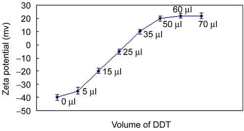
To check whether the reductive agent could change the zeta potential of Folate-chitosan−Sln from negative to positive value, the preformed Folate-chitosan−Sln was diluted into distilled water to form a final concentration of 1 mmol/ml of total lipid. One milliliter of the dilution was placed in eight tubes, respectively. While vortexing the test tubes, 0, 5, 15, 25, 35, 50, 60, and 70 μl of a disulfide reducing agent dithiothreitol (DTT, 0.5 mmol/ml in PBS pH 7.4) was added. The mixtures were then incubated for 30 min at room temperature and measured to get the zeta potential (). When adding 0 μl of DTT, the zeta potential is −40 mV, but adding 50 μl of DTT, it is +20 mV. Altogether when adding 60 μl and 70 μl of DTT, there is not too much difference with adding 50 μl of DTT, which indicates that the almost all of the disulfide bonds might have been cleaved by DTT.
Cell viability and transfection activity
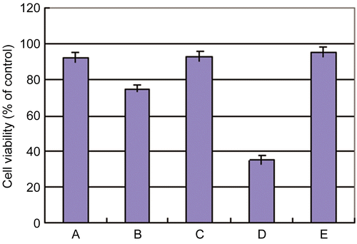
When HEK 293 cells are incubated with 15 μg of naked DNA, cell viability remains roughly the same (95%) as it is seen in control non-transfected cells (100%). There is no significant decrease in viability when the cells are incubated with 15 μg of Folate-chitosan−CHETA−Sln (92%) and CHETA−Sln (93%). On the other hand, 15 μg of DNA-Lipofectamine™ 2000 lipoplex drastically decreases the cellular viability to 30% ().
Figure 4. Viability of HEK293 cells determined in the MTT assay under different experimental conditions (15 mg/well): (A) Folate-chitosan-CHETA−Sln; (B) Folate-chitosan-Sln; (C) Cheta−Sln; (D) Lipofectamine™ 200; (E) Naked DNA. Results are presented as mean ± standard deviation (S.D.) (n = 5).
The results of eGFP and LacZ gene expression with or without the presence of serum are summarized in , respectively.
Figure 5. The flourescence images of eGFP gene expression in SKOV3 cells (folate receptor over-expressing, R+) and A549 cells (folate receptor deficient, R-) transfected by three different kinds of nanoparticles at the absence of serum. It was observed under the inverse fluorescence microscope (Olympas, japan).
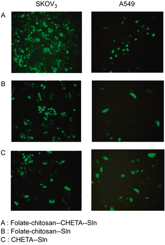
Figure 6. Transfectivities of Folate-chitosan-CHETA-Sln, Folate-chitosan−Sln, CHETA−Sln and naked DNA in SKOV3 cells (R+) and A549 cells (R-) at the absence of serum. The data represent the mean ± S.D of three wells and was representative of three independent experiments.
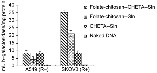
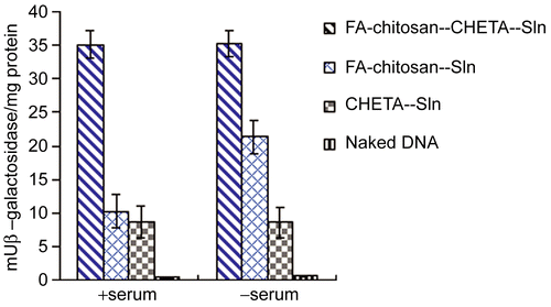
Figure 7. Transfectivities of Folate-chotosan-CHETA−Sln, Folate-chotosan−Sln, CHETA−Sln and naked DNA in SKOV3 cells (R+) at the presence or absence of serum (10%, v/v). The data represent the mean ± SD. of three wells and was representative of three independent experiments.
shows the green fluorescence protein expression in A549 cells (folate receptor deficient, R−) and SKOV3 cells (folate receptor over-expressing, R+) transfected with three different kinds of preformed nanoparticles without the presence of serum. This figure demonstrates that: (1) The transfectivity of both Folate-chitosan−CHETA−Sln and Folate-chitosan−Sln in SKOV3 cells is much stronger than that in A549 cells, which may be due to the folate receptor mediated intracellular gene delivery; (2) In SKOV3 cells (R+), the transfectivity of Folate-chitosan−Sln is lower than that of Folate-chitosan−CHETA−Sln, but much higher than that of CHETA−Sln, it indicates that the folate receptor targeting enhanced the transfectivity, meanwhile CHETA also contributed to it; and (3) In A549 cells (R−), the transfectivity of both Folate-chitosan−CHETA−Sln and CHETA−Sln is much higher than that of Folate-chitosan−Sln, this fact indicates that the cell infusion might have been enhanced due to the presence of CHETA, which resulted in increasing of the transfectivity.
To further prove the results of , LacZ was taken as another reporter gene to evaluate the transfectivity that was quantified as milliunits of β-galactosidase per milligram of total cell protein (mU β-galactosidase/mg protein). demonstrates the coincident results with .
The inhibitory effect of serum on transfectivity mediated by lipoplexes has been reported for different cell types and represents a serious limitation for their use in vivo. One important element in emulating in vivo conditions is the use of high concentrations of serum for transfection experiments in vitro, rather than serum-free medium, which optimizes transfection. So far, most of the studies on the effect of serum on transfection reported have been carried out at the presence of serum (CitationEscriou et al., 1998). indicates that the serum (10%, v/v) in culture did not affect the transfection efficiency of both Folate-chitosan−CHETA−Sln and CHETA−Sln, but severely inhibited that of Folate-chitosan−Sln, which also displays the contribution of CHETA.
Conclusions
The present investigation focuses on the preparation and characterization of the charge changing solid lipid nanoparticles mainly made of folate-chitosan and CHETA by the reverse micelle-double emulsion method. The resulted nanoparticle, Folate-chitosan−CHETA−Sln, has low cytotoxicity to HEK 293 cells and shows good transfectivity at the presence of serum. The results indicate that this strategy reported here is feasible and might be attractive to the gene delivery. Therefore, further efforts should be made to investigate the ability of Folate-chitosan−CHETA−Sln targeting therapeutic gene to animal tumors in vivo.
Acknowledgements
We are thankful for the help of Professor Shixiang Hou (Sichuan University) and his student Dr Yu Zheng on the synthesis of folate-chitosan and we also appreciate the financial support of the College Youth Science Foundation of Luzhou Medical College (No.685).
Declaration of interest: The authors report no conflicts of interest. The authors alone are responsible for the content and writing of the paper.
References
- Antony, A.C. (1996). Folate receptors. Annu Rev Nutr, 16, 501–21.
- Benns, J.M., Maheshwari, A., Furgeson, D.Y., Mahato, R.I., Kim, S.W. (2001). Folate-PEG-Folate-graft-polyethylenimine-based gene delivery. J Drug Target, 9, 123–39.
- Chan, P., Kurisawa, M., Chung, J.E., Yang, Y.Y. (2007). Synthesis and characterization of chitosan-g-poly(ethylene glycol)-folate as a non-viral carrier for tumor-targeted gene delivery. Biomaterials, 28, 540–9.
- Cheung, S.T., Tsui, T.Y., Wang, W.L., Yang, Z.F., Wong, S.Y., Ip, Y.C., Luk, J., Fan, S.T. (2002). Liver as an ideal target for gene therapy: expression of CTLA4Ig by retroviral gene transfer. J Gastroenterol Hepatol, 17, 1008–14.
- Cichon, G., Schmidt, H.H., Benhidjeb, T., Löser, P., Ziemer, S., Haas, R., Grewe, N., Schnieders, F., Heeren, J., Manns, M.P., Schlag, P.M., Strauss, M. (1999). Intravenous administration of recombinant adenoviruses causes thrombocytopenia, anemia and erythroblastosis in rabbits. J Gene Med, 1, 360–71.
- Corsi, K., Chellat, F., Yahia, L., Fernandes, J.C. (2003). Mesenchymal stem cells, MG63 and HEK293 transfection using chitosan-DNA nanoparticles. Biomaterials, 24, 1255–64.
- Dubé,; D., Francis, M., Leroux, J.C., Winnik, F.M. (2002). Preparation and tumor cell uptake of poly(N-isopropylacrylamide) folate conjugates. Bioconjug Chem, 13, 685–92.
- Dubes, A., Parrot-Lopez, H., Abdelwahed, W., Degobert, G., Fessi, H., Shahgaldian, P., Coleman, A.W. (2003). Scanning electron microscopy and atomic force microscopy imaging of solid lipid nanoparticles derived from amphiphilic cyclodextrins. Eur J Pharm Biopharm, 55, 279–82.
- Escriou V, Ciolina C, Lacroix F, Byk G, Scherman D, Wils P. (1998). Cationic lipid-mediated gene transfer: effect of serum on cellular uptake and intracellular fate of lipopolyamine/DNA complexes. Biochim Biophys Acta,1368, 276–88.
- Hejazi, R., Amiji, M. (2003). Chitosan-based gastrointestinal delivery systems. J Contr Rel, 89, 151–65.
- Jeong, J.H., Kim, S.W., Park, T.G. (2003). A new antisense oligonucleotide delivery system based on self-assembled ODN-PEG hybrid conjugate micelles. J Contr Rel, 93, 183–91.
- Jooss, K., Chirmule, N. (2003). Immunity to adenovirus and adeno-associated viral vectors: implications for gene therapy. Gene Ther, 10, 955–63.
- Ke, C.Y., Mathias, C.J., Green, M.A. (2003). The folate receptor as a molecular target for tumor-selective radionuclide delivery. Nucl Med Biol, 30, 811–7.
- Komatsu, H., Kitajima, A., Okada, S. (1995). Pharmaceutical characterization of commercially available intravenous fat emulsions: estimation of average particle size, size distribution and surface potential using photon correlation spectroscopy. Chem Pharm Bull, 43, 1412–5.
- Leamon, C.P. (2002). Fusogenic lipids and vesicles for delivery of pharmaceutical agents. US Patent 6,379,698.
- Lincoln, D.T., Ali, Emadi EM, Tonissen, K.F., Clarke, F.M. (2003). The thioredoxin-thioredoxin reductase system: over expression in human cancer. Anticancer Res, 23, 2425–33.
- Liu, W.G., Yao, K.D. (2002). Chitosan and its derivatives–a promising non-viral vector for gene transfection. J Contr Rel, 83, 1–11.
- Lu, Y., Low, P.S. (2003). Immunotherapy of folate receptor-expressing tumors: review of recent advances and future prospects. J Contr Rel, 91, 17–29.
- MacLaughlin, F.C., Mumper, R.J., Wang, J., Tagliaferri, J.M., Gill, I., Hinchcliffe, M., Rolland, A.P. (1998). Chitosan and depolymerized chitosan oligomers as condensing carriers for in vivo plasmid delivery. J Contr Rel, 56, 259–72.
- Mansouri, S., Cuie, Y., Winnik, F., Shi, Q., Lavigne, P., Benderdour, M., Beaumont, E., Fernandes, J.C. (2006). Characterization of folate-chitosan-DNA nanoparticles for gene therapy. Biomaterials, 27, 2060–5.
- Mao, S., Shuai, X., Unger, F., Simon, M., Bi, D., Kissel, T. (2004). The depolymerization of chitosan: effects on physicochemical and biological properties. Int J Pharm, 281, 45–54.
- Mehnert, W., Mäder, K. (2001). Solid lipid nanoparticles: production, characterization and applications. Adv Drug Deliv Rev, 47, 165–96.
- Müller, R.H., Jacobs, C., Kayser, O. (2001). Nanosuspensions as particulate drug formulations in therapy: rationale for development and what we can expect for the future. Adv Drug Deliv Rev, 47, 3–19.
- Müller, R.H., Mäder, K., Gohla, S. (2000). Solid lipid nanoparticles (SLN) for controlled drug delivery: a review of the state of the art. Eur J Pharm Biopharm, 50, 161–77.
- Nathwani, A.C., Davidoff, A.M., Hanawa, H., Hu, Y., Hoffer, F.A., Nikanorov, A., Slaughter, C., Ng, C.Y., Zhou, J., Lozier, J.N., Mandrell, T.D., Vanin, E.F., Nienhuis, A.W. (2002). Sustained high-level expression of human factor IX (hFIX) after liver-targeted delivery of recombinant adeno-associated virus encoding the hFIX gene in rhesus macaques. Blood, 100, 1662–9.
- Pack, D.W., Hoffman, A.S., Pun, S., Stayton, P.S. (2005). Design and development of polymers for gene delivery. Nat Rev Drug Discov, 4, 581–93.
- Qian, Z.M., Li, H., Sun, H., Ho, K. (2002). Targeted drug delivery via the transferrin receptor-mediated endocytosis pathway. Pharmacol Rev, 54, 561–87.
- Sapra, P., Allen, T.M. (2003). Ligand-targeted liposomal anticancer drugs. Prog Lipid Res, 42, 439–62.
- Turunen N, Karihtala P, Mantyniemi A, Sormunen R, Holmgren A, Kinnula VL, Soini Y. (2004). Thioredoxin is associated with proliferation, p53 expression and negative estrogen and progesterone receptor status in breast carcinoma. APMIS, 112, 123–32.
- Wang, S., Luo, J., Lantrip, D.A., Waters, D.J., Mathias, C.J., Green, M.A., Fuchs, P.L., Low, P.S. 1997. Design and synthesis of [111In] DTPA-folate for use as tumor targeted radiopharmaceutical. Bioconjug Chem, 8, 673–9.
- Zhong, Z.R., Liu, J., Deng, Y., Zhang, Z.R., Song, Q.G., Wei, Y.X., He, Q. (2007). Preparation and characterization of a novel nonviral gene transfer system: procationic-liposome-protamine-DNA complexes. Drug Deliv, 14, 177–83.
- Zhou, W., Yuan, X., Wilson, A., Yang, L., Mokotoff, M., Pitt, B., Li, S. (2002). Efficient intracellular delivery of oligonucleotides formulated in folate receptor-targeted lipid vesicles. Bioconjug Chem, 13, 1220–5.

